The midgut of Manduca sexta consists of a highly folded epithelium resting on a muscle and trachea framework (Cioffi, 1979). The cells of the epithelium are arranged in a characteristic pattern in which a single goblet cell is surrounded by 3–6 columnar cells. Each goblet cell is pear-shaped with a basally located nucleus, a central cavity (the goblet cavity) and a smooth apical surface that joins the goblet cavity through a valve. Columnar cells appear as cylinders in vivo, with a central nucleus, basal infoldings of the cell membrane and apical microvilli (Anderson and Harvey, 1966). Small spherical stem cells are located between the bases of the columnar and goblet cells (Hakim et al. 1988; Baldwin and Hakim, 1991). Stem cells greatly increase in number at the beginning of a molt and then differentiate and intercalate between existing mature epithelial cells in such a way as to perpetuate the typical larval midgut cell pattern (Baldwin and Hakim, 1991). In this way, the larval gut is enlarged at the molt to accommodate growth in the next instar.
Cultures of insect midgut epithelial cells can provide isolated cells for the physiological study of specific cell types, thus enabling the postulated role of goblet cells in V-ATPase-mediated primary H+transport and of columnar cells in amino
acid/K+ symport (Klein, 1992) to be verified, as well as providing isolated cells in which insect pathogen penetration can be studied (Federici, 1993a,b).
We have previously shown (Sadrud-Din et al. 1994) that, when pieces of midgut from pharate fourth-stage larvae were cultured in vitro for approximately 1 week, stem, goblet and columnar epithelial cells tended to fall away from the muscle and tracheal framework to lie in a random distribution on the bottom of the culture vessel. In mixed culture, in the presence of the insect molting hormone 20-hydroxyecdysone (20-HE), and of growth factors generated by co-cultured fat body (Loeb, 1994), stem cells in the culture entered mitosis and differentiated into new columnar and goblet cells.
In this work, we describe the isolation and culture of stem cells from the midgut of Manduca sexta. Midgut stem cell multiplication occurred in the presence of 20-HE and soluble fat body factors, but differentiation required factors present in conditioned medium from actively developing mixed midgut cultures. Thus, we were able to examine the culture conditions that enable midgut stem cells to replicate and to differentiate to mature epithelial forms, and we confirm that stem cells cultured in vitro give rise to cells that appear morphologically similar to mature goblet and columnar cells.
JEB9931
Isolated spherical stem cells from midguts of pharate fourth-instar larvae of Manduca sexta proliferated in vitro in the presence of 1 ng ml2120-hydroxyecdysone and
co-cultured fat body tissue or cell-free fat body extract from M. sexta, Lymantria dispar or Heliothis virescens. In this environment, the stem cells were able to undergo mitosis and increase in number. However, stem cells were only able to differentiate to mature goblet and columnar cells when cell-free conditioned medium, taken from midgut cell cultures containing mature cells as well as stem cells and differentiating forms, was introduced into the culture
medium. The presence of early and mature goblet cells, lying randomly on their sides, suggested that cell polarity developed in vitro as an intrinsic property of individual cells rather than with reference to an external inductive material. The differentiation factor (or factors) from the conditioned medium appears to include a heat-stable, peptide-like molecule of 10 kDa or less.
Key words: insect, Manduca sexta, growth factors, proliferative cells, gut.
Summary
IN VITRO DIFFERENTIATION OF ISOLATED STEM CELLS FROM THE MIDGUT
OF MANDUCA SEXTA LARVAE
SAKEENA SADRUD-DIN1,2, MARCIA J. LOEB1,* ANDRAZIEL S. HAKIM2
1Insect Neurobiology and Hormone Laboratory, US Department of Agriculture, Beltsville, MD 20705, USA and 2Department of Anatomy, Howard University School of Medicine, Washington DC 20059, USA
Accepted 21 September 1995
*Author for correspondence.
Materials and methods
Animals
Pharate fourth-instar Manduca sexta larvae were obtained from the colony maintained at the Insect Neurobiology and Hormone Laboratory, USDA, Beltsville, MD. Larvae were raised on an artificial diet and maintained at 28 ˚C under constant light.
Stem cell isolation
Prior to dissection, animals were surface-sterilized by consecutive immersion for approximately 2 min each in baths of 20 % detergent (Septisol, Vestal Laboratories, St Louis, MO), 0.1 % p-hydroxybenzoic acid methyl ester (Sigma, St Louis, MO) and 0.1 % sodium hypochlorite. Larvae were then transferred to sterile water and dissected immediately in a continuous-flow hood (Aerosafe, Bengton Associates, King of Prussia, PA). A lengthwise midline incision was made through the dorsal integument, exposing the midgut. Malpighian tubules were removed and, when present, the intact peritrophic membrane, containing gut contents, was also removed through a lengthwise incision in the midgut. During the molt, the peritrophic membrane is not visible. The midgut itself was then excised, rinsed twice in sterile Ringer (Loeb, 1991) containing 0.5 % gentamycin (Sigma) and 0.01 % antibiotic–antimycotic (Sigma) (modified Ringer). To obtain isolated stem cells, midgut tissue from three or four animals was cut into pieces of approximately 1 mm2 and placed into 35 mm wells of a six-well plate (Falcon) containing 1.5 ml of modified Grace’s medium (Gibco, Grand Island, NY) (Sadrud-Din et al. 1994) containing 1 ngml21 20-hydroxyecdysone (20-HE) (Calbiochem, La Jolla, CA) (culture medium). By gentle manual swirling of the plate every 15 min over a 45–60 min period, loosely associated stem cells were liberated into the medium. The gentler the swirling, the fewer non-stem cells were released. Non-stem cells comprised less than 0.3 % of the free cells observed after this process. Intact tissue fragments were then removed from each well using forceps. Free cells were centrifuged at 400 g for 5 min and, after removing the supernatants, cells were resuspended in approximately 100 ml of culture medium and distributed evenly into the several wells used in each experiment. The final volume of fluid in each six-well plate (Falcon) was 3 ml, although 300ml sufficed for each well of a 24-well plate (Falcon). Cultures were incubated at 25 ˚C in an air environment.
Cell counts
When cultures were prepared in 24-well plates, each well was scanned and the total number of differentiated cells of each type was recorded. The midgut cells tended to occupy the space just above the bottom of the dish and, although not attached to the dish or to each other, tended not to overlap each other. When the dish was moved slowly and systematically up and down the field of view with the aid of a joystick-driven movable stage (Nikon), the cell types in each well could be reliably counted and recorded. Since cells were initially distributed as equally as possible to each well, the total number
of cells (undifferentiated as well as differentiated) was counted in only one or two wells of the experimental and control sets at various times during the experiment and these counts were used to estimate the total number of cells in each of the wells. When cultures were prepared in six-well plates, cells were counted in four randomly chosen fields each measuring 0.5 mm2. The random fields included areas from the center and periphery of each well, since the cells tended to be unevenly distributed, with more cells in the center than at the periphery. The mean number of cells per field was calculated and used as the data point for each well at each observation. Every experiment was repeated at least three times. Thus, each data point in each figure represents the mean and S.E.M. of at least three independent experiments.
Source of growth factors Fat body and fat body extract
For several of the experiments, fat body was co-cultured with stem cells (Sadrud-Din et al. 1994). Fat body tissue was collected from 1- to 2-day-old developing pupae of both sexes of Lymantria dispar. A piece of doubly rinsed fat body, approximately 1 mm3, was either added directly to each culture well or isolated in a porous chamber in each well to prevent cell contact and yet allow passage of diffusible products (Millipore Corp., Marlborough, MA). Results were similar in both these co-culture situations. As an alternative to co-culture with living pupal fat body tissue, a cell-free extract was prepared from fat body tissue obtained from pupae of Heliothis virescens that had just been induced to break diapause (HFBX) (Loeb and Hayes, 1980). Green fat body from 40 pupae was incubated for 24 h in 2 ml of culture medium at 25 ˚C and then sonicated. Centrifugation for 5 min at 1600 g produced a layer of clear, cell-free fluid between the cell fragment pellet and a fatty overlayer; the clear fluid was removed and sterilized by passage through a 0.22mm filter (Gelman Scientific, Ann Arbor, MI) (Loeb, 1994). The sterile HFBX was frozen in 500ml samples and kept at 220 ˚C until used. Effective dosage was 10ml ml21of medium. MFBX was prepared from fat body dissected from freshly molted (partly green) pupae of Manduca sexta; abdominal fat body from 2.5 pupae was sonicated in 3 ml of modified Ringer and centrifuged, sterilized and stored as for HFBX. The optimal dosage was the same as that for HFBX, 10ml ml21of culture medium.
Conditioned medium
approximately 1000 cells at the start of each experiment. Alternatively, cells were equally distributed to each well of a six-well plate. Conditioned medium (or treated conditioned medium) was mixed with fresh medium (1:1 by volume). Each well of a 24-well plate contained 0.3 ml of fluid, while each well of a six-well plate held 3 ml. An equal number of control stem cell preparations was prepared in medium alone. To test for the effects of conditioned medium, stem cells were cultured in wells of 24- or six-well plates using similar concentrations of treated or untreated conditioned medium.
Properties of conditioned medium
Heat sensitivity. Cell-free conditioned medium placed in polypropylene tubes was heated for 10 min in a boiling water bath. Supernatants were removed after centrifugation for 3 min at 1600 g.
Enzyme treatments. Insoluble amylase from Bacillus sp., insoluble lipase from wheat germ, both attached to beaded agarose, and insoluble protease from bovine pancreas attached to carboxymethylcellulose (all from Sigma Chemical Co., St Louis, MO) were hydrated in modified Ringer according to the supplier’s instructions (Sigma). Enzyme slurries were distributed to sterile Eppendorf tubes to yield a final concentration of 0.5 units of enzyme activity (as specified by the manufacturer) when 1 ml of cell-free conditioned medium was added. Each tube was vortexed and incubated horizontally on a rotating table at 100 revs min21 for 3 h at room temperature. Reactions were stopped by centrifuging the tubes for 1 min at 1600 g to pellet the enzymes. Control samples of media were incubated alone or with hydrated, but enzyme-free, carboxymethylcellulose (Sigma). A 1 ml sample of supernatant was diluted with 2 ml of medium and applied to stem cells to assay for activity. After use, insoluble enzymes were washed three times with modified Ringer and stored at 4 ˚C for re-use according to the supplier’s instructions.
Molecular mass estimation. A 1 ml sample of cell-free conditioned medium was placed in the upper chamber of a Centricon-10 microconcentrator (Amicon, Danvers, MA) and centrifuged at room temperature for 20 min at 400 g. According to the manufacturer’s literature (Grace Co., publication no. 1-259D), the protein remaining in the upper chamber after centrifugation should have an Mr equal to or greater than 103103, while the molecules entering the lower chamber (filtrate) should have an Mrequal to or less than 103103. The resulting fluid in each of the chambers was diluted 1:1 by volume with fresh medium and mixed with stem cells as above. Ten such preparations were bioassayed.
Results
Multiplication of stem cells
M. sexta stem cells were not observed to multiply in medium alone. However, the number of stem cells quadrupled in 14 days, from approximately 20 to 80 cells per field, when co-cultured with fat body from developing L. dispar pupae
(Fig. 1). Although stem cells proliferated, little or no differentiation was detected. Similarly, proliferation without differentiation was observed when HFBX or MFBX was added to cultures in the absence of conditioned medium (results not shown).
Differentiation of stem cells
The cell-free supernatant of conditioned medium taken from mixed cultures was added to stem cells cultured in medium with a source of fat body factors. Stem cells cultured with this conditioned medium differentiated in 2–4 weeks (Fig. 2). Although the data shown in Fig. 2 were derived from cells grown with conditioned medium and HFBX, similar data were obtained using conditioned medium and MFBX or M. sexta fat body (Fig. 3) or L. dispar fat body (see also Figs 5, 6) as the source of fat body factors. Initially, only about 0.3 % of the cells in these cultures were non-stem cells, but, as each experiment progressed, 4–10 % of the cells were observed in various stages of differentiation. Control cultures with no added conditioned medium retained approximately the same number of non-stem cells as they did at the start of the experiment (Figs 2, 3). At least five kinds of morphologically different cells appeared in developing stem cell cultures, as previously discussed in mixed cell cultures (Sadrud-Din et al. 1994). Initially, only spherical stem cells of various sizes were observed. A day or two later, clear flattened cells with the edges thicker than the center were seen (spread out cells) (Fig. 4A, arrows). Within another day, spherical cells with
80
60
40
20
0 100
Number of cells per field
5 10 15
0
[image:3.609.338.548.71.275.2]Time (days)
tubular extensions of varying lengths were observed. There was a cavity in the extension (Fig. 4E). Clear pear-shaped cells, each with a large cavity which penetrated to the base of the cell, were also observed (Fig. 4D). These appeared to be mature goblet cells (Hakim et al. 1988). Granular spherical cells approximately the same size as stem cells but covered with microvilli were also seen (Fig. 4C). Within another day, granular cells of small-to-average size with extended microvilli at one end, resembling mature columnar cells (Hakim et al. 1988), were also seen in the culture (Fig. 4B). Previously, viability studies using Trypan Blue (Sadrud-Din et al. 1994) showed that 90 % of the cells in mixed cell cultures, including stem, spread out, differentiating and differentiated goblet and columnar cells, were healthy.
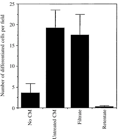
Properties of conditioned medium
Conditioned medium held in boiling water for 10 min retained its original activity (Fig. 5). There was some loss of activity (see Fig. 5) when conditioned medium was treated with amylase or lipase. However, stem cells cultured in the presence of conditioned medium treated with protease neither proliferated nor differentiated (Fig. 5). Control conditioned medium incubated with the insoluble protease carrier carboxymethylcellulose (CMC) was as effective in inducing differentiation as untreated conditioned medium (Fig. 5).
Centricon 10 filtration allowed all of the detectable differentiating activity of the conditioned medium to pass into the filtrate; none of the activity was detected in the retentate (Fig. 6).
Discussion
We have developed a unique method for collecting a highly enriched population of larval midgut stem cells from Manduca sexta. We simply shake them from newly dissected midgut fragments taken from molting larvae. Our approach was based on the ultrastructural observations made by Baldwin and Hakim (1991) that stem cells are not attached to other cells by septate junctions and that they rapidly proliferate to become the major cell type in the midgut early in the molt. In contrast, mature goblet and columnar cells remain anchored to one another by septate junctions at this time (Baldwin and Hakim, 1991) and, therefore, do not readily detach from the mass of midgut tissue during the short time used to isolate stem cells. Uptake of tritiated thymidine and deoxybromouridine by stem cells (Loeb and Hakim, 1995) indicates that these are the cells that proliferate in mixed cultures. Cultures enriched to approximately 99.7 % in stem cells gave rise to numerous maturing and mature goblet and columnar cells. After 3 weeks in culture, up to 20 % of the cells were observed in various stages of differentiation. Spread out cells (see arrows, Fig. 4A) were the earliest indication of differentiation of either cell type. The presence of spherical cells with a neck region preceded the appearance of a mature goblet cell
20
10
0 30
Number of cells per field
10 15 20 25 5
[image:4.609.331.539.67.328.2]Time (days) Columnar cells Goblet cells
Fig. 2. Differentiation of isolated midgut stem cells from Manduca
sexta cultured in the presence of a 1:1 mixture of fresh
medium:cell-free conditioned medium and HFBX (10ml ml21) (dotted lines) compared with fresh medium and HFBX (10ml ml21) (solid lines). Although many stem cells remained undifferentiated in these cultures, this figure only shows the numbers of cells that have differentiated to recognizable mature forms. Cells were cultured in 15 mm wells of 24-well plates. Error bars represent mean ±S.E.M. of seven experiments.
60
40
20
0 80
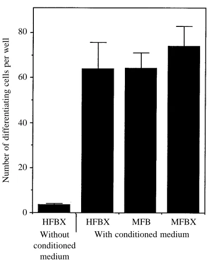
Number of differentiating cells per well
HFBX HFBX MFB MFBX With conditioned medium Without
[image:4.609.64.276.73.274.2]conditioned medium
Fig. 3. Differentiation of M. sexta midgut stem cells in a 1:1 mixture of fresh medium:conditioned medium supplemented with various sources of fat body factors. Newly isolated stem cells were introduced at the beginning of the experiment. HFBX without conditioned medium represents differentiation in the presence of HFBX without conditioned medium. HFBX and MFBX are cell-free extracts of H.
virescens and M. sexta fat body, respectively, while MFB represents
morphology. We also believe that the appearance of spherical cells with uniformly distributed microvilli preceded the appearance of mature columnar cells. The sequential appearance of these forms in the cultures clearly indicated that the stem cells were differentiating. Furthermore, the presence of early and mature goblet cells, lying randomly on
their sides, suggested that cell polarity developed in vitro as an intrinsic property of individual cells and not with reference to an external inductive material such as a morphologically defined basal lamina or another attached cell. Immature goblet cells from spruce budworm, maintained in culture, differentiate in a similar manner (D. Baines, personal communication). This result contrasts with the situation in vivo, where the goblet end of each goblet cell and the microvillar extensions of columnar cells are always oriented towards the midgut lumen. The nature of the orientation-inducing message in this epithelium is unknown. Stem cells are present at the bases of mature cells in vivo and then grow between them. This initial dorsolateral contact with mature cells might polarize the development of differentiating stem cells as part of the epithelial tissue in vivo. Alternatively,
A
B
C
D
[image:5.609.51.568.72.333.2]E
Fig. 4. Midgut cells in culture. (A) Isolated stem cells in culture. Arrows point to spread out cells; (B) two fully differentiated granular columnar cells in culture. Note microvilli at one end; (C) granule-containing spherical cell covered with microvilli; (D) fully differentiated goblet cell in culture; (E) developing goblet cells. The goblet vacuole is much smaller than those in the mature cells in D, the bases of cells are rounded rather than ‘knobbed’ and the necks of the cells appear to be curved. Note the small aggregate of developing cells of undetermined type above the scale bar. Scale bars, 5mm.
20
15
10
5
0 25
Number of differentiated cells per field
Conditioned medium
No CM
CMC
Protease Lipase Amylase Boiling
[image:5.609.73.276.507.738.2]feedback through cell adhesion molecules (Nathke et al. 1994) binding each cell to the basal lamina or to mature cells, or factors diffusing in from the hemolymph, may direct polarity in vivo. In culture, however, no organized basal lamina is present. In fact, laminin added to cultured midgut cells tended to inhibit proliferation and differentiation (Sadrud-Din et al. 1994). Another possibility is that the site at which a stem cell was last attached to its counterpart during division might serve as an alternative polarity initiation site (Nelson, 1992). There is as yet, however, no evidence for polarized mitosis in vivo during the molting process (Baldwin and Hakim, 1991).
There must be an internal stabilizing architecture in recognizable goblet and columnar cells. The structure of goblet cells appeared to be essentially the same as that seen in vivo. Perhaps the polar organization of the cells in our cultures is intrinsically determined and stabilized by a rigid cytoskeleton. Unlike the uniform columnar cells seen in vivo (Hakim et al. 1988), cells of the columnar type observed in our cultures had a variety of shapes and widths, and the microvilli-covered areas were of various dimensions (Fig. 4). The presence of intermediate conditions in the columnar cell where the crown of the cell, covered by microvilli, can extend variable distances
around the cell is consistent with the idea that, in the absence of contact with neighboring cells, the apical domains are extensible. We speculate that the cell attachments that bind adjacent cells in vivo limit the potential breadth at the apex of these cells.
We have shown that proliferation of isolated midgut stem cells can occur in medium supplemented with 20-HE and either fat body tissue from developing pupae or a cell-free extract of fat body tissue prepared from developing H. virescens, M. sexta or L. dispar pupae. Although fat body is a tissue with numerous metabolic functions, such as fat, nitrate, protein and carbohydrate metabolism, waste accumulation and detoxification (Chapman, 1971), it also produces growth factors or cytokines. Fat body, or medium exposed to fat body cells, has induced growth and development of Galleria mellonella wing imaginal discs (Oberlander and Tomblin, 1972; Benson et al. 1974), induced meiosis in H. virescens spermatocysts (Giebultowicz et al. 1987), promoted mitosis and development of genital tract tissues from H. virescens (Loeb, 1991; Loeb and Hakim, 1991; Loeb and Lynn, 1993) and induced germ band formation in eggs of a braconid wasp, Microplitis croceipes, parasitic on Heliothis spp. (Ferkovich et al. 1991). Fat body extracted from H. virescens contains at least nine growth factors with different molecular masses that cause mitosis and differentiation of H. virescens genital tract (Loeb, 1994). Since fat body from M. sexta, L. dispar and H. virescens all had a similar effect on M. sexta midgut cell proliferation, the growth factor(s) responsible may be common to all these insects. It is possible that at least some similar fat body growth factors are secreted by the three lepidopteran species used in this work. This result is not unexpected. In vertebrates, factors chemically recognizable as, for instance, epidermal growth factor or platelet-derived growth factor are highly conserved and have been isolated from tissues in species ranging from mouse to man (Deuel, 1987).
Fat body or its derivatives, HFBX and MFBX, only induced proliferation of stem cells. Differentiation required the addition of medium conditioned by mixed cultures containing stem cells, differentiating cells and mature goblet and columnar cells. Therefore, another factor, or set of factors, produced by differentiating and/or mature forms of M. sexta midgut cell, is required in synergy with fat body factors to induce differentiation of midgut stem cells in vitro. This dependency shows that the mature larval midgut epithelium regulates differentiation of the stem cells. Such dual regulatory mechanisms are typical of stem cell systems in which cells replicate but also commit some of their daughter cells to differentiate to other forms. Hematopoietic stem cells require erythropoietin to multiply, but specific colony-stimulating growth factors are needed to regulate the types of cells which differentiate from stem cells (Burgess and Nicola, 1983). Negative control is exerted by growth factors such as transforming growth factor b (TGFb) from stromal cells or mature hemopoietic cells (Haig, 1992). In vertebrate gut epithelium, ordinary stem cell division is regulated by
20
15
10
5
0 25
Number of differentiated cells per field
No CM
Untreated CM
Filtrate
[image:6.609.65.272.70.313.2]Retentate
circulating epidermal growth-factor-like or gastrin-like molecules. However, when lesions develop, local soluble factors direct both stem cell division and differentiation (Burgess and Nicola, 1983; Potten and Chadwick, 1994).
The differentiation factor(s) in midgut conditioned medium was not destroyed by heating to 100 ˚C for 5 min. Since the carrier of the insoluble protease used, carboxymethylcellulose (CMC), had no effect on the activity of conditioned medium, the destructive effect of the insoluble protease preparation was undoubtedly due to protease and not to CMC. Amylase and lipase had intermediate deleterious effects on conditioned medium effectiveness. Data from Amicon microconcentrators indicated that the Mrof Manduca sexta midgut differentiation factor (or factors) was 103103or less, thus implicating a small peptide (or peptides). Vertebrate differentiation factors, such as the colony-stimulating factors necessary for vertebrate hematocyte differentiation, are peptides of 23–28 kDa (Burgess and Nicola, 1983), while TGFb has a molecular mass of approximately 25 kDa (Deuel, 1987). Thus, the factor or factors responsible for insect midgut differentiation appears to be smaller than the peptides commonly associated with this function in vertebrates.
We have demonstrated that isolated larval midgut stem cells proliferate in culture in the presence of molting hormone (20-HE) and growth factors from fat body. However, in order to differentiate, stem cells must also receive soluble chemical messages from mature or maturing cells in the midgut epithelium. Proliferation and differentiation in vitro did not proceed at the same rapid rate reported in vivo (Baldwin and Hakim, 1991). Therefore, there are more controlling factors yet to be discovered in this system. However, it is evident that the development of Manduca sexta larval midgut is, in part, self-regulated.
Thanks to Drs Carol Sheppard and Kate Baldwin for helpful comments on the manuscript and to Kimberly Jones for excellent technical assistance. Mention of commercial products in this manuscript does not imply endorsement by the US Department of Agriculture.
References
ANDERSON, E. ANDHARVEY, W. R. (1966). Active transport by the
Cecropia midgut. II. Fine structure of the midgut epithelium. J. Cell Biol. 31, 107–134.
BALDWIN, K. M. ANDHAKIM, R. S. (1991). Growth and differentiation of the larval midgut epithelium during molting in the moth,
Manduca sexta. Tissue & Cell 23, 411–422.
BENSON, J., OBERLANDER, H., KOREEDA, M. AND NAKANISHI, K. (1974). Isolation of a fat body factor which stimulates evagination of Galleria mellonella wing discs in vitro. Wilhelm Roux Arch.
EntwMech. Org. 175, 327–338.
BURGESS, A. ANDNICOLA, N. (1983). Growth Factors and Stem Cells. New York: Academic Press. pp. 1–22, 93–124.
CHAPMAN, R. F. (1971). The Insects: Structure and Function. New York: Elsevier. 819pp.
CIOFFI, M. (1979). The morphology and fine structure of larval midgut
of a moth (Manduca sexta) in relation to active ion transport. Tissue
& Cell 11, 467–479.
DEUEL, T. F. (1987). Polypeptide growth factors: roles in normal and abnormal cell growth. A. Rev. Cell Biol. 3, 443–492.
FEDERICI, B. A. (1993a). Insecticidal bacterial proteins identify the midgut epithelium as a source of novel target sites for insect control. Archs Insect Biochem. Physiol. 22, 367–371.
FEDERICI, B. A. (1993b). Viral pathobiology in relation to insect control. In Parasites and Pathogens of Insects, vol. 2 (ed. N. E. Beckage, S. N. Thompson and B. A. Federici), pp. 81–101. New York: Academic Press.
FERKOVICH, S. M., DILLARD, C. AND OBERLANDER, H. (1991). Stimulation of embryonic development in Microplitis croceipes (Braconidae) in cell culture media preconditioned with a fat body cell line derived from a non-permissive host, Gypsy moth,
Lymantria dispar. Archs Insect Biochem. Physiol. 18, 169–175.
GIEBULTOWICZ, J. M., LOEB, M. J. ANDBORKOVEC, A. B. (1987). In
vitro spermatogenesis in lepidopteran larvae: role of the testis
sheath. Invert. Reprod. Dev. 11, 211–226.
HAIG, D. M. (1992). Haematopoietic stem cells and the development of the blood cell repertoire. J. comp. Path. 106, 121–136. HAKIM, R. S., BALDWIN, K. M. AND BAYER, P. (1988). Cell
differentiation in the embryonic midgut of the tobacco budworm moth, Manduca sexta. Tissue & Cell 20, 51–62.
KLEIN, U. (1992). The insect V-ATPase, a plasma membrane proton pump energizing secondary active transport: immunological evidence for the occurrence of a V-ATPase in insect ion-transporting epithelia. J. exp. Biol. 172, 345–354.
LOEB, M. J. (1991). Growth and development of spermducts of the tobacco budworm moth Heliothis virescens, in vivo and in vitro.
Invert. Reprod. Dev. 20, 67–73.
LOEB, M. J. (1994). Characterization of genital tract growth factor-like activity from testis sheaths and fat body of Heliothis virescens.
Archs Insect Biochem. Physiol. 26, 263–277.
LOEB, M. J. AND HAKIM, R. S. (1991). Development of genital imaginal discs of Heliothis virescens cultured in vitro with 20-hydroxyecdysone and fat body or testis sheaths. Invert. Reprod.
Dev. 20, 237–242.
LOEB, M. J. ANDHAKIM, R. S. (1995). Insect midgut cells in culture: a typical stem cell system. In Vitro 31, 85A.
LOEB, M. J. AND HAYES, D. K. (1980). Critical periods in the regulation of the pupal molt of the tobacco budworm, Heliothis
virescens. Ann. ent. Soc. Am. 73, 679–682.
LOEB, M. J. AND LYNN, D. E. (1993). Genital tract growth and development-promoting activity from insect cell lines. In Vitro Cell
dev. Biol. 29A, 633–635.
NATHKE, I. S., HINCK, L. E. ANDNELSON, W. J. (1994). Epithelial cell adhesion and development of cell surface polarity: possible mechanisms for modulation of cadherin function, organization and distribution. J. Cell Sci. (Suppl.) 17, 137–145.
NELSON, W. J. (1992). Regulation of cell surface polarity from bacteria to mammals. Science 259, 948–955.
OBERLANDER, H. AND TOMBLIN, C. (1972). Cuticle deposition in imaginal disks: effects of juvenile hormone and fat body in vitro.
Science 177, 441–442.
POTTEN, C. S. ANDCHADWICK, C. A. (1994). Small intestine growth regulatory factors extracted by simple diffusion from intact irradiated intestine and tested in vivo. Growth Factors 10, 63–75. SADRUD-DIN, S. Y., HAKIM, R. S. AND LOEB, M. J. (1994).
Proliferation and differentiation of midgut cells from Manduca