H. P. Niendorf'
M.
Laniado2 W. Semmler2,3 W. Schbrner2 R. Felix2Dedicated to Professor W. Frommhold on the occasion of his 65th birthday.
Received May 30,1986; accepted after revision March 9,1987.
Presented in part at the annual meeting of the American Society of Neuroradiology, San Diego, January 1986.
This work was supported by grant 01 VF 142, Bundesministerium fur Forschung und Technologie, Bonn.
1 Department of Radiology, FB Medizin,
Scher-ing AG, P.O. Box 65 03 11, 0-1000 Berlin 65, W. Germany. Address reprint requests to H. P. Nien-dort.
2 Department of Radiology, Klinikum Charlotten-burg, Freie Universitat Berlin, Spandauer Damm 130,0-1000 Berlin 19, W. Germany.
3 Present address: Deutsches Krebsforschungs-zentrum, Institut fUr Nuklearmedizin, 1m Neuenhei-mer Feld 280,0-6900 Heidelberg 1, W. Germany.
AJNR 8:803-815, September/October 1987
0195-6108/87/0805-0803
© American Society of Neuroradiology
Dose Administration of
Gadolinium-DTPA in MR Imaging
of Intracranial Tumors
803
Eleven patients with intracranial tumors were investigated with MR imaging at different dose levels of gadolinium-DTPA to determine a safe and effective dose for imaging intracranial tumors. The patients were divided into two groups. Baseline spin-echo images were obtained with a repetition time of 800 msec and an echo time of 35 msec, and a total of 0.1 mmol of DTPA/kg (six patients) or 0.2 mmol gadolinium-DTPA/kg (five patients) was injected according to a fractionated incremental dose regime (0.025, 0.025, and 0.05 mmol/kg and 0.05, 0.05, and 0.1 mmol/kg, respectively). Postcontrast MR was performed after each injection. In group 1 the best visualization was achieved after the third injection in four cases. In one glioblastoma and in a pituitary adenoma tumor margins were well defined at lower dose levels. In group 2, with five patients, the total dose of 0.2 mmol of gadolinium-DTPA/kg (0.05, 0.05, and 0.1) significantly improved tumor visualization after the third injection in only one patient with multiple metastases. No short-term side effects were encountered. In a range of parameters measured in both serum and whole blood, slight transient elevation of serum iron levels was the only appreciable change.
As a result of our investigation we conclude that 0.1 mmol of gadolinium-DTPA/kg is a safe and suitable dose for brain-tumor imaging. In selected cases 0.2 mmol/kg may increase the diagnostic yield.
The results of preclinical studies and of the first use of gadolinium-DTPA in
humans (20 healthy male volunteers) suggest that 0.1 mmol of gadolinium-DTPAf kg is a well-tolerated and effective dose for MR imaging [1-4]. In its first clinical
use in patients, a favorable result was obtained with this dose in the contrast enhancement of brain tumors [4-12). However, there is no clinical proof as to whether doses other than 0.1 mmol/kg may be even more suitable with respect to tolerance and efficacy. With regard to safety, the lowest effective dose must be established. On the other hand, detection and characterization of a lesion may be further improved with higher doses. The purpose of the present study of 11 patients was to determine the optimum dose of gadolinium-DTPA for visualization of brain tumors on contrast-enhanced MR.
Subjects and Methods
We studied six women and five men 41-74 years old who had intracranial tumors. Histologic confirmation was available in six cases. In the other cases the diagnoses were based on the clinical findings and on the results of plain and contrast-enhanced CT (Table 1). A contrast-enhanced CT scan (300 mg I/kg) of the lesion was a precondition for enrollment in the study.
CT examinations were performed within 1 week before the MR investigations.
As gadolinium (Gd)-DTPA is an investigational drug, a strict protocol was established for the examination. Each patient was given detailed information, both oral and written, on the purpose of the study. Written informed consent was obtained in all cases.
804
NIENDORF ET AL. AJNR:8, September/October 1987TABLE 1: Summary of Patients with Intracranial Tumors and Tumor Changes by Dose of Gd-DTPA
% ChangeajTumor Demarcation by Dose (in mmol/kg) Group: Case No. Age Gender Diagnosis Histology
0.1 0.2 Preenhancement 0.025 0.05
1, Maximum dose of 0.1 mmol/kg:
1 56 F Glioblastoma (grade Confirmatory O/Poor 13/None BfPoor 9/Good
IV)
2 52 M Glioblastoma Not done O/Poor 16/Poor 12/Fair 2/Good
3 41 M Glioblastoma (grade Confirmatory O/None 20/None 1/Poor 22/Good
IV)
4 51 F Glioblastoma Not done O/Fair 22/Poor 16/Good 3/Good
5 74 M Metastasis (bron- Confirmatory O/None 10/None 16/Fair 6/Good
chial carcinoma)
6 47 M Pituitary adenoma Confirmatory O/Fair 12/Good 9/Good 16/Good
2, Maximum dose of 0.2 mmol/kg:
7 43 M Glioblastoma Not done O/None 25/Poor 6/Good 11/Good
B 45 F Multiple metastases Not done O/None 16/None 4/Fair 12/Goodb
(unknown pri-mary)
9 67 F Acoustic neuroma Confirmatory O/Fair 73/Good 6/Good 6/Good
10 60 F Meningioma Confirmatory O/Poor 7/Fair 5/Good 6/Good
11 73 F Lymphoma Not done O/Poor 14/Fair 7/Good 15/Good
• Percentage change is based on the difference in signal intensity between two consecutive scans. b The increasing number of enhancing lesions was included in the assessment.
scanning planes were selected. The sagittal plane was used for the pituitary adenoma.
To define the representative slice position, a double-echo multiple-slice spin-echo (SE) sequence was used with a pulse-repetition time (TR) of 1600 msec and echo-delay times (TEs) of 35 and 70 msec (SE 1600/35, 70). In the representative slice, pre- and postcontrast scans (TR
=
BOO msec, TE=
35 msec) were obtained. Two averages were acquired on a 256 x 256 matrix. Scanning time for the SE BOO/ 35 sequence was about 7 min.At the time of investigation only one slice could be obtained by the multislice technique per 200 msec of TR. For efficient patient care a TR of BOO msec, which provides four slices at a time, was chosen rather than a TR of 400 msec (only two slices). A TE of 35 msec was the shortest TE available for multislice sequences. Selection of the SE BOO/35 pulse sequence was supported by an estimation of signal intensity (SI) for Gd-DTPA in a dose range up to 1.0 mmol/L. Esti-mations were based on the relation of relaxivity and concentration of Gd-DTPA [2]. No major disadvantages were expected from the chosen TR of BOO msec as opposed to a shorter TR.
Gd-DTPA was IV injected according to a fractionated incremental dose regime (Fig. 1). In the first group (cases 1-6) Gd-DTPA was initially administered at a dose of 0.025 mmol/kg. Fifteen minutes after this first injection another 0.025 mmol/kg was injected, and 15 min later 0.05 mmol/kg more was injected. The recording of contrast-enhanced scans (SE BOO/35) began 5 min after each injection; that is, at dose levels of 0.025, 0.05, and 0.1 mmol of Gd-DTPAjkg,
respectively. The same protocol was used in the second group (cases 7-11). However, at each injection twice the amount of Gd-DTPA was given; that is, 0.05 mmol/kg for the first and second injections and 0.1 mmol/kg for the third injection. The imaged levels therefore were 0.05 mmol/kg on the first postcontrast scan, 0.1 mmol/kg on the second postcontrast scan, and 0.2 mmol/kg on the third postcontrast scan. In both groups total postcontrast investigation time starting with the first injection was 42 min per patient. In this article the postcontrast scans are referred to as the first, second, or third postcontrast scan.
All patients were observed during the MR investigation and
ques--15
Precontrast
I
- 5
1 st -+
I
o
B.I 5 I 153 rd -+ Injection
I
I
23.5 30
Postcontrast
Q§J
L::J
I
I38.5 45
t (min) Fig. 1.-lmaging and dose protocol for both groups of patients. Each postcontrast SE 800/35 scan was started 5 min after respective injection. t= time.
tioned about side effects at the end of the examination and 24-4B hr later. Blood samples were taken immediately before the first injection of Gd-DTPA and 2 or 4 hr after it. Additional blood samples were taken at 24 and 4B hr after injection. Blood samples were analyzed for a variety of parameters including serum creatinine, blood urea, SGOT, SGPT, lactic dehydrogenase, blood screening and coagulation tests, and serum iron and bilirubin.
The contrast medium used was an aqueous, stable solution of the di-N-methylglucamine salt of the DTPA complex of gadolinium (Scher-ing AG, Berlin) in a concentration of 0.5 mol/L. Gd-DTPA was IV injected into the antecubital vein via a plastic indwelling cannula at an injection rate of about 10 ml/min. To ensure complete administra-tion of Gd-DTPA the catheter was flushed with 5 ml saline immediately after each injection.
[image:2.612.59.562.108.307.2] [image:2.612.318.560.352.469.2]AJNR:8, September/October 1987 Gd-DTPA DOSE-FINDING STUDY 805
For quantitative evaluation, data processing was used as described elsewhere [13]. The 51s of tumor tissue, of presumably necrotic portions of the tumor, or perifocal edema, and of normal brain tissue were measured by the region-of-interest (ROI) technique or, pre-and postcontrast 5E 800/35 images. The 51 measurements of tumor
tissue were done in the enhancing portion of the lesion. When the
tumor tissue was not directly visible on precontrast scans, the ROI
was determined from the postcontrast scans by using anatomic
structures and/or matrix coordinates as a guide. The 51 of "necrotic" tissue in MR was measured in those portions of the tumor that showed no or only minimum enhancement on CT and exhibited inhomogeneously high 51 on precontrast T2-weighted images as well as only minimal increases of 51 after injection of Gd-DTPA. 51 meas-urements of edema were done in those areas that were identified as high-51 regions on the 5E 1600/70 scans and that displayed no visually appreciable 51 increase on postcontrast scans (5E 800/35). The 51 of normal brain tissue was measured in the white matter of both hemispheres.
In each patient 51 values were obtained in the 5E 800/35 sequence at various dose levels. For all four measurements receiver and transmitter attenuation values were adjusted automatically. To ac-count for slightly different settings of these values an intraindividual correction factor was obtained. Therefore, with each brain scan an external standard solution (1.5 mmol of Gd-DTPAfL) was imaged simultaneously in a cylindrical plastic tube (2.5-cm diameter, 8-cm length). This tube was attached to the inside of the head coil. Correction factors for the 51 values of tumor, "necrotic" tissue, edema, and normal brain in each scan were obtained as follows. The respec-tive 51 value of the external standard at each scanning time was divided by the arithmetic mean of the four 51 values measured on the external standard for all four 5E 800/35 scans of each patient. 51s for tumor, "necrotic" tissue, edema, and normal brain were measured on each corresponding scan and were then multiplied by the respec-tive correction factor. The result was the corrected 51 value, which was used for quantitative evaluation.
Results
Group 1 (0.1 mmol Gd-DTPA/kg)
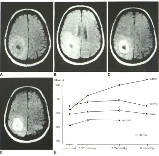
Visual assessment.-Of six tumors, a doughnut-shaped glioblastoma (case 4) and a pituitary adenoma (case 6) were differentiated from adjacent tissue on precontrast SE 800/35 scans (Fig. 2A).ln the other cases an abnormality was definite, but the exact tumor outline could not be established (Fig. 3A). At a dose level of 0.025 mmol of Gd-DTPA/kg slight (cases 1 and 5), moderate (cases 2 and 3), and strong (cases 4 and 6) SI increases were observed in two cases each (Figs. 2B and 3B). The strong SI increase provided improved demar-cation of the pituitary adenoma (case 6). In one glioblastoma (Fig. 2B) the strong enhancement of the primarily hypointense lesion resulted in roughly the same degree of contrast
be-tween tumor and edema. Tumor was poorly differentiated
from edema, although there had been fair tumor demarcation before administration of contrast material.
At a dose level of 0.05 mmol of Gd-DTPA/kg the pituitary adenoma (case 6) displayed a slight increase in SI when compared with the preceding dose level. In four cases (cases 1-3 and 5) SI increases were scored as moderate (Fig. 3C). One glioblastoma (case 4) showed a strong SI increase (Fig. 2C). Except for this case and for the pituitary adenoma, tumor
margins still were poorly defined relative to surrounding tissue
at a dose level of 0.05 mmol Gd-DTPAJkg. In one glioblastoma
(case 1) a second area anterior to the first showed slight enhancement (Fig. 3C).
At a dose level of 0.1 mmol of Gd-DTPA/kg slight increases
in SI occurred in two cases (cases 4 and 6) when compared with the dose level of 0.05 mmol/kg (Fig. 2D). A moderate SI
increase was observed in a cerebral metastasis. In the other
cases (cases 1-3) SI increases were scored as strong. The
moderate and strong SI increases, respectively, resulted in
well-defined tumor margins (cases 1-3 and 5) at the dose
level of 0.1 mmol Gd-DTPAJkg (Fig. 3D). In case 1 the second enhancing region was verified and clearly identified as a lesion (Fig. 3D). The slight SI increases in cases 4 and 6 did not improve tumor demarcation further (Fig. 2D).
Quantitative evaluation.-In all six cases the SI curves showed a marked increase in SI values of tumor tissue on all
postcontrast measurements as compared with precontrast
scans (Figs. 2E, 3E, and 4). A continuous increase of SI in
tumor tissue was recorded with each injection, even though the percentage increase from one dose level to the next was small in some cases (Table 1). In three (cases 1, 2, and 4) of four glioblastomas the highest increase in SI was measured after the first injection. This increase in SI grew smaller with each of the subsequent injections in cases 2 and 4. In case 1 the incremental increases were almost equal after the second
(8%) and third (9%) injections. In case 3 the SI increases were
almost equal after the first (20%) and third (22%) injections, with only a very slight increase after the second injection
(1 %). In the cerebral metastasis (case 5) and pituitary
ade-noma (case 6) the greatest SI increases between two scans were recorded after the second and third injections,
respec-tively.
In five cases (cases 1-5) "necrotic" tumor tissue was
de-tected. Three SI measurements (case 3, pre- and first
post-contrast scan; case 4, third postcontrast scan) could not be
obtained because of positional changes exceeding the extent
of "necrosis." In all cases the SI of "necrotic" tissue on postcontrast scans was higher than the SI on the precontrast scan. However, with the exception of the first dose level of 0.025 mmol/kg, the magnitude of SI increases was markedly less than that of enhancing tumor tissue. Thus the difference
between the SI of enhancing tumor tissue and of tumor
"necrosis" grew remarkably larger after the second injection,
with further improvement after the third injection (Fig. 4).
In five cases perifocal edema was detected (cases 1-5). As
opposed to SI curves of tumor tissue, the SI of edema showed only insignificant changes after the injection of Gd-DTPA (Fig. 4). A slight increase in SI was always measured after the first
injection and also after the second injection; however, after
the third injection, SI values showed a slight decrease as compared with the second injection in all but one case (case
3).
In all cases and at all dose levels the postcontrast SI values of normal brain tissue were minimally higher as compared
with those on precontrast scans. There were only insignificant
806
NIENDORF ET AL. AJNR:8. September/October 1987A
o
B
SI (a.U.)
1200
1000
800
600
400
c
______ A - - - _ A necrosis
A
---,- I
precontrast 0.025 mmollkg
I
0.05mmollkg
E
Fig. 2.-Case 4: 51-year-old woman with presumed right parietooccipital glioblastoma. SE 800/35 images.
tumor
edema
brain
SE 800/35
I 0.1 mmollkg
A, Before enhancement. Area of very low signal intensity in right occipital region is well differentiated from doughnut-shaped ring of intermediate signal
intensity. This ring in tum is surrounded by roughly differentiable area of higher signal intensity, confirmed as perifocal edema on unenhanced T2-weighted
image.
B, 0.025 mmol of Gd-DTP/kg. Strong increase in signal intensity but no improvement of contrast vs perifocal edema. Slightly different slice position
resulted from patient movement.
C, 0.05 mmol of Gd-DTPA/kg. Strong increase in signal intensity relative to previous dose level results in well-defined tumor margins with good
differentiation of tumor tissue from central "necrosis" and from perifocal edema.
D, 0.1 mmol of Gd-DTPA/kg. Further increase in signal intensity of tumor tissue but no further increase of diagnostically useful information or contrast
enhancement. Slightly different slice position may at least partly account for now poorly defined margin between tumor and "necrosis."
E, Signal-intensity (SI) values of tumor tissue, "necrotic" tissue, edema, and normal brain. Due to positional changes signal-intensity measurement of
[image:4.612.56.562.102.600.2]AJNR:8, September/October 1987
A
o
Gd-DTPA DOSE-FINDING STUDY
B
51 (aU.) -,
700
807
c
600
~.
,"m"
~~!::;;:---;?-::;:::::-~~~--
---===::
~
~d~~a
. ; : ; .
- - - 4
necrosis500
...
5E 800/35
I I I
precontrast 0.025 mmol/ky 0.05 mmollkg 0.1 mmollkg
[image:5.612.57.558.74.574.2]E
Fig. 3.-Case 1: 56-year-old woman with right parietooccipital glioblastoma. SE 800/35 images.
A, Before enhancement. Lateral area of inhomogeneous signal intensity lower than that of normal brain tissue is seen in right occipital region. There is
compression of posterior horn of lateral ventricle and effacement of sulci. Area of edema identified on T2-weighted unenhanced scan (arrows).
B, 0.025 mmol of Gd-DTPA/kg. Suspected area has slight increase in signal intensity with poorly defined border of enhancing tissue caused by
isointensity .
C, 0.05 mmol of Gd-DTPA/kg. Enhancing portions of suspected area show moderate enhancement relative to normal brain tissue with poorly defined
posterior and medial borders. In temporoparietal region an additional area now shows slight increase in signal intensity.
D, 0.1 mmol of Gd-DTPA/kg. Occipital tumor is now well enhanced with good demarcation vs central "necrotic" area and vs surrounding brain tissue.
Second area shows definite enhancement and is identified as additional lesion.
E, Signal-intensity (51) values of tumor tissue, "necrotic" tissue, edema, and normal brain. Signal-intensity values are in arbitrary units (a.U.).
Group 2 (0.2 mmol Gd-DTPA/kg)
Visual assessment.-Results obtained with the fractional
doses of 0.05 mmol/kg and 0.1 mmol/kg in group 2 were
808 NIENDORF ET AL. AJNR:8, September/October 1987
normalized SI
1.40
.~.
'"mo'
.
/
~
.. _ _ _ _ _ _ " necroSIS-
..
---~ " edema
~t)====== v brain
o
1.20 1.00
SE 800/35
I I I
IJrecontrast 0.025 mmol/kg 0.05 mmal/kg 0.1 mlTIal/kg Fig. 4.-00se-dependent signal-intensity (Sl) changes on SE 800/35
images measured in tumor tissue, tumor "necrosis," perifocal edema, and
normal brain in six patients (cases 1-6). Signal-intensity values are nor
-malized to precontrast measurements.
At the dose level of 0.2 mmol Gd-DTPAJkg a further SI
increase in a glioblastoma (case 7) was observed in a small
area not suspected as tumor tissue at lower dose levels (Fig.
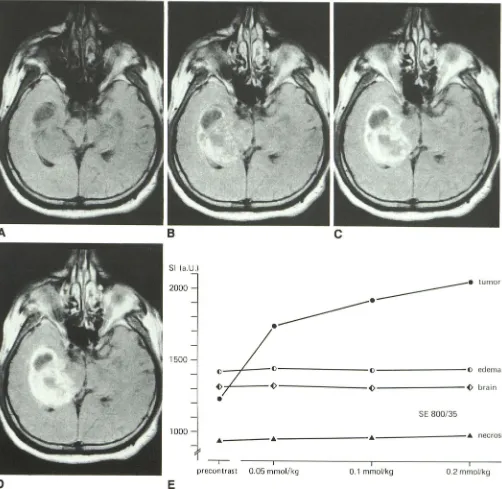
5D). In the case of multiple metastases (case 8) SI increases were seen in three lesions when compared with the dose level of 0.1 mmol Gd-DTPA/kg. Also two other lesions were
displayed at 0.2 mmol/kg (Fig. 6D). In two cases (cases 10
and 11) slight increases in SI were observed, whereas in an acoustic neuroma (case 9) no further enhancement could be seen (Fig. 7D).
Quantitative eva/uation. - The third injection, which doubled
the image dose from 0.1 to 0.2 mmol Gd-DTPAJkg, produced
a further increase in the SI of tumor tissue of 6-15% in all
cases (Figs. 5E, 6E, 7E, and 8 and Table 1).
The only case in which a central necrosis was detected was case 7, a glioblastoma. After some decrease in SI was
measured on the first (-8%) and second (-3%) postcontrast scans SI showed some increase (6%) on the third post-contrast scan.
In three cases perifocal edema was detected (cases 7-9). Case 7 showed a slight SI increase (5%) after the third
injection, but the SI value remained below baseline. In cases 8 and 9 the recorded values were not significantly different from values obtained at the dose level of 0.1 mmol Gd-DTPA/ kg.
When compared with the dose level of 0.1 mmol Gd-DTPAJ kg, SI values of normal brain tissue remained almost constant
except for a slight increase (4%) in case 7 (Figs. 5E and 8). Tolerance. -In both groups the fractionated injections of Gd-DTPA were well tolerated by all patients. No side effects such as venous pain, nausea, or vomiting were recorded. The only appreciable changes measured were slight transient elevations of serum iron. In most cases serum iron levels were back to baseline 24 hr after injection. At 48 hr after
injection baseline levels were reached in all cases.
Discussion
In 1984 the paramagnetic contrast agent Gd-DTPA became available for clinical research studies in MR [3,4]. Reports of its use in patients with intracranial tumors showed favorable results at an IV injected dose of 0.1 mmol/kg [4-12]. Since the LDso for Gd-DTPA determined in several species after IV injection is about 10 mmol/kg body weight [2] a safety factor of about 100 can be assumed. By comparison, the safety factor of iodinated contrast agents such as the well-known diatrizoates was determined to be about 10, depending on the injection volume and the iodine concentration [14]. Gd-DTPA dosages below 0.1 mmol/kg may also be effective for contrast enhancement, which, in turn, would further increase safety. On the other hand, the high safety factor of more than 100 allows for the use of higher doses of Gd-DTPA for possibly improved detection and characterization of lesions. Both aspects were evaluated in the present brain tumor study with Gd-DTPA injections in a dose range of 0.025-0.2 mmol/ kg. The intraindividual rather than the interindividual study design was chosen because of better comparability and higher reliability of intraindividual data and because fewer
patients are necessary to generate such data.
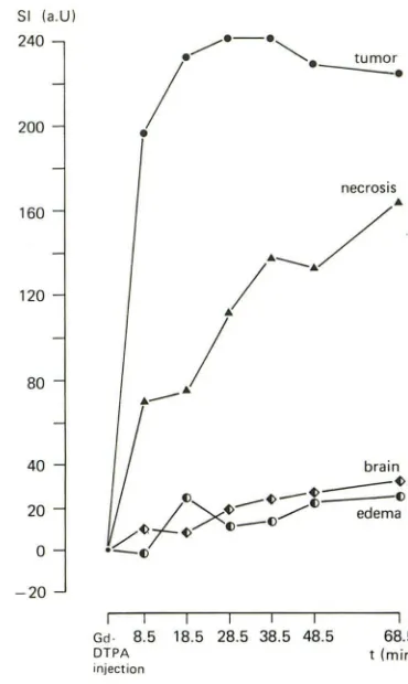
In a brain-tumor study of 15 patients it was shown that tumor tissue displays marked enhancement 5 min after IV injection of 0.1 mmol of Gd-DTPA/kg without significant fur-ther changes for at least 45 min after injection [13]. Although
the time-SI curves of individual tumors differed somewhat from the curve of median values given in Figure 9, these time-course data are principally in keeping with data published by Graif et al. [15]. With 0.1 mmol of Gd-DTPA/kg and with the SE 1500/44 sequence they found SI increases in seven low
-grade primary lesions about 22 min after injection. Slight decreases were observed with a second measurement at 50 min after injection. In metastatic lesions (n = 4) further en-hancement was displayed at about 60 min after injection when compared with the preceding measurement (SE 1500/ 44) at about 40 min. Total postcontrast investigation time in our present study, however, was only 42 min (Fig. 1). The
time-course data reported by Graif et al. [15] are compatible with the data of Schorner et al. [13], which served to set up
the design of our present study.
It was assumed for tumor tissue that each of the three
injections produced time-SI curves similar to those obtained after a single injection of 0.1 mmol of Gd-DTPA/kg. On the basis of the aforementioned course of the time-SI curve, the SI increases in tumor tissue after the second and third injec-tions were regarded to be in the range of maximum SI values obtainable at the respective dose levels. The SI increases measured in enhancing tumor tissue after the second and third injections, therefore, can be attributed predominantly to
the additive effect of additional Gd-DTPA dose fractions and not to delayed SI enhancement from previous dose fractions. SI in MR does not show a linear dependence on the
[image:6.612.56.301.79.254.2]AJNR:8. September/October 1987 Gd-DTPA DOSE-FINDING STUDY 809
B
c
SI (a.U.)
2000 _ _ _ _ _ _ _ - tumor
---c---c
edema ---~ ---~ brainSE 800/35
______________ 4 _ _ _ _ _ _ _ _ _ _ _ _ 4 necrosis
I I
0.1 mmol/kg 0.2 mmollkg
Fig. 5.-Case 7: 42-year-old man with presumed right temporal glioblastoma. SE 800/35 images.
A, Before enhancement. Compression of ventricle, effacement of sulci, and shift of midline structures, as well as large hypo intense area, suggest mass
in right medial temporal lobe.
B, 0.05 mmol of Gd-DTPA/kg. Moderate increase in signal intensity displays areas of disturbed blood-brain barrier in large region with inhomogeneous
signal intensity and poorly defined margins.
C, 0.1 mmol of Gd-DTPA/kg. Good demarcation of garland-shaped tumor with "necrotic" tissue.
D, 0.2 mmol of Gd-DTPA/kg. Adjacent to medial margin of tumor, additional small area of enhancement is seen in region that at lower dose levels was
not identified as enhancing tumor tissue.
E, Signal-intensity (SI) values of tumor tissue, "necrotic" tissue, edema, and normal brain. Signal intensity of edema was measured on adjacent section
obtained by multi slice technique. Signal-intensity values are in arbitrary units (a.U.). T2 shortening decreases SI. Up to a certain concentration of Gd-DTPA the T1 effect continues to increase SI. At higher concentrations, however, the T1 effect levels off and the
influence of a shortened T2 predominates attenuating SI. This
is caused by rapid dephasing of spins in the x-y plane, which
in turn results in loss of signal at the time of sampling, even
at short TE intervals.
[image:7.612.56.558.88.577.2]810 NIENDORF ET AL. AJNR:8. September/October 1987
A
B
SI (a.U.)
1800
c
~.
tumor1600
---~~
_________
~
___________________
v
___________________
v
edema1400
~---- _ _ _ _ _ _ _ _ _ _ _ ~ brain
SE 800/35
I I I
o
E
0.05 mmol/kg 0.1 mmol/kg 0.2 mmol/kg
precontrast
Fig. S.-Case 8: 45-year-old woman with presumed multiple metastases. SE 800/35 images.
A, Before enhancement. Asymmetry in signal intensity of right vs left postsylvian region caused suspicion of lesion. Area of edema (arrow) was seen on T2-weighted precontrast scan.
B, 0.05 mmol of Gd-DTPA/kg. Three poorly defined areas show slight enhancement in signal intensity (arrows).
C, 0.1 mmol of Gd-DTPA/kg. The three small areas have increased in signal intensity. Two lesions in right postsylvian region show improved demarcation. D, 0.2 mmol of Gd-DTPA/kg. All three areas of enhancement show further increase in signal intensity and are now well demarcated relative to surrounding tissue. Two additional lesions are seen (arrow).
E, Signal-intensity (SI) values of tumor tissue, edema, and normal brain. Signal-intensity values of tumor tissue are arithmetic mean of three lesions.
Values are in arbitrary units (a.U.).
values of tumor tissue, suggesting tissue concentrations of
Gd-DTPA compatible with predominantly T1 effect. The in-creasing T2 effect, however, produced less dramatic rises in dose-SI curves at higher doses. With total doses of both 0.1 and 0.2 mmol Gd-DTPA/kg the summary curves showed a
steeper increase in SI values after the first injection than after
the second and third administration of Gd-DTPA (Figs. 4 and
[image:8.613.57.563.79.574.2]AJNR:8. September/October 1987 Gd-DTPA DOSE-FINDING STUDY 811
A
B
c
SI (a.U.)
1800 _ _ _ _ _ _ _ • tumor
---
.
•
1600
1400
SE 800/35
1200
()---()- - - -() edema
~---~---
-~---~ brain1000
o
E
1
- , -
•
precontrastI I I
[image:9.614.53.560.76.591.2]0.05 mmol/kg 0.1 mmol/kg 0.2 mmol/kg
Fig. 7.-Case 9: 67-year-old woman with right acoustic neuroma. SE 800/35 images.
A, Before enhancement. Well-defined area of decreased signal intensity in cerebellopontine angle. Compression and displacement of fourth ventricle. Area of edema (arrows) was identified on T2-weighted precontrast scan.
B, 0.05 mmol of Gd-DTPA/kg. Strong contrast enhancement with very good demarcation of tumor margins, whereas tumor itself shows some
inhomogeneities in signal intensity.
C, 0.1 mmol of Gd-DTPA/kg. Moderate increase in signal intensity. Tumor now displays more homogeneous signal intensity, but there is no further diagnostic information.
D, 0.2 mmol of Gd-DTPA/kg. A slight increase in signal intensity is seen, but no change in tumor demarcation.
E, Signal-intensity (51) values of tumor tissue and normal brain. Signal-intensity values are in arbitrary units (a.U.).
With regard to diagnostic yield the dose of 0.2 mmoljkg showed two lesions in a patient with multiple metastases that had not been detected at a dose level of 0.1 mmol Gd-DTPAJ kg. Three other lesions in the same patient were shown to
812 NIENDORF ET AL. AJNR:8, September/October 1987
normalized SI
1.40
1.20
/"
----.
~"
'"-'
SE 800/35
1.00
o
==::::::::::~-_---o- - -_ _ _ _ - 0 '---o _ _ - - - - -0 ""em, b"in----,- I
precontr<lst 0.05 mmol/kg 0.1 mmoI l/kg O.2mmol/I kg
Fig. 8.-Dose-dependent changes in signal intensity (51) on SE 800/35
images (TR
=
800 msec, TE=
35 msec) measured in tumor tissue and normal brain in five patients (cases 7-11). Perifocal edema was detected in three patients and tumor "necrosis" in only one. Data for tumor "necro-sis" are not in curve. Signal-intensity values are normalized to precontrastmeasurements.
impact on patient management in these two cases; however,
they may have clinical impact in selected cases. In the other
three cases in group 2 (0.2 mmol Gd-DTPAfkg), even when
an SI increase was measured, the area of enhancement did not change. Thus, for routine administration, no justifiable reasons for doses higher than 0.1 mmol Gd-DTPAfkg emerged from our study.
The other objective of our study was to determine whether the dose of 0.1 mmol/kg could be reduced without loss of diagnostic information. In six cases (cases 1-6) contrast-enhanced MR was performed with 0.025 mmol of Gd-DTPA/ kg. Only in case 6 (pituitary adenoma) was contrast-enhanced MR at 0.025 mmol/kg diagnostically useful, showing tumor extension. In this case the tumor could already be delineated before injection of Gd-DTPA. After injection of 0.025 mmol of Gd-DTPAfkg the increase in SI resulted in increased contrast between tumor and adjacent structures with further improve-ment of differentiation between both. However, with respect
to therapy planning, Gd-DTPA did not contribute
indispensa-ble information in this case.
In all 11 cases, contrast-enhanced MR was performed with
a dose of 0.05 mmol Gd-DTPAfkg. In addition to the pituitary adenoma, a glioblastoma (Fig. 2) and an acoustic neuroma (Fig. 7) could be delineated at this dose level. Both tumors
were hYPointense on precontrast SE 800/35 scans and
there-fore were differentiable from surrounding tissue.
As with the dose level of 0.05 mmol/kg all 11 cases in
groups 1 and 2 were evaluated with 0.1 mmol/kg. In eight of
the 11 cases this dose was indispensable for diagnostic
images. Our results, therefore, strongly suggest that 0.1 mmol
Gd-DTPAfkg rather than lower doses are required for
brain-tumor imaging.
To assess the validity of our results for short TR/short TE
SE pulse sequences, an experimental study was performed
in one brain-tumor-bearing dog. After precontrast SE 800/35
51 (a.U) 240
200
160
120
80 40 20
o
-20 I Gd· DTPA/
./.-.
~
.
--.
tumor•
necrosis
I I I I I I
8.5 18.5 28.5 38.5 48.5 68.5
t (min)
injection
Fig. 9.- Time-dependent changes in image contrast in 15 patients with intracranial tumors. Differences between pre- and postcontrast signal-intensity (51) values are calculated for tumor, "necrotic" tissue, edema, and normal brain. Differences in signal intensity are given as median values. t = time. (Adapted from [13].)
and SE 500/16 images had been obtained, a total dose of 0.3 mmol Gd-DTPAfkg was IV injected according to the fractionated incremental dose regime (Fig. 1, 0.1 mmolfkg with each injection). Pre- and postcontrast imaging was per-formed with the pulse sequences SE 500/16 (5 min postinjec-tion) and SE 800/35 (8 min postinjecpostinjec-tion) with single signal averaging in order to meet the experimental protocol for dose administration (Fig. 1). Generally, a greater SI increase of enhancing tumor tissue was found with SE 500/16 as com-pared with SE 800/35 (Figs. 10E and 11 E). The SI difference (contrast) between enhancing tumor tissue and normal brain after injection of 0.1 mmol/kg, however, was greater with SE 800/35. According to the quantitative results of this experi-ment a somewhat higher dose of contrast medium was nec-essary to obtain a comparable contrast between enhancing tumor tissue and normal brain with SE 500/16. Visual evalu-ation of SE 500{16 and SE 800/35 images supported the quantitative results (Figs. 10 and 11).
[image:10.612.60.305.70.251.2] [image:10.612.341.526.80.390.2]AJNR:8, September/October 1987 Gd-DTPA DOSE-FINDING STUDY 813
A
B
c
51 (a.U.)
1000 _ _ tumor
---800
---~ brain
600
-400 5E 500/16
I I I
precontrast 0.1 mmol/kg 0.2 mmol/kg 0.3 mmol/kg
o
E
Fig. 10.-Canine brain. Tumor suspected clinically because of history of repeated seizures. SE 500/16 images. Examination was done according to
protocol in Figure 1, except total dose was 0.3 mmol of Gd-DTPA/kg.
A, Before enhancement. Area of low signal intensity is displayed in rhinencephalic region of brain.
B, 0.1 mmol of Gd-DTPA/kg. Signal-intensity increase within tumor tissue with fair demarcation.
C, 0.2 mmol of Gd-DTPA/kg. Slight increase of tumor signal intensity and good demarcation of enhancing tumor.
D, 0.3 mmol of Gd-DTPA/kg. Slightly increasing contrast enhancement in tumor tissue.
E, Signal-intensity (SI) values of tumor tissue and normal brain. Signal-intensity values are in arbitrary units (a.U.).
patients showed only slight transient elevations of serum iron in some patients, which usually lasted for only 24 hr and was in no case observed to persist after 48 hr. This has been reported by other investigators also, but has been found to be of no clinical relevance [19, 20].
[image:11.614.55.559.83.582.2]814 NIENDORF ET AL. AJNR:8. September/October 1987
B
c
SI (a.U.1
1000
_e
_________
-
e
tumor800
_ _ _ _ _ _ _ _ _ ~ brain
600
400
SE 800/35
I I
precontrast 0.1 mmol/kg 0.2 mmol/kg 0.3mmolI /kg
D
E
Fig. 11.-Same canine as in Figure 10 studied at SE 800/35. Examination was done according to protocol in Figure 1, but total dose was 0.3 mmol of
Gd-DTPA/kg and scanning was performed 8 min after injection.
A, Before enhancement. Poorly defined area of slightly hypointense signal intensity in rhinencephalic region.
B, 0.1 mmol of Gd-DTPA/kg. Increase in signal intensity provides fair demarcation relative to surrounding tissue.
C, 0.2 mmol of Gd-DTPA/kg. Further contrast enhancement and good demarcation of enhancing tumor.
D, 0.3 mmol of Gd-DTPA/kg. Slight increase in signal intensity.
[image:12.612.55.559.93.596.2]AJNR:8, September/October 1987 Gd-DTPA DOSE-FINDING STUDY 815 mmol Gd-DTPAjkg may yield additional information and can
be given safely.
In the meantime the evaluated optimum dose of 0.1 mmol Gd-DTPAjkg has proven to be appropriate for clinical pur
-poses, also for more T1-weighted sequences, with a shorter TR (TR ~ 500 msec) and shorter TE (TE ~ 30 msec) yielding comparable and good diagnostic results [6-8, 20-23]. ACKNOWLEDGMENTS
We thank Renate Kentenich, Jochen Kasbohm, and Hossein
Zol-faghari for technical assistance; Martina Licht for help in manuscript
preparation; and Dr. Schwartz-Porsche for Figures 10 and 11.
REFERENCES
1. Brasch RC, Weinmann HJ, Wesbey GE. Contrast-enhanced NMR imaging:
animal studies using gadolinium-DTPA complex. AJR 1984;142:625-630
2. Weinmann HJ, Brasch RC, Press WR, Wesbey GE. Characteristics of gadolinium-DTPA complex: a potential NMR contrast agent. AJR
1984;142:619-624
3. Laniado M, Weinmann HJ, Schorner W, Felix R, Speck U. First use of Gd
-DTPA/dimeglumine in man. Physiol Chern Phys Med NMR 1984;16:
157-165
4. Schorner W, Felix R, Laniado M, et al. Prufung des kernspintomogra -phischen Kontrastmittels Gadolinium-DTPA am Menschen: Vertraglichkeit,
Kontrastbeeinflussung und erste klinische Ergebnisse. ROFO
1984;140:493-500
5. Schorner W, Felix R, Claussen C, et al. Kernspintomographische Diagnos -tik von Hirntumoren mit dem Kontrastmittel Gadolinium-DTPA. ROFO
1984;141 :511-516
6. Claussen C, Laniado M, Kazner E, Schorner W, Felix R. Application of
contrast agents in CT and NMR: their potential in imaging of brain tumors. Neuroradio/ogy 1985;27: 164-171
7. Claussen C, Laniado M, Schorner W, et al. The use of gadolinium-DTPA
in magnetic resonance imaging of glioblastomas and intracranial metas
-tases. AJNR 1985;6:669-674
8. Felix R, Schorner W, Laniado M, et al. Brain tumors: MR imaging with gadolinium-DTPA. Radiology 1985;156:681-688
9. Runge V, Schorner, W, Niendorf HP, et al. Initial evaluation of gado
linium-DTPA for contrast-enhanced magnetic resonance imaging. Magnetic Res·
onance Imag 1985;3:27-35
10. Carr DH, Brown J, Bydder GM, et al. Intravenous chelated gadolinium as
a contrast agent in NMR imaging of cerebral tumours. Lancet 1984;1:
484-486
11. Carr DH, Brown J, Bydder GM, et al. Gadolinium-DTPA as a contrast agent in MRI: initial clinical experience in 20 patients. AJR 1984;143:
215-224
12. Bydder GM, Kingsley OPE, Brown J, Niendorf HP, Young IR. MR imaging of meningiomas including studies with and without gadolinium-DTPA. J
Comput Assist Tomogr 1985;9:690-697
13. Schorner W, Laniado M, Niendorf HP, Schubert C, Felix R. Time-dependent changes of image contrast in brain tumors after gadolinium-DTPA. AJNR
1986;7: 1 013-1 020
14. Wolf GL, Burnett KR, Goldstein EJ, Joseph PM. Contrast agents for
magnetic resonance imaging. In: Kressel HY, ed. Magnetic resonance
annual 1985. New York: Raven, 1985:247
15. Graif M, Bydder GM, Steiner RE, Niendorf HP, Thomas DGT, Young IR.
Contrast-enhanced MR imaging of malignant brain tumors. AJNR
1985;6:855-862
16. Brasch RC. Work in progress. Methods of contrast enhancement for NMR
imaging and potential applications. Radiology 1983;147:781-788
17. Runge VM, Clanton JA, Lukehart CM, Partain CL, James AE. Paramagnetic
agents for contrast-enhanced NMR imaging: a review. AJR 1983;
141:1209-1215
18. Niendorf HP, Semmler W, Laniado M, Felix R. Some aspects of the use of contrast agents in magnetic resonance imaging. Diagn Imag Clin Med
1986;55:25-36
19. Bradley WG, Brant-Zawadzki M, Brasch RC, et al. Initial clinical experience with Gd-DTPA in North America: MR contrast enhancement of brain tumors
(abstract). Radiology 1985;157(P): 125
20. Runge VM, Price AC. Gd-DTPA: an i.v. contrast agent for MRI investigation of intracranial neoplastic disease. Presented at the annual meeting of the
American Society of Neuroradiology, San Diego, January 1986 21. Laniado M, Niendorf HP, Schorner W, Felix R. Spin-echo and inversion
-recovery sequences for gadolinium-DTPA-enhanced MRI of intracranial
tumors. Acta Radio/1986;369:469-471
22. Brant-Zawadzki M, Berry I, Osaki L, Brasch R, Murovic J, Norman D.
Gd-DTPA in clinical MR of the brain: 1. Intraaxiallesions. AJNR 1986;7:78
1-788
23. Berry I, Brant-Zawadzki M, Osaki L, Brasch R, Murovic J, Newton TH.


