Printed in Great Britain
HEAT EXCHANGE IN RELATION TO BLOOD FLOW
BETWEEN THORAX AND ABDOMEN IN BUMBLEBEES
BY BERND HEINRICH
Department of Entomological Sciences, University of California, Berkeley, California 94720
{Received 2 October, 1975)
SUMMARY
1. The narrow passage within the petiole between thorax and abdomen is anatomically constructed so that counter-current exchange should retain heat in the thorax despite blood flow to and from the cool abdomen.
2. However, the counter-current heat exchanger can be physiologically circumvented. Exogenously heated bumblebees prevented overheating of the thorax by shunting heat into the abdomen. They also regurgitated fluid, which helped to reduce head temperature but had little effect on thoracic temperature.
3. Temperature increases in the ventrum of the abdomen occurred in steps exactly coinciding with the beats of the ventral diaphragm, and with the abdominal 'ventilatory' pumping movements when these were present. The ability to prevent overheating of the thorax by transport of heat to the abdomen was abolished when the heart was made inoperative.
4. At low thoracic temperatures the ventral diaphragm beat at a wide range of rates and with varying interbeat intervals, while the heart beat at a high frequency relative to the ventral diaphragm, but at a very low ampli-tude. However, when thoracic temperature exceeded 43 °C the amplitudes of both were high, and the interbeat intervals as well as the beating fre-quencies of the two pulsatile organs became identical in any one bee. Furthermore, heated bees engaged in vigorous abdominal pumping at the same frequency as that of their heart and ventral diaphragm pulsations.
5. The results indicate that the anatomical counter-current heat ex-changer is reduced or eliminated during heat stress by 'chopping' the blood flow into pulses, and the blood pulses are shunted through the petiole alternately by way of a switch mechanism.
INTRODUCTION
The metabolic rate of bumblebee queens during free flight varies from about 63 to 126 joules/g thorax.min, depending on the load. However, metabolic rate is rela-tively independent of ambient temperature from 10 to 30 °C, under which conditions thoracic temperature remains near 39-42 °C (Heinrich, 1975). The above data strongly suggest that the flight metabolism alone, at ambient temperatures above 10 °C, produces more than ample heat to elevate thoracic temperature to observed levels and that thoracic temperature is stabilized by regulating heat loss alone. The
562 BERND H E I N R I C H
metabolic rate of incubating (stationary) bees, on the other hand, is inversely related to ambient temperature, varying from approximately 54 joules/g thorax. min at 5 °C, to near resting levels at 35 °C (Heinrich, 1974a). In contrast to flying bees, incubating bees maintain a high abdominal temperature by transferring more heat into the abdomen at low than at high ambient temperatures.
It has not been elucidated how heat loss is regulated. Mechanisms whereby heat is dissipated from the thorax and transported into the abdomen by the blood, or retained in the thorax despite blood flow to the abdomen, are here examined in terms of heart-ventilatory cycles and anatomy.
MATERIALS AND METHODS
Unless otherwise indicated, all of the experimental animals were queens of Bombus vosnesenskii Radoszkowski, which weigh approximately o*5-o*7 g. Most of the bees were derived from colonies maintained in the laboratory.
Heat transfer to the abdomen was investigated in bees fastened on to a styrofoam pad. The bees were held fast by pins placed along the sides of the thorax and petiole. A microscope lamp was used to focus heat on to the thorax (or abdomen) while the abdomen (or thorax) was shielded with tin foil. The present study concerns the physiological mechanisms for heat dissipation that are activated when thoracic tem-perature is made to approach lethal levels. Whether the same or other mechanisms of heat transfer are used under different circumstances (for example, during activity) remains conjectural.
In most of the heating and cooling experiments, body temperatures were recorded with a Honeywell multichannel potentiometric recorder, using 36 gauge copper-constantan thermocouples that were enamel-insulated except for the tips. In other experiments body temperatures were recorded together with heart and ventral diaphragm activity on the same chart using a Beckman R411 Dynograph recorder. Thoracic temperature in these experiments was measured with a Veco 32A130 thermistor (having a diameter of 0-25 mm and a time constant of 0-5 s) and a thermistor coupler. Abdominal temperatures and rapid temperature fluctuations in the petiole and abdomen were measured with 46-gauge copper-constantan thermocouples insulated, except at the tip, with cotton. The temperatures were measured with an Omega T-4 thermocouple read-out meter and recorded concurrently with other measurements by a Dynograph recorder.
Mechanical activities of the heart and ventral diaphragm were each measured from paired electrodes inserted one on each side of the respective organs. Electrical resist-ance changes, resulting from mechanical disturbresist-ance or fluid flow between the paired 36 gauge copper electrodes, were measured using Biocom model 2991 Impedence converters and recording with the Dynograph. In dissected animals pen excursions of the Dynograph coincided exactly with the visually apparent pulsations of these organs. The electrodes and thermocouples in the abdomen were inserted between the tergites/sternites by piercing the intersegmental membrane. They were fixed into place using a mixture of melted beeswax and resin applied from the head of a pin.
were observed visually and recorded manually, or they were measured electronically using the impedance converter and recorded on the Dynograph recorder. Attaching both electrodes on different parts of the abdomen did not register the generally low amplitude abdominal movements. Instead, one of the enamel-insulated electrode leads was bent double, and the 1800 bend was attached with wax on to the tip of the abdomen. The uninsulated tips of both electrodes (one stationary and the other moved by the abdomen) were immersed in water close to each other. Amplitude of the pen excursions corresponded with amplitude of the visually observed abdominal pumping movements, but no systematic efforts were made in this study to calibrate the movements in terms of absolute distances.
All operative procedures were done while the animals were under light CO2
-narcosis. The animals were usually narcotized for several periods of 3-4 min before all of the probes had been inserted and fastened.
In some experiments, the heart was ligated through the intersegmental membrane at the second abdominal segment using human hair and a surgeon's eye needle.
Bees used for the tongue-lashing experiments had recently emerged in a captive colony. Few of them flew as vigorously as do most queens that are active in early spring. They were flown in a 3-9 1 jar. Temperatures in the jar were held within 0-5 °C by conducting the experiments in a temperature controlled room at a relative humidity of 50-60 %. The tongue-lashing behaviour was obvious, but it could not always be ascertained whether the proboscis also held a droplet of fluid. In many cases, however, such a droplet was visible.
Most of the dissections were of living animals so that the activity of the ventral diaphram and heart could be observed. Trypan blue in cockroach saline was used for staining.
RESULTS
(A) Circulatory anatomy
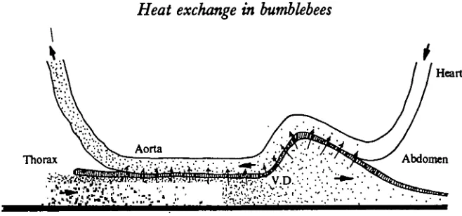
As in other insects, the main features of the circulatory system in bumblebees consists of a dorsal vessel or heart (referred to later as H) and a ventral diaphragm (V.D.). The heart continues into the thorax as the aorta (Fig. 1). The heart generally pumps blood anteriorly, while the ventral diaphragm propels it posteriorly.
The following description applies specifically to B. vosnesenslm, but dissections of Bombus edwardsii Cresson revealed no obvious differences in any major details. The salient features from the standpoint of thermoregulation (discussed later) is that the heart is attached close to the dorsal surface of the abdominal wall. It bends down sharply along an air sac at the anterior end of the abdomen, and forms a loop that is loosely attached to the same air-sac as it droops down loosely near the petiole on to the V.D. The ventral horizontal portion of the heart is in direct contact with the ventral diaphragm. The ventral diaphragm has its origin within the petiole, and the heart, the tracheae and the oesophagus pass above it (Fig. 2). After traversing the petiole the heart (now called the aorta) curves dorsally, entering between the right and the left dorsal longitudinal muscles. The aortae of both B. vomesenskii and B. edwardsii then make a sharp loop between these muscles before entering the head (Fig. 1).
The V.D. is a thin transparent sheet of primarily transverse muscle that overlies
5
64
BERND HEINRICHAorta
Insulation
D.L.M. Heart
-.Ventral diaphragm
Thermal window Regurgitated fluid
Fig. I. Anatomy of Bombus votnetetukii (and B. ahoardsii) relevant to thermoregulation. The top view shows regional differences in depth of insulation. Note also droplet of fluid on proboscis (see text). Arrows indicate movement. The sagittal view shows main internal features. D.L.M. = dorsal longitudinal muscle. N.C. = nerve cord.
the ventral surface of the abdomen. The nerve cord is surrounded by haemolymph beneath the membrane, and is clearly visible through it. In the vicinity of the petiole at least, the V.D. can seal off the lumen of the petiole above it.
(B) Heat transfer between thorax and abdomen
Bees transferred exogenously applied heat from thorax to abdomen, or vice versa. When sufficient heat focused on to the thorax (with the incandescent lamp) to elevate thoracic temperature (TTJ,), the entire abdomen increased in temperature relatively uniformly (Figs. 3, 4). Similarly, when the abdomen of live bees was heated, the temperature of the thorax increased. Animals killed in place (by injecting 10 fil of ether into the thorax) and heated as before had only minor increases in abdominal (or thoracic) temperature when heated on the thorax (or abdomen) (Fig. 3).
V— 4
Fig. 2. Sagittal section of petiole area (left), showing anatomical arrangements of heart (i), air sacs (A.S. and 2), ventral diaphragm (V.D. and 5), tracheae (3), oesophagus (4), nerve cord (N.C.), and blood channel (6). Diagram at right shows cross-section through abdomen viewing anteriorly and into the aperture in the petiole (broadly heart-shaped). Pointers between the two diagrams (drawn to different scale) connect the same structures and areas. Large arrows show direction of blood (indicated by stippling) flow. Small arrows (near V.D.) indicate direction of movement of this organ during that portion of the pumping cycle depicted.
and electrodes simultaneously inserted into their bodies generally did not generate sufficient endothermic heat to maintain a high body temperature on their own for more than a few minutes). In exogenously heated animals, the rate of heat transfer to the abdomen was usually low, but it increased greatly as T>rb began to approach lethal levels. Thus, at a high ambient temperature (TA) the bees generated a greater temperature excess of the abdomen than at a low TA, even though the input of thermal energy into the thorax was nearly identical at both temperatures (Figs. 4, 5). Regardless of the difference between rT h and the temperature in various areas of
the abdomen (TAb), all internal body temperatures became nearly identical soon after thoracic heating was stopped. That is, TTh initially cooled rapidly (while TAXt may have been rising), and then all parts of the body cooled at the same rate. In dead animals, on the other hand, the cooling of thorax and abdomen were often independent of each other. It is apparent that heat was being transferred within the body of live animals.
566
44
42
40
38
36
34
32
30
28
26
24
BERND H E I N R I C H
Abdomen
Alive Dead
10 0 5
Time(min)
[image:6.451.47.380.65.328.2]10 10
Fig. 3. Body temperature changes of the abdomen during thoracic heating of the live bee (left) and during abdominal heating in the live (middle) and in the dead bee (right). Body tempera-tures were simultaneously measured at four locations ( • , thorax, O, ventrally in and abdominal segment, A, ventrally in 4th abdominal segment, + , dorsally in and abdominal segment). The thermocouples were not moved during the three heating experiments. Heat input (with an incandescent lamp) was reduced in this experiment (at arrows) in order to prevent body temperature from exceeding 44 °C.
(C) Activity of the heart
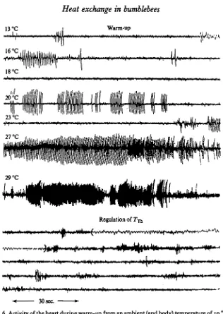
The mechanical activity of the heart consisted either of rapid low amplitude beats or of less rapid high amplitude beats. The distinction between the two was often not clear at low thoracic temperatures. The low amplitude beats were commonly observed in resting bees, during the following endothermic warm-up (Fig. 6), and during exogenous heating of the thorax before TTh reached approximately 42 °C. They increased in frequency from about 300/min at a TTh of 30 °C, to a frequency of
600/min at 40 °C (Fig. 15). During vigorous shivering the beats were sometimes absent for several consecutive minutes (Fig. 6). The rate of both the low and the high amplitude heart-beats had a Q10 of about 2 from a TTb of 3o°-4O °C.
35
30
j>
15
10
7 °C
21 °C Dead
21 °C
7°C
Dead
10 Time (min)
[image:7.451.93.335.48.432.2]15 20
Fig. 4. Changes in thoracic (TjiJ and abdominal (JVt) temperature excess in a bee (thorax = 244 mg, abdomen 0290 mg) during heating of the thorax at ambient temperatures of 7 °C ( •—), and ai °C ( ) when alive, and after being killed ( ). The position of the thermocouples was not changed during the three heating experiments. Heat input was the same throughout each run, and from one run to the next. Abdominal temperatures indicated show range recorded from three thermocouples (see Fig. 3). Nine other bees investigated showed qualitatively similar behaviour.
(D) Activity of the ventral diaphragm
The ventral diaphragm consists ofa sheet of muscle with transverse fibres spanning the lower portion of the abdomen. When relaxed the sheet sags ventrally and lies upon the ventral nerve cord and the lower portion of the abdominal wall (Fig. 1). During contraction (which always proceeded in a posterior direction in dissected animals) the ventral diaphragm rises and creates a fluid-filled perineurial cavity underneath. As the contraction proceeds posteriorly along the muscular sheet, the blood issuing from the thorax is moved posteriorly.
568
BERND HEINRICH 36 -32 28 24 g 20 8.K
12 42 38 34 30 26 22 50 46 42 38 34 30 26 Tied-off heart 10 10 Time(min) 10 Fig. 5. Effect of ligation of the heart on thoracic ( # ) and abdominal (O) temperature (measured in the ventrum of the third abdominal segment) during thoracic heating. The operated bee died in less than 4 min of heating. Heating curves of the same bee at ambient temperatures (TA) of about 8 °C and 21 °C are given for comparison. Heat input to thethorax was the same during each of the three heating cycles and the thermocouples remained in place throughout The thorax of the bee weighed 197 mg, and the abdomen weighed 35a mg.
short periods of inactivity (Fig. 8). During and following endothermic warm-up (shivering) the frequency and amplitude of diaphragm pulsations increased above resting levels. Application of COa resulted in almost immediate cessation of both
heart and V.D. activity and a concomittantly greatly reduced thoracic cooling rate (Fig-
9)-Transfer of heat to the abdomen was, beat for beat, correlated with diaphragm activity (Fig. 10), provided that the heart was beating (Fig. 14) and that the difference between thoracic and abdominal temperatures was sufficiently high (Fig. 12). Simul-taneously with each diaphragm beat, the temperature rose sharply up to 0-2 °C above previous levels and then declined to near, but slightly above, the previous abdominal temperature (Fig. 10). As a consequence, beats occurring in rapid succession caused a step-wise rise in TAb (Fig. 11). Abdominal cooling followed cessation of the beats
presumably because blood (and heat) no longer issued from the thorax. The tem-perature pulses, concomittant with the diaphragm beats, were absent or small when rT h was near 7"A (and T^), but they increased in amplitude as the difference between
13 °C
1 r 10
"A*
18 °C
Warm-up
f**"+<'
29 °C
[image:9.451.69.385.41.486.2]Regulation of r-rj,
Fig. 6. Activity of the heart during warm-up from an ambient (and body) temperature of 10 °C, and during the regulation of thoracic temperature at 33-35 °C at an ambient temperature of 19 °C. The bee initiated cool-down immediately after heating to 30 °C. The position of the recording electrodes was not changed during the two continuous recordings (subdivided into arbitrary a min sections) totalling nearly 24 min. A small portion of the record (in paren-theses) was expanded by using a higher chart speed. Thoracic temperatures at the beginning of the 2 min sections of warm-up are given at left. The two centre strips (27 °C, 29 °C) show transition from 'slow' to 'rapid' beats. Abdominal pumping movements were continuous throughout both warm-up and regulation, and synchronous with the large-amplitude heart pulsations when these were present during warm-up.
heat dissipation that are related to high temperature excess; they would also be observed at a constant blood flow.
570 BERND HEINRICH
Fig. 7. Activity of the heart during three representative heating cycles (top) and one cooling cycle (bottom). Lower traces indicate simultaneously recorded thoracic temperatures in °C. Observe change from high frequency low amplitude beats, to low frequency high amplitude beats during heating above 41 °C, and the reverse during cooling.
iA^^$(^^
Fig. 8. The variable mechanical activity of the ventral diaphragm of a bee at rest. The two strips are from a 4 min sequence recorded at 25 °C.
activity in any obvious way, but it did stop, or greatly reduce, the amplitude of the abdominal temperature pulses (Fig. 14) and the associated rise of abdominal temperature (Fig. 5).
2 8
-Fig. 9. Activity of the ventral diaphragm during endothermic warm-up, and during post warm-up thoracic cooling with and without ventral diaphragm activity (abolished with CO|). Thoracic cooling is retarded when the activity of the ventral diaphragm (and also that of the heart) is abolished.
Heart
jffj—f ftfihMf
Diaphragm
'Ab
I I I 1 I I I I I I I M I 1 I I I I I I I I I I
0-5 °G
[image:11.451.43.414.48.232.2]34 °C 30 26
Fig. 10. Interrelationship of the activity of the heart, ventral diaphragm and temperature pulses in the ventrum of the first abdominal segment. Thoracic temperature, also concurrently recorded, is indicated. Recordings were taken at three different speeds. The time-mark at the bottom indicates seconds.
inter-pulse intervals of the heart increased until matching those of the V.D. (Figs. 16, 17).
(E) Abdominal pumping (' ventilatory' movements)
The 'ventilatory' movements in bees, as in some other Hymenoptera, consist of rapid in-out telescoping of the abdomen. Inward telescoping of the abdomen in honeybees creates positive pressure, causing partial collapse of the giant abdominal air sacs, and driving air into the thorax (Bailey, 1954) in the manner of a bellows.
[image:11.451.42.416.289.478.2]572 BERND HEENJUCH
-29-0
-28-5
Fig. I I . Summation of temperature pulses in the second abdominal segment in relation to V.D. activity (top), during a a-min interval when Tn declined from ji-b'C-^o-g °C. Abdominal
temperature also shows an over-all decrease.
Diaphragm
YrrwY^^^
30 29 28. 40 35 30 25
Diaphragm
• * m r t ' ' »
y^ ^
30
29
284 40
35 30 25
TAI
Fig. ia. Continuous record (here subdivided for convenience into two 40-s sections) of thoracic and abdominal temperatures during thoracic heating with an incandescent lamp, showing the initiation of temperature pulses, associated with V.D. activity ia the abdomen and their increase in amplitude as Tm rose above
heating the metabolic rate of the thoracic muscles for the most part probably did not rise above resting levels, and the abdominal pumping movements were usually absent or of a very low amplitude (Fig. 18). However, when the bees were exogenously heated to above 43 °C they usually engaged in continuous high amplitude abdominal pumping at rates near 300/min. The initiation of the abdominal pumping was often abrupt at high TTh, and it was inferred that the pumping movements, which wete
i
4
1
o
Io o o CE>
s
o
o o
o o o o
CD
Ob
00 O
o a o
[image:13.451.44.392.300.515.2]8 9 10 11 12
Fig. 13. Rates of thoracic cooling immediately after warm-up (O), at least 30 s after cessation of warm-up (O), and while under COg narcosis (#), as a function of thoracic temperature
45
40
35
30
25 -•0V0V000O000O00..00 t
i i i i
Heart ligated
K-6S-H
8
00000
V.D.
0 1 2 3 0 1 2
Tirae(min)
Fig. 14. Effect of heart ligation on thoracic and abdominal temperatures during thoracic heating and on temperature pulsations in the petiole (Tp,) as a function of V.D. activity. Arrows on temperature records indicate the time when the V.D. and temperature pulsations (shown in insets) were recorded.
appeared to be deeper at high TTh, had significance in thermoregulation in the heated bees.
574
BERND HEINRICH700 •
600
500
a
1
40a
300
200
100
0+
o 9 oo ' *
4 *V . *
25. 30 35 40 45 Thoracic temperature (°Q
50
Fig. 15. Rate of beating of the ventral diaphragm ( + ), and rates of low amplitude ( • ) and high amplitude (O) heart beats in relation to thoracic temperature.
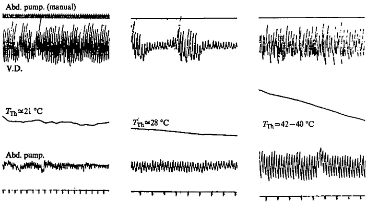
intermittent abdominal pumping movements were observed they were usually, but not always, in synchrony with V.D. beats (Fig. 18). Therefore, the high temperature pulses of blood recorded in the abdomen coincided not only with the ventral diaphragm beats, but also with the externally visible abdominal pumping. In sum, at high thoracic temperature with each in-out movement of the abdomen there was one ventral diaphragm beat, one high temperature pulse in the ventrum of the abdomen, and one beat of the heart. These results will be discussed later in terms of their probable significance to the regulation of blood flow and heat exchange between thorax and abdomen.
(F) Temperatures in the petiole
In queens of B. vosnesemkii, the aperture inside the petiole (through which pass the oesophagus, two large tracheal trunks, the dorsal vessel, the nerve cord and a channel for blood flow into the abdomen) is approximately heart-shaped (Fig. 2) with dimensions of 2 x 1 mm. Depending upon its position within these narrow confines, a thermocouple ought to be able to measure both the cool blood entering the thorax and the warm blood leaving it. From such data (provided the thermo-couples are correctly positioned and their response-time and sensitivity are high enough) it should be possible to observe some aspects of the fluid mechanics and heat flow.
H. 25 °C
V.D.
WyrtAftiw^
30 °C
V.D.
40 °C H.
V.D.
44 °C H.
[image:15.451.47.331.72.469.2]V.D.
lli
Fig. 16. Concurrent activity of the heart (H.) and the ventral diaphragm (V.D.) at four different thoracic temperatures (indicated at left) during heating of the thorax in a bee. Five second intervals are indicated by horizontal lines. The electrodes, and amplification, were not changed throughout the experiment.
576
BERND HEINRICH4-3
20
•Z 1-5
10
0-5
40
Ventral diaphragm
10 h
Heart
35 40 4545 40 35
40
40
28
| High ampl.
24 24 28 Low amplitude 40
39 39 _„_-•$•
25 30 35 40 45 45 40 Thoracic temperature (°Q
35 30 25
Fig. 17. Interpulse intervals of the heart (right) and the ventral diaphragm (left) as a function of thoracic temperature in two bees (A & B). Both bees had highly variable V.D. pulse intervals at low Txfc, but at Tn of 44 °C the interpulse intervals of the heart and the V.D. of both bees
became the same. Bars represent 2 s.D. on each side of the mean (horizontal lines). The vertical lines show the range. The numbers indicate N.
Abd. pump, (manual)
V.D.
7^=42-40 °C
[image:17.451.49.410.59.257.2]Abd. pump.
Fig. 18. Relationship between ventral diaphragm activity and abdominal pumping movements. Thoracic temperature concurrently recorded is indicated at the centre of each of the three samples from the same bee. The time marker indicates seconds. Sometimes the abdominal pumping movements were at a different frequency from the V.D. (right), but usually the two were at the same frequency (left and centre), even though the diaphragm pulsations varied in amplitude (centre). Manually recorded abdominal pumping movements (top, left) corre-spond in frequency with those electronically recorded.
rapid succession during one interval having no V.D. beats, then there should be one corresponding large low temperature pulse. Similarly, if two V.D. pulses are inserted within one heart interpulse, there should be one large high temperature pulse. When a high and a low temperature pulse pass the thermocouple simultaneously then it should register little or no temperature change. In one bee where the heart pulse intervals were regular, whereas the V.D. pulse intervals varied from approximately 200-500 ms, seemingly erratic high and low temperature pulses at relatively low frequency (relative to heart pulsations) were measured in the petiole (Fig. 21). These results suggest that the commonly observed sine-wave temperature changes in the petiole, which occurred in synchrony with the heart and V.D. pulsations, represented the passage of alternate fluid pulses.
(G) Tongue-lashing
Bees heated above 44-55 °C sometimes struggled violently and regurgitated fluid. Extruded liquid was usually held in the partially extended proboscis (Fig. 1). In-and-out flexing of the proboscis (tongue-lashing) maintained the droplet in motion. The same behaviour is well-known in honeybees as a mechanism that concentrates dilute nectar during honey production and aids in nest temperature regulation as a result of evaporative cooling (Lindauer, 1954). Resting bumblebees that have fed on large amounts of dilute sugar syrup also occasionally lashed their tongues.
In order to determine if tongue-lashing was related to the regulation of body temperature of bumblebees in flight, the animals were forced to fly at both high and low ambient temperatures. Only the bees flown at high ambient temperatures engaged
578
BERND HEINRICHV.D.
t t f t t ^
Abdominal pump.
TTTT"TT~rT~rTT~r
T T I I I I I I MM TFig. 19. Minute temperature fluctuations (approximately 0-05 °C) in the thorax (centre) in relation to V.D. activity (top) and abdominal pumping (bottom) in a bee. At low thoracic temperature (left) the abdominal pumping movements were of low amplitude though still at the same frequency as those of the V.D. However, these pumping movements were at a much lower frequency than the minute temperature fluctuations in the thorax (which were presumably a function of heart activity). At high thoracic temperature (right) the abdominal pumping movements were at a high amplitude, and the thoracic temperature fluctuations, at least to the degree of available resolution, were near or at the aame frequency as the V.D. and the pumping movements of the abdomen. The largest pen excursions on the abdominal pumping record were registered during the greatest observed abdominal elongations. In this record the abdominal pumping movements at 7TI, = 2 40C correspond to a maximum of 0-5 mm, while those at 7TA1=4S °C varied from approximately 2-4 mm (determined by holding a
ruler underneath the abdomen). Time marker in seconds.
in the behaviour (Table 1). In order to estimate the amount of cooling that could occur, recently killed bees were placed under a heat lamp in the air stream of a wind tunnel. A fluid droplet placed on to the proboscis caused head temperature to decline by nearly 2 °C, but TTb declined by less than 0-5 °C (Fig. 22).
DISCUSSION
(A) Anatomy and physiology of heat conservation
B
V.D.
'Pn
[image:19.451.51.409.42.240.2]H.
Fig. 20. Temperature pulses in the petiole (TpJ as a function of simultaneously recorded ventral diaphragm (V.D.) and heart (H) activity. A. Relatively indistinct temperature peaks at high
Tm (43 °C), where the H. and the V.D. are at the same frequency. B. Low temperature pulses
during abdominal heating. C. High temperature pulses during thoracic heating, with 60-cycle noise added. Note the indistinct temperature steps (indicated by arrows) at low interbeat intervals of the V.D., and lack of obvious temperature correspondence with heart activity. The three records are from the same animals and from electrodes unchanged in position.
H.
'Pe
V.D.
Fig. ai. Erratic temperature pulses in the petiole (centre) as a function of heart (H) and ventral diaphragm (V.D.) activity in an animal exhibiting similar H. and V.D. frequency, but variable V.D. interpulse intervals. Left: 7 ^ = 37 °C, Right: 7 ^ = 43 °C.
produced by shivering (Kammer & Heinrich, 1972; Heinrich & Kammer, 1973) and subsequently lost without heating the thorax represents food calories wasted (Heinrich, 19746; Heinrich, 1975).
One of the primary, and immediately most obvious, features of heat conservation is the insulating layer of pile on the thorax, which Church (i960) found to account for approximately 65% of the temperature excess. Heat flow from the posterior portion of the thorax into the abdomen is presumably retarded by the three air sacs insulating the entire anterior portion of the abdomen (Fig. 1).
[image:19.451.50.408.321.504.2]BERND HEINRICH
Table i . Occurrence of tongue-lashing during and immediately after continuous flight
as a function of ambient temperature
(The bees had been fed ad Ubidum on 72% honey solution. They were flown inside a 3-9 1 jar. Durations of flight ranged from 2 to 7 min. The bees that tongue-lashed during flight appeared to be the most vigorous flyers.)
No. of bees tongue-lashing Ambient 1tCIlipCiULU
CC) 25 31
35 4*
re 1—
During flight
0 0 2
3
After flight
0 0
4 8
N 8
1 2 1 1 1 1
20 25 Tune (min)
30 35 40 45
[image:20.451.63.368.157.556.2]Heart
Fig. 23. Schematic drawing of anatomical arrangement of the heart (aorta) and the ventral diaphragm in the petiole, and the probable counter-current heat exchange which would return heat (represented by dots) to the thorax when blood is flowing simultaneously in both directions.
The active thoracic muscles are supplied with nutrients carried by the circulating haemolymph from the relatively cool abdomen. The circulating blood could cause cooling of the muscles that must be maintained at a high temperature so that flight and the high rate of heat production can continue. During flight at 5 °C with a TTh of 35 °C, for example, the circulation of only 0-5 ml of fluid could potentially remove 3-6 of the 4-8 joules produced per min and cause TTh to plummet so that further
flight would not be possible. However, the anatomical arrangement of the heart with respect to the ventral diaphragm in the narrow confines of the petiole conforms to a counter-current heat exchanger (Figs. 2, 23). An air sac pushes an elongated horizontal portion of the heart onto the ventral diaphragm. Thus, the blood flows in opposite directions are brought within several cell-layers of each other. At least some of the heat in the blood leaving the thorax and entering the abdomen must return to the thorax after entering the dorsal vessel; counter-current heat exchange is inevitable as long as blood is flowing in both directions at the same time. The heart beats rapidly but very weakly before thoracic overheating occurs, and the fluid will therefore probably be traversing the petiole in the heart in a nearly steady but thin stream, rather than in pulses, thus facilitating counter-current exchange.
In the Apoidea there is a great variety of sizes and activity patterns, and hence different thermal problems, and there is a great variety in circulatory anatomy (Wille, 1958) which is possibly related to these thermal problems. Honeybees, for example, have a highly convoluted aorta in the petiole (Snodgrass, 1925). The greatly increased surface area of the vessel in this area should ensure counter-current heat exchange. Honeybees are relatively small and would have much less occasion to overheat in flight, and they do not incubate their brood with their abdomen. Thus they have little occasion to transfer heat from the thorax.
(B) Amount of heat transfer by the blood
582
BERND HEINRICHInspiration
Expiration
Fig. 24. Schematic representation of probable air and blood flow cycle through the petiole during inspiration and expiration at high T-n,, showing elimination of the counter-current heat exchanger. In part, due to the synchronized pulsation of the V.D. that acts as a 2-way valve, blood (heavy stippling) passes as a pulse through the petiole from the thorax to the abdomen during inspiration. During expiration blood pulses in the opposite direction as the V.D. valve at least partially closes the fluid passage out of the thorax, while at the same time enlarging both the fluid and air passages into it. e£= abdominal movements; ^ = movement of blood/air; -*• = direction of heat flow.
4-3 °C/min, heat input to the thorax was (4-3 °C/min) (3-35 joules/g°C) (0-24 g) = 3^46 joules/min, so that the total heat input to the thorax was approximately 5-66 joules/min (equivalent to about 23 joules/g). At a difference of 9 °C between thoracic and abdominal temperatures (Fig. 4), the transport of 2-2 joules/min (or 40 % of applied heat) could be accomplished by (2*2 joules)/(4-io, joules/ml °C) (o.°C) = 0-058 ml blood. Heart pulsations at TT h = 41 °C averaged 300/min (Fig. 15) so that
each pulse contained approximately (58/300) ~ 0-2 fil of blood. In unrestrained bees, rates of convection and/or conduction of heat from the body are necessarily greater, even at the same differences between body and ambient temperatures. Secondly, unrestrained bees can generate nearly twice the temperature excess of the example cited above, and the same amounts of blood flow calculated above would accomplish more heat transfer.
(C) Anatomy and physiology of heat dissipation
Bumblebees must dissipate heat from the thorax during flight at relatively modest ambient temperatures in order to prevent the heat produced as a by-product of muscular contractions from causing overheating. Secondly, they transfer heat from the thorax into the abdomen with which they incubate their brood (Heinrich, 1974 a, b). At least two morphological features facilitate heat transfer to the abdomen. Firstly, the aorta makes a hair-pin loop through the thoracic musculature thus pro-viding a greater surface area for heat exchange than a straight tube. The elongation of the vessel in the centre of heat production ensures transfer of heat from muscles to blood. The loop is also present in sphinx moths which regulate Trb in flight by transferring heat from the thoracic muscles to the abdomen (Heinrich, 1971), but it is absent in the much smaller uninsulated honeybees (Snodgrass, 1925).
The heated blood enters the head. Presumably head temperature can never rise above TTh, but it may approach it closely at high rates of blood flow, as when the
bees begin to overheat at high ambient temperature. 'Tongue-lashing' (Fig. 1), however, can reduce head temperature by 2 °C or more (Fig. 22), and should readily prevent head temperatures from exceeding potential high-temperature danger points (Table 1) that the thoracic muscles may generate.
While the loop of the aorta in the centre of the thorax facilitates thoracic cooling with the relatively cool blood from the abdomen, the sparseness of insulation on the ventral surface of the abdomen facilitates the loss of this heat to the exterior of the animal. During flight, heat from the venter of the abdomen is lost by convection to the air, and during brood incubation heat travels by conduction into the brood to which the abdomen is applied.
(D) The circulation-ventilation cycle during heat loss
584 BERND HEINRICH
counter-current heat exchanger in the petiole, which can occur only when blood is traversing the petiole in alternate pulses rather than simultaneously in both directions. Examination of the above possibility involves determining when blood is entering and leaving the thorax relative to the heart, ventral diaphragm and abdominal pump-ing cycles. High temperature pulses coincided beat for beat with the ventral diaphragm beats. Thus, blood leaves the thorax in relatively discrete pulses. The ventral dia-phragm in effect 'chops' the blood stream, momentarily damming it and then releasing blood in pulses through the petiole. The flow of blood through the petiole into the abdomen must occur at that instant when the diaphragm is raised (when it is in contraction) leaving a channel under it. Simultaneously the aperture above it through which pass the heart and the two large tracheae is reduced (see Fig. 2). This suggests that the flow of air from the abdominal air sacs into the thorax (and possibly blood pulses passing through the heart) cannot occur simultaneously with blood flow out of the thorax; air and blood are presumably entering the abdomen simultaneously during abdominal expansion (Fig. 24). Air movement into the abdomen depends on the negative pressure created during expansion, and the blood flow may be aided by it. If the negative pressure created during abdominal expansion acts on the blood as well as on the air, then the rapid abdominal pumping movements observed in exogenously heated bees, which have relatively minor respiratory needs, may have its primary function in augmenting blood flow into (and possibly out of) the abdomen. The above observations also suggest that air is pumped into the thorax from the abdomen when the abdomen contracts, which is known for honeybees (Bailey, 1954).
How and when does the blood return to the thorax relative to the pulse that leaves it during abdominal expansion? Firstly, it can be deduced that during thoracic heating at high thoracic temperature the blood enters the thorax in pulses rather than as a continuous flow. (Heat exchange within the thorax occurs nevertheless because of a compensation - a great elongation of the vessel within the thoracic muscles.) The heart's beats change from very weak and rapid movements to large amplitude pulsations occurring at lower frequency during overheating. To establish that counter-current heat exchange has been abolished or reduced it must be shown that the blood pulses, travelling in opposite directions and each occurring at equal intervals, are not traversing the petiole at the same instant.
There are at least three reasons why the pulses probably alternate: (1) Rapid tem-perature changes measured directly in the petiole indicate that there is alternate flow when the two pulses are at the same frequency. (2) The space mutually occupied by the tracheae and the anteriorly pumping heart is partially occluded by the anterior portion of the ventral diaphragm when it is contracted and allowing a pulse of blood to cross the petiole into the abdomen. (3) While expanding, the abdomen takes in blood from the thorax; this suggests that blood flow is at least in part subject to negative hydrostatic pressures in the abdomen. If so it may also be influenced by positive pressure during abdominal contraction, helping to augment blood flow into the thorax by way of the dorsal vessel.
this rhythm of the ventral diaphragm and thereby augment volume and control of blood flow. As a consequence, the counter-current heat exchanger (Fig. 23) is physio-logically bypassed, permitting increased heat flow from thorax to abdomen (Fig. 24).
Supported by NSF grants GB-31542 and BMS 74-18897. I thank Kathryn Freistadt who was helpful in the laboratory, Howell Daly who gave assistance and invaluable encouragement in the anatomical work, Tracy Allen who parted with some of his queens, Jack Jones who gave many helpful comments and criticisms on a draft of the manuscript, and Terry Lashutka who patiently bore a lot of typing.
REFERENCES
BAILEY, L. (1954). The respiratory currents in the tracheal system of the adult honeybee. J. exp.
Biol. 31, 589-93.
CHURCH, N. S. (i960). Heat loss and body temperatures of flying insects. II. Heat conduction within the body and its loss by radiation and convection. J. exp. Biol. 37, 187-212.
HBINRICH, B. (1971). Temperature regulation of the sphinx moth, Manduca sexta. II. Regulation of heat loss by control of blood circulation. J'. exp. Biol. 54, 153-66.
HBINRICH, B. (19740). Thermoregulation in bumblebees: I. Brood incubation by Bornbut votnesenMi queens. J. comp. Phytiol. 88, 129-40.
HBINRICH, B. (19746). Thermoregulation in endothermic insects. Science, N.Y. 185, 747-56. HBINRICH, B. (1975). Thermoregulation in bumblebees. II. Energetics of warm-up and free flight.
J. comp. Phytiol. 96, 155—66.
HBINRICH, B. & KAMMER, A. E. (1973). Activation of the fibrillar muscles in the bumblebee during warm-up, stabilization of thoracic temperature and flight J. exp. Biol. 58, 677-88.
KAMMKR, A. E. & HBINRICH, B. (1973). Neural control of bumblebee fibrillar muscles during shivering.
J. comp. Pkysiol. 78, 337-45.
LINDAUER, M. (1954). Temperaturregulierung und Wasserhaushalt im Bienenstaat. Z. vergl. Pkytiol. 36,
391-432-SNODGRABS, R. E. (1925). Anatomy and Phytiology of the Honeybee, p. 180. New York and London: McGraw-Hill.