Diiodidobis(1-methylimidazole-
j
N
3)-cadmium(II)
Juan Zhao
College of Mechanical Engineering, Qingdao Technological University, Qingdao 266033, People’s Republic of China
Correspondence e-mail: zhaojuanqd@163.com
Received 13 September 2008; accepted 19 September 2008
Key indicators: single-crystal X-ray study;T= 298 K; mean(C–C) = 0.020 A˚; Rfactor = 0.063;wRfactor = 0.178; data-to-parameter ratio = 20.2.
In the title compound, [CdI2(C4H6N2)2], each Cd atom is
coordinated by two N atoms from two 1-methylimidazole and two iodido ligands. The Cd atom has a distorted tetrahedral coordination. Intermolecular C—H I hydrogen bonds link the monomeric units, generating a one-dimensional supra-molecular chain along theaaxis.
Related literature
For a related structure, see: Chandet al.(2003).
Experimental
Crystal data
[CdI2(C4H6N2)2] Mr= 530.43
Orthorhombic,Pbca
a= 13.5570 (9) A˚
b= 14.5615 (14) A˚
c= 14.9585 (19) A˚
V= 2953.0 (5) A˚3 Z= 8
MoKradiation
= 5.64 mm1 T= 298 K
0.100.100.10 mm
Data collection
Bruker SMART 1K CCD area-detector diffractometer Absorption correction: multi-scan
(SADABS; Sheldrick, 2004)
Tmin= 0.574,Tmax= 0.579
2888 measured reflections 2768 independent reflections 1811 reflections withI> 2(I)
Rint= 0.013
Refinement
R[F2> 2(F2)] = 0.063 wR(F2) = 0.178 S= 0.98 2768 reflections 137 parameters
40 restraints
H-atom parameters constrained
max= 1.18 e A˚3
min=0.85 e A˚ 3
Table 1
Hydrogen-bond geometry (A˚ ,).
D—H A D—H H A D A D—H A
C5—H5B I1i
0.96 3.03 3.9797 169
Symmetry code: (i)x1 2;yþ
3 2;zþ2.
Data collection:SMART(Bruker, 2001); cell refinement:SAINT (Bruker, 2001); data reduction:SAINT; program(s) used to solve structure: SHELXTL (Sheldrick, 2008); program(s) used to refine structure:SHELXTL; molecular graphics:SHELXTL; software used to prepare material for publication:SHELXTLand local programs.
This work was supported by the National Natural Science Foundation of China (grant No. 20601015) and the Natural Science Foundation of Shandong Province (Y2006B12).
Supplementary data and figures for this paper are available from the IUCr electronic archives (Reference: BQ2097).
References
Bruker (2001).SMARTandSAINT. Bruker AXS Inc., Madison, Wisconsin, USA.
Chand, B. G., Ray, U. S., Mostafa, G. M., Lu, T., Falvello, L. R., Soler, T., Toma`s, M. & Sinha, C. (2003).Polyhedron,22, 3161–3169.
Sheldrick, G. M. (2004).SADABS. University of Go¨ttingen, Germany. Sheldrick, G. M. (2008).Acta Cryst.A64, 112–122.
Acta Crystallographica Section E Structure Reports
Online
supporting information
Acta Cryst. (2008). E64, m1320 [doi:10.1107/S1600536808030225]
Diiodidobis(1-methylimidazole-
κ
N
3)cadmium(II)
Juan Zhao
S1. Comment
In the title compound (I) (Fig. 1), each Cd atom is tetrahedrally surrounded showing a CdN2Cl2 coordination sphere. Each
Mim(Mim = N-methylimidazole) acts as a monodentate-N(imidazole) donor ligand. The two imidazole rings are planar
and make a dihedral angle of 69.46 (3)°. The Cd—N(imidazole) distance [Cd—N2, 2.238 (9); Cd—N4, 2.201 (10)°] is
comparable with reported data (Chand, et al., 2003). The Cd—I bond distances are 2.7248 (13)Å and 2.7358 (13)Å. The
angles extended in tetrahedral CdN2I2 geometry are I1—Cd—I2 119.20 (5)°, N4—Cd—N2, 112.2 (4)° and suggest a
small distortion. All other angles are within the limits of distorted Td-geometry. Intermolecular C—H···I hydrogen bonds
link the monomeric units to produce a one-dimensional supramolecular chain along the a-axis.
In the corresponding copper compound [Cd(HaaiMe)2Cl2] (Chand, et al., 2003), the CdII has a distorted tetrahedron
coordination environment.
S2. Experimental
N-Methylimidazole (32.8 mg, 0.4 mmol) in MeOH (10 ml) was added in dropwise to a stirred methanolic solution (10
ml) of CdI2 (366.2 mg, 0.1 mmol) at room temperature (298 K). The colorless solution was left undisturbed for 2 weeks.
Colorless crystals were obtained. These were then washed with water and finally, dried in vacuo.
S3. Refinement
H atoms were positioned geometrically (C—H = 0.93Å or 0.96 Å) and allowed to ride on their parent atoms with Uiso(H)
Figure 1
[image:3.610.150.466.109.562.2]The molecular structure of (I), showing 50% probability displacement ellipsoids and the atom-numbering scheme.
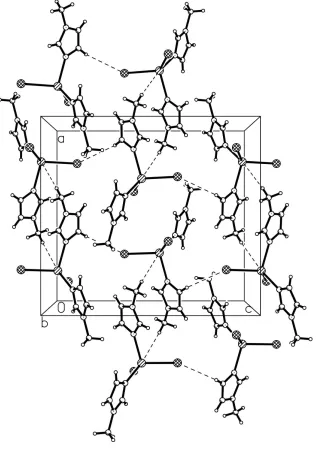
Figure 2
The packing of (I), viewed down the b-axis.
Diiodidobis(1-methylimidazole-κN3)cadmium(II)
Crystal data
[CdI2(C4H6N2)2]
Mr = 530.43
Orthorhombic, Pbca
Hall symbol: -P 2ac 2ab
a = 13.5570 (9) Å
b = 14.5615 (14) Å
V = 2953.0 (5) Å3
Z = 8
F(000) = 1936
Dx = 2.386 Mg m−3
µ = 5.64 mm−1
T = 298 K
Block, colorless 0.10 × 0.10 × 0.10 mm
Data collection
Bruker SMART 1K CCD area-detector diffractometer
Radiation source: fine-focus sealed tube Graphite monochromator
Thin–slice ω scans
Absorption correction: multi-scan (SADABS; Sheldrick, 2004)
Tmin = 0.574, Tmax = 0.579
2888 measured reflections 2768 independent reflections 1811 reflections with I > 2σ(I)
Rint = 0.013
θmax = 26.0°, θmin = 2.5°
h = 0→16
k = 0→17
l = 0→18
Refinement
Refinement on F2 Least-squares matrix: full
R[F2 > 2σ(F2)] = 0.063
wR(F2) = 0.178
S = 0.99 2768 reflections 137 parameters 40 restraints
Primary atom site location: structure-invariant direct methods
Secondary atom site location: difference Fourier map
Hydrogen site location: inferred from neighbouring sites
H-atom parameters constrained
w = 1/[σ2(F
o2) + (0.1P)2 + 1P] where P = (Fo2 + 2Fc2)/3 (Δ/σ)max = 0.001
Δρmax = 1.18 e Å−3 Δρmin = −0.85 e Å−3
Extinction correction: SHELXTL (Sheldrick, 2001), Fc*=kFc[1+0.001xFc2λ3/sin(2θ)]-1/4 Extinction coefficient: 0.0017 (2)
Special details
Geometry. All e.s.d.'s (except the e.s.d. in the dihedral angle between two l.s. planes) are estimated using the full covariance matrix. The cell e.s.d.'s are taken into account individually in the estimation of e.s.d.'s in distances, angles and torsion angles; correlations between e.s.d.'s in cell parameters are only used when they are defined by crystal symmetry. An approximate (isotropic) treatment of cell e.s.d.'s is used for estimating e.s.d.'s involving l.s. planes.
Refinement. Refinement of F2 against ALL reflections. The weighted R-factor wR and goodness of fit S are based on F2, conventional R-factors R are based on F, with F set to zero for negative F2. The threshold expression of F2 > σ(F2) is used only for calculating R-factors(gt) etc. and is not relevant to the choice of reflections for refinement. R-factors based on F2 are statistically about twice as large as those based on F, and R- factors based on ALL data will be even larger.
Fractional atomic coordinates and isotropic or equivalent isotropic displacement parameters (Å2)
x y z Uiso*/Ueq
Cd 1.20251 (6) 0.53829 (6) 1.04405 (6) 0.0520 (3)
I1 1.21055 (7) 0.59157 (7) 0.86882 (6) 0.0621 (3)
C1 1.5970 (10) 0.6525 (10) 1.0696 (12) 0.080 (5)
H1A 1.5798 0.7004 1.0284 0.120*
H1B 1.6475 0.6146 1.0439 0.120*
H1C 1.6208 0.6792 1.1242 0.120*
N1 1.5119 (6) 0.5977 (7) 1.0881 (7) 0.052 (3)
I2 1.13534 (7) 0.36699 (7) 1.08491 (8) 0.0707 (4)
N2 1.3612 (7) 0.5502 (7) 1.0836 (7) 0.052 (3)
C2 1.5085 (12) 0.5226 (11) 1.1438 (10) 0.070 (4)
H2A 1.5602 0.4954 1.1750 0.084*
C3 1.4143 (12) 0.4979 (10) 1.1428 (10) 0.072 (4)
H3A 1.3877 0.4512 1.1778 0.087*
N4 1.1068 (8) 0.6296 (7) 1.1229 (7) 0.052 (2)
C4 1.4208 (9) 0.6106 (9) 1.0530 (9) 0.054 (3)
H4A 1.4040 0.6566 1.0126 0.065*
C5 0.8536 (8) 0.6817 (10) 1.2037 (9) 0.061 (4)
H5A 0.8204 0.6280 1.1819 0.092*
H5B 0.8242 0.7355 1.1779 0.092*
H5C 0.8479 0.6844 1.2676 0.092*
C6 1.0338 (10) 0.7448 (10) 1.1951 (9) 0.066 (3)
H6A 1.0230 0.8005 1.2238 0.080*
C7 1.1210 (11) 0.7135 (11) 1.1622 (11) 0.078 (4)
H7A 1.1810 0.7443 1.1658 0.093*
C8 1.0131 (9) 0.6115 (9) 1.1374 (8) 0.054 (3)
H8A 0.9841 0.5563 1.1206 0.064*
Atomic displacement parameters (Å2)
U11 U22 U33 U12 U13 U23
Cd 0.0379 (5) 0.0590 (5) 0.0591 (6) 0.0005 (4) 0.0060 (4) 0.0008 (5)
I1 0.0644 (6) 0.0656 (6) 0.0564 (5) −0.0062 (4) −0.0039 (4) 0.0073 (4)
C1 0.046 (8) 0.069 (9) 0.125 (14) −0.023 (7) −0.017 (8) −0.024 (9)
N1 0.024 (5) 0.064 (6) 0.068 (7) −0.011 (4) 0.009 (5) −0.022 (6)
I2 0.0632 (6) 0.0605 (6) 0.0883 (8) −0.0060 (4) 0.0029 (5) 0.0166 (5)
N2 0.037 (5) 0.052 (6) 0.067 (7) 0.003 (5) −0.008 (5) −0.009 (5)
C2 0.068 (10) 0.081 (11) 0.062 (9) 0.013 (8) −0.010 (8) −0.012 (8)
N3 0.041 (4) 0.050 (5) 0.049 (5) 0.002 (4) 0.001 (4) −0.001 (4)
C3 0.081 (11) 0.064 (8) 0.073 (10) 0.027 (8) 0.003 (8) 0.016 (8)
N4 0.052 (5) 0.056 (5) 0.050 (5) 0.000 (4) 0.003 (4) 0.002 (4)
C4 0.038 (6) 0.065 (8) 0.059 (8) −0.022 (6) 0.010 (6) 0.006 (6)
C5 0.040 (7) 0.076 (9) 0.068 (9) 0.014 (6) −0.001 (6) 0.009 (7)
C6 0.060 (6) 0.056 (5) 0.083 (8) −0.001 (5) −0.007 (6) −0.013 (5)
C7 0.069 (6) 0.071 (6) 0.094 (8) −0.022 (5) 0.015 (6) −0.009 (6)
C8 0.052 (5) 0.055 (5) 0.054 (6) 0.002 (4) 0.007 (5) −0.010 (5)
Geometric parameters (Å, º)
Cd—N4 2.201 (10) N3—C8 1.313 (14)
Cd—N2 2.238 (9) N3—C6 1.392 (16)
Cd—I2 2.7248 (13) N3—C5 1.544 (14)
Cd—I1 2.7358 (13) C3—H3A 0.9300
C1—N1 1.429 (16) N4—C8 1.315 (16)
C1—H1A 0.9600 N4—C7 1.369 (18)
C1—H1B 0.9600 C4—H4A 0.9300
C1—H1C 0.9600 C5—H5A 0.9600
N1—C4 1.355 (16) C5—H5B 0.9600
N1—C2 1.377 (19) C5—H5C 0.9600
N2—C3 1.372 (16) C6—H6A 0.9300
C2—C3 1.33 (2) C7—H7A 0.9300
C2—H2A 0.9300 C8—H8A 0.9300
N4—Cd—N2 112.2 (4) C2—C3—N2 111.2 (14)
N4—Cd—I2 103.7 (3) C2—C3—H3A 124.4
N2—Cd—I2 109.4 (3) N2—C3—H3A 124.4
N4—Cd—I1 111.4 (3) C8—N4—C7 104.1 (11)
N2—Cd—I1 101.1 (3) C8—N4—Cd 122.5 (9)
I2—Cd—I1 119.20 (5) C7—N4—Cd 133.3 (9)
N1—C1—H1A 109.5 N2—C4—N1 110.0 (12)
N1—C1—H1B 109.5 N2—C4—H4A 125.0
H1A—C1—H1B 109.5 N1—C4—H4A 125.0
N1—C1—H1C 109.5 N3—C5—H5A 109.5
H1A—C1—H1C 109.5 N3—C5—H5B 109.5
H1B—C1—H1C 109.5 H5A—C5—H5B 109.5
C4—N1—C2 108.3 (10) N3—C5—H5C 109.5
C4—N1—C1 125.7 (12) H5A—C5—H5C 109.5
C2—N1—C1 126.0 (12) H5B—C5—H5C 109.5
C4—N2—C3 106.3 (12) C7—C6—N3 106.8 (12)
C4—N2—Cd 124.4 (9) C7—C6—H6A 126.6
C3—N2—Cd 129.2 (10) N3—C6—H6A 126.6
C3—C2—N1 103.9 (13) C6—C7—N4 109.3 (12)
C3—C2—H2A 128.0 C6—C7—H7A 125.3
N1—C2—H2A 128.0 N4—C7—H7A 125.3
C8—N3—C6 104.7 (10) N3—C8—N4 114.9 (12)
C8—N3—C5 129.7 (10) N3—C8—H8A 122.5
C6—N3—C5 125.6 (10) N4—C8—H8A 122.5
Hydrogen-bond geometry (Å, º)
D—H···A D—H H···A D···A D—H···A
C5—H5B···I1i 0.96 3.03 3.9797 169