Role of splice variants in the metastatic
progression of prostate cancer
Rachel M. Hagen1and Michael R. Ladomery1
Faculty of Health and Life Sciences, University of the West of England, Coldharbour Lane, Bristol BS16 1QY, U.K.
Abstract
AS (alternative splicing) and its role in disease, especially cancer, has come to forefront in research over the last few years. Alterations in the ratio of splice variants have been widely observed in cancer. Splice variants of cancer-associated genes have functions that can alter cellular phenotype, ultimately altering metastatic potential. As metastases are the cause of approximately 90% of all human cancer deaths, it is crucial to understand how AS is dysregulated in metastatic disease. We highlight some recent studies into the relationship between altered AS of key genes and the initiation of prostate cancer metastasis.
Introduction
Splicing was discovered in the late 1970s by scientists who were comparing the adenoviral mRNA sequence with that of its genome. They observed that particular genomic sequences were unable to hybridize to the mRNA, looping out. They had discovered the so-called intervening sequences now called ‘introns’. For this pioneering work, Phillip Sharp and Richard Roberts received the Nobel Prize in Medicine in 1993. In 1980, it was observed that the single immunoglobulin gene could produce two different protein products: a membrane-bound antibody and an antibody that could be secreted [1,2]. This was one of the first examples of AS (alternative splicing). The sequencing of the human genome has highlighted the fact that the number of genes does not explain the observed transcriptomic and proteomic complexity [3]. This apparent paradox can be explained through AS; it allows multiple mRNA variants to be produced from a single gene. Recent studies that use high-throughput sequencing indicate that up to 95% of human genes can generate multiple splice variants from a single pre-mRNA [4,5]. AS is widespread across eukaryotes, and understanding its nature and regulation has become a key question in molecular biology.
AS and its regulation
Pre-mRNA splicing is achieved by a set of ribonucleoprotein complexes, the snRNPs (small nuclear ribonucleoproteins), which together form the spliceosome when fully assembled on to pre-mRNAs. The U1 snRNP binds to 5!donor sites (at
Key words:alternative splicing, central alternative exon, epidermal growth factor, metastasis, prolactin, prostate cancer.
Abbreviations used:AS, alternative splicing; CAE, central alternative exon; EGFR, epidermal growth factor receptor; EMT, epithelial–mesenchymal transition; ERG, E26-related gene; hnRNP, heterogeneous nuclear ribonucleoprotein; MMP, matrix metalloproteinase; PP, protein phosphatase; PRLR, prolactin receptor; RBP, RNA-binding protein; SF, short form; snRNP, small nuclear ribonucleoprotein; SR, serine/arginine.
1Correspondence may be addressed to either author (email [email protected] or
the 3!-end of exons). The 3!splice acceptor site is preceded by a
polypyrimidine tract at the 3!-end of introns; this tract follows
the branchpoint A. U2AF65 binds the polypyrimidine tract facilitating the binding of U2 snRNP across the branchpoint A; the U4/5·6 tri-snRNP then associates, the spliceosome is fully formed and two transesterification reactions then occur. In the first reaction, the branchpoint A attacks the 5! splice site effectively cutting the 5!-end of the intron off,
forming a lariat; in the second, the upstream exon then joins the downstream exon and the intron is fully detached [6–8]. Several types of AS exist, including (i) exon skipping; (ii) the use of alternative 5!or 3!splice sites; (iii) mutually exclusive
exons; (iv) intron retention; and (v) alternative promoters or 3! processing sites [8]. The degree to which splice sites
conform to the consensus can contribute to the regulation of AS. Auxiliary sequences help the recruitment of the snRNPs; these are known as ESE/ESS/ISE/ISS (exonic or intronic splice enhancers or silencers). These are recognized by RBPs (RNA-binding proteins) known as splice factors. The most extensively researched classes of splice factors include the SR (serine/arginine)-rich proteins and the hnRNPs (heterogeneous nuclear ribonucleoproteins) [9]. Apoptosis is a well-studied example of a pathway that is regulated through AS. Apoptotic genes, such as those for Bcl-2, Bcl-x and caspase 9, can express pro-apoptotic or anti-apoptotic variants [10].
It is increasingly clear that the dysregulation of AS can produce huge consequences in relation to disease progression [11,12]. Alterations in AS have been found in numerous cancers, including lung, breast, ovarian and prostate [13– 16]. Although the extent of AS differs between tissue type, collectively in cancer cells a number of key pathways, including cell growth, apoptosis, cell signalling and cell motility, have altered AS patterns when compared with normal or benign samples. Thus it has become apparent that aberrant AS patterns may play a crucial role in the initiation and progression of cancer. However, it should also be noted that, due to the complex nature of cancer progression, AS
patterns and regulation can differ between primary tumours, circulating tumour cells and metastatic sites.
The use of bioinformatic approaches coupled with the analysis of expressed sequence tags, microarray data, and more recently next generation sequencing has dramatically increased the rate of discovery of new splice isoforms and particularly cancer-specific AS [6,8,14,16–20]. Kim et al. [16] demonstrated that cancers generally experience a reduction in exon skipping but an increase in the use of alternative 5!
and 3!splice sites and intron retention. The reduction in exon
skipping may be explained in part due to the observation that SR proteins are more prone to nonsense-mediated degradation in cancer [21]. Alterations in AS events may also occur as a result of changes in the expression and activity of RBPs. SR proteins are phosphorylated by kinases such as Akt, Clk1, DYRK1a (dual-specificity tyrosine-phosphorylated and -regulated kinase 1a) and SRPK1 (serine/arginine-rich protein-specific kinase 1) or dephosphorylated by PPs (protein phosphatases) such as PP1 and PP2A modulating both intracellular localization and activity [22]. Interestingly the EMT (epithelial–mesenchymal transition) programme has been associated with a number of alterations in splice isoform ratios and in the expression of splice factors. A recent study identified several RBPs as being altered in cancer cells, including the ESRPs (epithelial splicing regulatory proteins) and members of the RBFOX, CELF, MBNL and hnRNP families [23].
Metastasis in prostate cancer
The treatment of localized prostate cancer largely results in excellent survival outcome. In contrast, metastatic prostate cancer is associated with a decreased survival rate of 33% [24]. Metastasis occurs as a result of a number of events [25– 27]. In the initial stages, tumour cells can undergo phenotypic transformation changing from an epithelial to a mesenchymal cellular phenotype. This is associated with the loss of E-cadherin (a hallmark of epithelial cells that modulates cell– cell adherence) and the gain of N-cadherin (expressed in mesenchymal cells). This switch occurs in the so-called EMT that is crucial in the process of development. EMT results in loss of cell–cell adhesion and, as such, the cells can invade the surrounding stroma and enter surrounding blood vessels (intravasation). The degradation of the ECM (extracellular matrix) surrounding the tumour is initiated by the MMP (matrix metalloproteinase protein), and migratory cells often have increased MMP expression. The mesenchymal nature of the tumour cells increases the survival of the cells and their ability to resist apoptosis in response to chemotherapy through the release of fatty acids that protect the tumour cell from undergoing apoptosis [25]. Motile mesenchymal-like cancer cells are free to migrate and adhere to distinct sites of metastasis. Prostate cancer cells most commonly adhere to bone to form metastases, since the bone marrow and bone microenvironment provide optimum conditions to support the proliferation of prostate cancer cells [28,29]. A number of factors appear to cause a high proportion of prostate cancer
cells to migrate to bone, including high expression of BMPs (bone morphogenetic proteins) and TGFβ (transforming
growth factor β). Additionally, prostate cancer cells are
thought to bind to the bone marrow vasculature through specific cell–cell interactions via integrinαvβ3 and
protease-activated receptors, allowing the tumour cells to invade the bone marrow (extravasation) [30].
A number of cancer-specific splice variants [e.g. androgen receptor, FGF (fibroblast growth factor) receptor, CD44, pyruvate kinase, VEGF (vascular endothelial growth factor)] involved in the metastatic programme have been discovered of relevance to prostate cancer. We now highlight three recently discovered gene variants that can modify metastasis and possibly EMT (via E-cadherin) in prostate cancer and their potential use as therapeutic targets.
Examples of AS in metastatic prostate
cancer
ERG (E26-related gene)
ERG is a member of the ETS family of transcription factors and is the most consistently overexpressed gene in prostate cancer [31]. ERG has the potential to regulate multiple cellular pathways such as cell proliferation, differentiation, inflammation and bone formation. ERG expression is largely dysregulated as a result of a gene fusion event with the adjacent androgen-regulated TMPRSS2 promoter on chromosome 21 [32]. The presence of the TMPRSS2–ERG fusion and ERG overexpression results in increased tumour growth and invasive properties [33]. ERG overexpression down-regulates β1 integrin and E-cadherin expression in
prostate cancer cell line, suggesting that ERG may play a role in phenotypic alteration of cells to a more mesenchymal and motile state [34]. In addition, ERG also has the potential to directly regulate MMPs, the urokinase plasminogen activator (PLAU) and osteopontin, all involved in metastasis [33,35]. This pinpoints ERG as a potentially important driving factor in the progression of prostate cancer towards metastatic disease.
Although most research has concentrated on total ERG mRNA expression, recent studies suggest that the AS of ERG may play a role in altering cellular phenotypes. The
The PRLR (prolactin receptor)
Prolactin is synthesized in the pituitary gland and other tissues such as the breast and prostate in which it acts in an autocrine/paracrine manner. PRL initiates a signalling cascade via the PRLR that in turn can regulate many cellular pathways including cell growth, cell metabolism, angiogenesis and apoptosis [39]. ThePRLRgene comprises ten exons and is alternatively spliced to express multiple variants [40]. The commonest PRLR variants produced differ in the length of the signal transducing intracellular domain. As a result they are named LF (long form), IF (intermediate form) and SFs (short forms; SF1a and SF1b) [40,41]. Studies have shown that the SF1b variant amplifies signalling through ERK (extracellular-signal-regulated kinase) pathway and results in increased expression of the vitamin D receptor and the cell cycle regulatory protein p21, leading to a reduction in proliferation and cell cycle arrest [42]. A PRLR-specific antagonist, S179D, decreases cell growth and cell proliferation of prostate cancer cells in vitro and
in vivo via increased production of the SF1b variant [43,44]. Constitutive overexpression of the SF1b variant in prostate cancer cells, comparative to a long-term treatment plan using S179D, results in decreased cell proliferation and increased cell–matrix and cell–cell aggregation of cells; this correlates with increased E-cadherin and decreased MMP9 mRNA expression in SF1b-overexpressing cells. Consequently S179D-treated prostate cancer cells (via up-regulation of SF1b variant) show decreased invasive and migratory properties. Therefore overexpressing SF1b variant as a result of S179D treatment in the early stages of prostate cancer disease may prevent EMT initiation and metastasis.
The EGFR (epidermal growth factor receptor)
[image:3.595.320.549.178.416.2]EGFR is a member of the RTKs (receptor tyrosine kinases) family of growth factor receptors involved in proliferation, motility and cell survival. The EGFR gene is composed of 28 exons and can produce numerous variants. Variants with deletion in the extracellular domain result in increased proliferation of cancer cells and increased malignancy and subsequently correlate with a poor prognosis [45]. The most common variant with an altered extracellular domain is EGFRvIII, which is produced as a result of skipping of exons 2–7 and is ligand-independent and thus constitutively active. This variant is specific for tumour cells. Inhibition of EGF (epidermal growth factor) signalling in cancer cells restores E-cadherin levels [46]. Clinical trials using monoclonal antibodies and vaccines specifically directed against the EGFRvIII isoform are in progress [47]. Preliminary results using an EGFRvIII-targeted vaccine against malignant glioma show increased survival in patients that received the vaccine compared with control patients [47].In vivomurine models of cancer also show the efficacy of the vaccine in decreasing tumour growth [47]. Therefore this vaccine may be relevant in a number of cancers, including prostate. Recently a new variant of EGFR in which exon 4 is skipped (de4 EGFR) was discovered in glioma, ovarian and prostate cancer
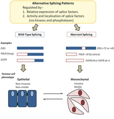
Figure 1 Aberrant splicing patterns can affect cellular phenotypes
Alterations in AS can affect the inclusion of exons within genes. For example the inclusion of 72 bp exon 11 ofERGand exon skipping in thePRLRandEGFRgenes can confer a more mesenchymal phenotype in tumour cells (potentially via a reduction in E-cadherin expression) resulting in increased potential for migration and invasion. Studies suggest that cells may return to a more epithelial phenotype (with decreased metastatic potential) if aberrant splicing patterns can be restored to wild-type patterns.
[48]. As with the EGFRvIII variant, the de4 EGFR variant appears to be cancer-specific and is not found in normal or tumour adjacent samples [48].In vitroandin vivostudies show that the de4 EGFR variant has increased metastatic potential compared with wild-type EGFR [48]. Skipping of exon 4 disrupts EGFR-binding activity. As a result the de4 EGFR variants self-dimerize, leading to constitutive tyrosine phosphorylation and activation of the receptor and downstream signalling events. One effect is the up-regulation of the MAPK (mitogen-activated protein kinase) pathway and the down-regulation of E-cadherin; this is inversely related to cell invasion. The increase in metastatic potential observed in that study warrants further research to assess the potential of the de4 EGFR variant in prognosis and as a novel therapeutic target in metastatic prostate cancer.
Conclusion
highly attractive therapeutic targets, since only cancer cells will be targeted, decreasing toxicity towards normal cells. Importantly, as shown in the examples given here, there are already promising therapies against cancer-specific variants in metastatic prostate cancer. An attractive possibility with the PRLR-specific antagonist and the EGFR therapies is the potential ability to increase E-cadherin expression in mesenchymal cells by changing the splicing patterns of regulatory genes such asPRLRandEGFR(Figure 1). This may then reverse the cells to a more epithelial phenotype which is non-motile in nature. The concept of EMT reversal has been described previously [50]. The reversal to a more epithelial cellular phenotype may result in preventing initial stages of the metastatic programme. In addition, manipulating AS to alter the mesenchymal characteristics of prostate cancer cells may also result in the increase susceptibility of these cells to undergo apoptosis as a result of chemotherapy. Thus combining these treatments may result in a better clinical outcome for patients.
Funding
R.M.H. was supported by a QR-funded grant (University of the West of England, Bristol, U.K.).
References
1 Alt, F.W., Bothwell, A.L., Knapp, M., Siden, E., Mather, E., Koshland, M. and Baltimore, D. (1980) Synthesis of secreted and membrane-bound immunoglobulin mu heavy chains is directed by mRNAs that differ at their 3!ends. Cell20, 293–301
2 Early, P., Rogers, J., Davis, M., Calame, K., Bond, M., Wall, R. and Hood, L. (1980) Two mRNAs can be produced from a single immunoglobulinµ
gene by alternative RNA processing pathways. Cell20, 313–319 3 International Human Genome Sequencing Consortium (2004) Finishing
the euchromatic sequence of the human genome. Nature431, 931–945 4 Pan, Q., Shai, O., Lee, L.J., Frey, B.J. and Blencowe, B.J. (2008) Deep
surveying of alternative splicing complexity in the human transcriptome by high-throughput sequencing. Nature Genet.40, 1413–1415 5 Wang, E.T., Sandberg, R., Luo, S., Khrebtukova, I., Zhang, L., Mayr, C.,
Kingsmore, S.F., Schroth, G.P. and Burge, C.B. (2008) Alternative isoform regulation in human tissue transcriptomes. Nature456, 470–476 6 David, C.J. and Manley, J.L. (2010) Alternative pre-mRNA splicing
regulation in cancer: pathways and programs unhinged. Genes Dev.24, 2343–2364
7 Wahl, M.C., Will, C.L. and Luhrmann, R. (2009) The spliceosome: design principles of a dynamic RNP machine. Cell136, 701–718
8 Blencowe, B.J. (2006) Alternative splicing: new insights from global analyses. Cell126, 37–47
9 Nilsen, T.W. and Graveley, B.R. (2010) Expansion of the eukaryotic proteome by alternative splicing. Nature463, 457–463
10 Schwerk, C. and Schulze-Osthoff, K. (2005) Regulation of apoptosis by alternative pre-mRNA splicing. Mol. Cell19, 1–13
11 Blencowe, B.J. (2000) Exonic splicing enhancers: mechanism of action, diversity and role in human genetic diseases. Trends Biochem. Sci.25, 106–110
12 Black, D.L. (2003) Mechanisms of alternative pre-messenger RNA splicing. Annu. Rev. Biochem.72, 291–336
13 Venables, J.P., Klinck, R., Koh, C., Gervais-Bird, J., Bramard, A., Inkel, L., Durand, M., Couture, S., Froehlich, U., Lapointe, E. et al. (2009) Cancer-associated regulation of alternative splicing. Nat. Struct. Mol. Biol.
16, 670–676
14 Skotheim, R.I. and Nees, M. (2007) Alternative splicing in cancer: noise, functional, or systematic? Int. J. Biochem. Cell Biol.39, 1432–1449 15 Srebrow, A. and Kornblihtt, A.R. (2006) The connection between splicing
and cancer. J. Cell Sci.119, 2635–2641
16 Kim, E., Goren, A. and Ast, G. (2008) Insights into the connection between cancer and alternative splicing. Trends Genet.24, 7–10 17 Lee, C. and Wang, Q. (2005) Bioinformatics analysis of alternative
splicing. Brief. Bioinform.6, 23–33
18 Kechris, K., Yang, Y.H. and Yeh, R.F. (2008) Prediction of alternatively skipped exons and splicing enhancers from exon junction arrays. BMC Genomics9, 551
19 Barash, Y., Calarco, J.A., Gao, W., Pan, Q., Wang, X., Shai, O., Blencowe, B.J. and Frey, B.J. (2010) Deciphering the splicing code. Nature465, 53–59
20 Fackenthal, J.D. and Godley, L.A. (2008) Aberrant RNA splicing and its functional consequences in cancer cells. Dis. Models Mech.1, 37–42 21 Ni, J.Z., Grate, L., Donohue, J.P., Preston, C., Nobida, N., O’Brien, G., Shiue,
L., Clark, T.A., Blume, J.E. and Ares, Jr, M. (2007) Ultraconserved elements are associated with homeostatic control of splicing regulators by alternative splicing and nonsense-mediated decay. Genes Dev.21, 708–718
22 Chen, M. and Manley, J.L. (2009) Mechanisms of alternative splicing regulation: insights from molecular and genomics approaches. Nat. Rev. Mol. Cell Biol.10, 741–754
23 Shapiro, I.M., Cheng, A.W., Flytzanis, N.C., Balsamo, M., Condeelis, J.S., Oktay, M.H., Burge, C.B. and Gertler, F.B. (2011) An EMT-driven alternative splicing program occurs in human breast cancer and modulates cellular phenotype. PLoS Genet.7, e1002218
24 Jemal, A., Siegel, R., Ward, E., Murray, T., Xu, J. and Thun, M.J. (2007) Cancer statistics, 2007. Ca-Cancer J. Clin.57, 43–66
25 Hugo, H., Ackland, M.L., Blick, T., Lawrence, M.G., Clements, J.A., Williams, E.D. and Thompson, E.W. (2007) Epithelial–mesenchymal and mesenchymal–epithelial transitions in carcinoma progression. J. Cell. Physiol.213, 374–383
26 Bozzuto, G., Ruggieri, P. and Molinari, A. (2010) Molecular aspects of tumor cell migration and invasion. Ann. Ist. Super. Sanita46, 66–80 27 Karayi, M.K. and Markham, A.F. (2004) Molecular biology of prostate
cancer. Prostate Cancer Prostatic Dis.7, 6–20
28 Kingsley, L.A., Fournier, P.G., Chirgwin, J.M. and Guise, T.A. (2007) Molecular biology of bone metastasis. Mol. Cancer Ther.6, 2609–2617 29 Bonfil, R.D., Chinni, S., Fridman, R., Kim, H.R. and Cher, M.L. (2007)
Proteases, growth factors, chemokines, and the microenvironment in prostate cancer bone metastasis. Urol. Oncol.25, 407–411 30 Morrissey, C. and Vessella, R.L. (2007) The role of tumor
microenvironment in prostate cancer bone metastasis. J. Cell. Biochem.
101, 873–886
31 Petrovics, G., Liu, A., Shaheduzzaman, S., Furusato, B., Sun, C., Chen, Y., Nau, M., Ravindranath, L., Chen, Y., Dobi, A. et al. (2005) Frequent overexpression of ETS-related gene-1 (ERG1) in prostate cancer transcriptome. Oncogene24, 3847–3852
32 Tomlins, S.A., Rhodes, D.R., Perner, S., Dhanasekaran, S.M., Mehra, R., Sun, X.W., Varambally, S., Cao, X., Tchinda, J., Kuefer, R. et al. (2005) Recurrent fusion ofTMPRSS2andETStranscription factor genes in prostate cancer. Science310, 644–648
33 Tomlins, S.A., Laxman, B., Varambally, S., Cao, X., Yu, J., Helgeson, B.E., Cao, Q., Prensner, J.R., Rubin, M.A., Shah, R.B. et al. (2008) Role of the TMPRSS2–ERGgene fusion in prostate cancer. Neoplasia10, 177–188 34 Gupta, S., Iljin, K., Sara, H., Mpindi, J.P., Mirtti, T., Vainio, P., Rantala, J., Alanen, K., Nees, M. and Kallioniemi, O. (2010) FZD4 as a mediator of ERG oncogene-induced WNT signaling and epithelial-to-mesenchymal transition in human prostate cancer cells. Cancer Res.70, 6735–6745 35 Flajollet, S., Tian, T.V., Flourens, A., Tomavo, N., Villers, A., Bonnelye, E.,
Aubert, S., Leroy, X. and Duterque-Coquillaud, M. (2011) Abnormal expression of the ERG transcription factor in prostate cancer cells activates osteopontin. Mol. Cancer Res.9, 914–924
36 Wang, J., Cai, Y., Yu, W., Ren, C., Spencer, D.M. and Ittmann, M. (2008) Pleiotropic biological activities of alternatively splicedTMPRSS2/ERG fusion gene transcripts. Cancer Res.68, 8516–8524
37 Owczarek, C.M., Portbury, K.J., Hardy, M.P., O’Leary, D.A., Kudoh, J., Shibuya, K., Shimizu, N., Kola, I. and Hertzog, P.J. (2004) Detailed mapping of theERG–ETS2interval of human chromosome 21 and comparison with the region of conserved synteny on mouse chromosome 16. Gene324, 65–77
38 Jhavar, S., Reid, A., Clark, J., Kote-Jarai, Z., Christmas, T., Thompson, A., Woodhouse, C., Ogden, C., Fisher, C., Corbishley, C. et al. (2008) Detection ofTMPRSS2–ERGtranslocations in human prostate cancer by expression profiling using GeneChip Human Exon 1.0 ST arrays. J. Mol. Diagn.10, 50–57
40 Hu, Z.Z., Meng, J. and Dufau, M.L. (2001) Isolation and characterization of two novel forms of the human prolactin receptor generated by alternative splicing of a newly identified exon 11. J. Biol. Chem.276, 41086–41094
41 Trott, J.F., Hovey, R.C., Koduri, S. and Vonderhaar, B.K. (2003) Alternative splicing to exon 11 of human prolactin receptor gene results in multiple isoforms including a secreted prolactin-binding protein. J. Mol. Endocrinol.30, 31–47
42 Wang, H., Zhou, M., Shi, B., Zhang, Q., Jiang, H., Sun, Y., Liu, J., Zhou, K., Yao, M., Gu, J. et al. (2011) Identification of an exon 4-deletion variant of epidermal growth factor receptor with increased metastasis-promoting capacity. Neoplasia13, 461–471
43 Wu, W., Ginsburg, E., Vonderhaar, B.K. and Walker, A.M. (2005) S179D prolactin increases vitamin D receptor and p21 through up-regulation of short 1b prolactin receptor in human prostate cancer cells. Cancer Res.
65, 7509–7515
44 Huang, K.T. and Walker, A.M. (2010) Long term increased expression of the short form 1b prolactin receptor in PC-3 human prostate cancer cells decreases cell growth and migration, and causes multiple changes in gene expression consistent with reduced invasive capacity. Prostate70, 37–47
45 Huang, P.H., Miraldi, E.R., Xu, A.M., Kundukulam, V.A., Del Rosario, A.M., Flynn, R.A., Cavenee, W.K., Furnari, F.B. and White, F.M. (2010) Phosphotyrosine signaling analysis of site-specific mutations on EGFRvIII identifies determinants governing glioblastoma cell growth. Mol. BioSyst.6, 1227–1237
46 Yates, C.C., Shepard, C.R., Stolz, D.B. and Wells, A. (2007) Co-culturing human prostate carcinoma cells with hepatocytes leads to increased expression of E-cadherin. Br. J. Cancer96, 1246–1252
47 Sampson, J.H., Heimberger, A.B., Archer, G.E., Aldape, K.D., Friedman, A.H., Friedman, H.S., Gilbert, M.R., Herndon, II, J.E., McLendon, R.E., Mitchell, D.A. et al. (2010) Immunologic escape after prolonged progression-free survival with epidermal growth factor receptor variant III peptide vaccination in patients with newly diagnosed glioblastoma. J. Clin. Oncol.28, 4722–4729
48 Wang, H., Shi, B., Zhang, Q., Jiang, H., Hu, S., Kong, J., Yao, M., Yang, S. and Li, Z. (2012) Growth and metastasis suppression of glioma xenografts expressing exon 4-deletion variant of epidermal growth factor receptor by monoclonal antibody CH12-mediated receptor degradation. FASEB J.26, 73–80
49 Xu, Q. and Lee, C. (2003) Discovery of novel splice forms and functional analysis of cancer-specific alternative splicing in human expressed sequences. Nucleic Acids Res.31, 5635–5643
50 Das, S., Becker, B.N., Hoffmann, F.M. and Mertz, J.E. (2009) Complete reversal of epithelial to mesenchymal transition requires inhibition of both ZEB expression and the Rho pathway. BMC Cell Biol.10, 94