(
E
)-2-Acetylpyrazine
4-nitrophenyl-hydrazone
Shang Shan,* Yu-Liang Tian, Shan-Heng Wang, Wen-Long Wang and Ying-Li Xu
College of Chemical Engineering and Materials Science, Zhejiang University of Technology, People’s Republic of China
Correspondence e-mail: [email protected]
Received 7 June 2008; accepted 10 June 2008
Key indicators: single-crystal X-ray study;T= 295 K; mean(C–C) = 0.003 A˚;
Rfactor = 0.043;wRfactor = 0.150; data-to-parameter ratio = 15.8.
In the title compound, C12H11N5O2, the molecule adopts anE configuration, with the benzene and pyrazine rings located on opposite sides of the N C double bond. The face-to-face separations of 3.413 (14) and 3.430 (8) A˚ , respectively between parallel benzene rings and between pyrazine rings indicate the existence of – stacking between adjacent molecules. The crystal structure also contains N—H N and C—H O hydrogen bonding.
Related literature
For general background, see: Okabe et al. (1993); Huet al.
(2001); Chenet al.(2007). For a related structure, see: Shanet al.(2008).
Experimental
Crystal data
C12H11N5O2
Mr= 257.26 Monoclinic,P21=n a= 8.0101 (6) A˚
b= 12.5154 (11) A˚ c= 12.1506 (12) A˚ = 98.564 (2)
V= 1204.51 (18) A˚3
Z= 4
MoKradiation = 0.10 mm1
T= 295 (2) K 0.400.380.26 mm
Data collection
Rigaku R-AXIS RAPID IP diffractometer
Absorption correction: none 11633 measured reflections
2747 independent reflections 1446 reflections withI> 2(I) Rint= 0.033
Refinement
R[F2> 2(F2)] = 0.042
wR(F2) = 0.149
S= 1.08 2747 reflections
174 parameters
H-atom parameters constrained
max= 0.20 e A˚
3 min=0.19 e A˚3
Table 1
Hydrogen-bond geometry (A˚ ,).
D—H A D—H H A D A D—H A
N2—H2N N5i
0.91 2.30 3.185 (2) 164
C11—H11 O1ii 0.93 2.60 3.300 (3) 133
Symmetry codes: (i)xþ1 2;yþ
1 2;zþ
1
2; (ii)x1;y1;z.
Data collection: PROCESS-AUTO (Rigaku, 1998); cell refine-ment:PROCESS-AUTO; data reduction:CrystalStructure(Rigaku, 2002); program(s) used to solve structure:SIR92 (Altomareet al., 1993); program(s) used to refine structure:SHELXL97(Sheldrick, 2008); molecular graphics:ORTEP-3 for Windows(Farrugia, 1997); software used to prepare material for publication:WinGX(Farrugia, 1999).
The work was supported by the Natural Science Foundation of Zhejiang Province of China (No. M203027).
Supplementary data and figures for this paper are available from the IUCr electronic archives (Reference: SG2248).
References
Altomare, A., Cascarano, G., Giacovazzo, C. & Guagliardi, A. (1993).J. Appl. Cryst.26, 343–350.
Chen, Z.-Y., Wu, G.-Q., Jiang, F.-X., Tian, Y.-L. & Shan, S. (2007).Acta Cryst. E63, o1919–o1920.
Farrugia, L. J. (1997).J. Appl. Cryst.30, 565. Farrugia, L. J. (1999).J. Appl. Cryst.32, 837–838.
Hu, W., Sun, N. & Yang, Z. (2001).Chem. J. Chin. Univ.22, 2014–2017. Okabe, N., Nakamura, T. & Fukuda, H. (1993).Acta Cryst.C49, 1678–1680. Rigaku. (1998).PROCESS-AUTO. Rigaku Corporation, Tokyo, Japan. Rigaku. (2002).CrystalStructure. Rigaku/MSC, The Woodlands, Texas, USA. Shan, S., Tian, Y.-L., Wang, S.-H., Wang, W.-L. & Xu, Y.-L. (2008).Acta Cryst.
E64, o1153.
Sheldrick, G. M. (2008).Acta Cryst.A64, 112–122.
Acta Crystallographica Section E Structure Reports
Online
supporting information
Acta Cryst. (2008). E64, o1265 [doi:10.1107/S1600536808017479]
(
E
)-2-Acetylpyrazine 4-nitrophenylhydrazone
Shang Shan, Yu-Liang Tian, Shan-Heng Wang, Wen-Long Wang and Ying-Li Xu
S1. Comment
Hydrazone and its derivatives have attracted much attention because of their potential application in biology (Okabe et
al., 1993; Hu et al., 2001). As part of an ongoing investigation into anti-cancer compounds (Chen et al., 2007), the title
compound has recently been prepared in our laboratory and its crystal structure is presented here.
The molecular structure of the title compound is shown in Fig. 1. The molecule adopts an E-configuration, with the
benzene and pyrazine rings located on the opposite positions of the N3=C7 double bond, similar to that found in a related
structure, (E)-2-Furyl methyl ketone 2,4-dinitrophenylhydrazone (Shan et al., 2008). The pyrazine plane is twisted with
respect to the benzene ring by a smaller dihedral angle of 14.25 (10)°.
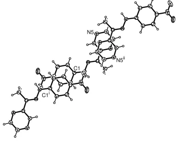
The partially overlapped arrangement is observed between parallel benzene rings and between parallel pyrazine rings
(Fig. 2), face-to-face separations of 3.413 (14) [for benzene rings] and 3.430 (8) Å [for pyrazine rings] are significantly
shorter than van der Waals thickness of the aromatic ring (3.70 Å), and indicate the existence of π-π stacking between the
adjacent molecules. Intermolecular N—H···N and weak C—H···O hydrogen bondings are present in the crystal structure
(Table 1).
S2. Experimental
4-Nitrophenylhydrazine (0.31 g, 2 mmol) was dissolved in ethanol (10 ml), then H2SO4 solution (98%, 0.5 ml) was added
slowly to the ethanol solution with stirring. The solution was heated at about 333 K for several minutes until the solution
cleared. An ethanol solution (5 ml) of acetylpyrazine (0.24 g, 2 mmol) was dropped slowly into the above solution with
continuous stirring, and the mixture solution was kept at about 333 K for 0.5 h. When the solution had cooled to room
temperature, yellow microcrystals appeared. They were separated and washed with cold water three times to get the
product 0.40 g. Single crystals of the title compound were obtained by recrystallization from an absolute ethanol solution.
S3. Refinement
Methyl H atoms were placed in calculated positions with C—H = 0.96 Å and torsion angle was refined to fit the electron
density, Uiso(H) = 1.5Ueq(C). Imino H atom was located in a difference Fourier map and refined as riding in its as-found
relative position, Uiso(H) = 1.2Ueq(N). Aromatic H atoms were placed in calculated positions with C—H = 0.93 and
Figure 1
The molecular structure of (I) with 30% probability displacement ellipsoids (arbitrary spheres for H atoms), dashed line
indicates hydrogen bonding.
Figure 2
[image:3.610.123.489.358.653.2](E)-2-Acetylpyrazine 4-nitrophenylhydrazone
Crystal data
C12H11N5O2
Mr = 257.26
Monoclinic, P21/n
Hall symbol: -P 2yn a = 8.0101 (6) Å b = 12.5154 (11) Å c = 12.1506 (12) Å β = 98.564 (2)° V = 1204.51 (18) Å3
Z = 4
F(000) = 536 Dx = 1.419 Mg m−3
Melting point: 498 K
Mo Kα radiation, λ = 0.71069 Å Cell parameters from 4236 reflections θ = 3.2–25.0°
µ = 0.10 mm−1
T = 295 K Prism, yellow
0.40 × 0.38 × 0.26 mm
Data collection
Rigaku R-AXIS RAPID IP diffractometer
Radiation source: fine-focus sealed tube Graphite monochromator
Detector resolution: 10.00 pixels mm-1
ω scans
11633 measured reflections
2747 independent reflections 1446 reflections with I > 2σ(I) Rint = 0.033
θmax = 27.4°, θmin = 3.0°
h = −10→10 k = −16→16 l = −15→15
Refinement
Refinement on F2
Least-squares matrix: full R[F2 > 2σ(F2)] = 0.042
wR(F2) = 0.149
S = 1.09 2747 reflections 174 parameters 0 restraints
Primary atom site location: structure-invariant direct methods
Secondary atom site location: difference Fourier map
Hydrogen site location: inferred from neighbouring sites
H-atom parameters constrained w = 1/[σ2(F
o2) + (0.0615P)2 + 0.2787P]
where P = (Fo2 + 2Fc2)/3
(Δ/σ)max = 0.001
Δρmax = 0.20 e Å−3
Δρmin = −0.19 e Å−3
Extinction correction: SHELXL97 (Sheldrick, 2008), Fc*=kFc[1+0.001xFc2λ3/sin(2θ)]-1/4
Extinction coefficient: 0.024 (3)
Special details
Geometry. All e.s.d.'s (except the e.s.d. in the dihedral angle between two l.s. planes) are estimated using the full covariance matrix. The cell e.s.d.'s are taken into account individually in the estimation of e.s.d.'s in distances, angles and torsion angles; correlations between e.s.d.'s in cell parameters are only used when they are defined by crystal symmetry. An approximate (isotropic) treatment of cell e.s.d.'s is used for estimating e.s.d.'s involving l.s. planes.
Refinement. Refinement of F2 against ALL reflections. The weighted R-factor wR and goodness of fit S are based on F2,
conventional R-factors R are based on F, with F set to zero for negative F2. The threshold expression of F2 > σ(F2) is used
only for calculating R-factors(gt) etc. and is not relevant to the choice of reflections for refinement. R-factors based on F2
are statistically about twice as large as those based on F, and R- factors based on ALL data will be even larger.
Fractional atomic coordinates and isotropic or equivalent isotropic displacement parameters (Å2)
x y z Uiso*/Ueq
N1 0.9003 (3) 0.68785 (16) 0.3418 (2) 0.0684 (6)
N2 0.6332 (2) 0.36085 (12) 0.57394 (13) 0.0448 (4)
N3 0.5365 (2) 0.28334 (12) 0.51838 (13) 0.0425 (4)
N4 0.3197 (2) 0.04376 (14) 0.55830 (14) 0.0518 (5)
N5 0.2175 (2) 0.07812 (15) 0.33069 (14) 0.0571 (5)
O1 0.9806 (3) 0.75846 (16) 0.3955 (2) 0.1041 (8)
O2 0.8729 (3) 0.68826 (16) 0.2397 (2) 0.1012 (8)
C1 0.6976 (2) 0.43994 (15) 0.51344 (16) 0.0412 (5)
C2 0.7855 (3) 0.52394 (16) 0.57162 (17) 0.0490 (5)
H2 0.7989 0.5251 0.6490 0.059*
C3 0.8519 (3) 0.60439 (16) 0.51558 (18) 0.0521 (5)
H3 0.9109 0.6601 0.5544 0.062*
C4 0.8306 (3) 0.60198 (15) 0.40117 (18) 0.0488 (5)
C5 0.7461 (3) 0.51920 (17) 0.34168 (17) 0.0513 (5)
H5 0.7339 0.5187 0.2644 0.062*
C6 0.6803 (3) 0.43744 (17) 0.39758 (16) 0.0482 (5)
H6 0.6246 0.3809 0.3583 0.058*
C7 0.4778 (2) 0.20837 (14) 0.57459 (15) 0.0408 (5)
C8 0.5120 (3) 0.19727 (18) 0.69835 (17) 0.0593 (6)
H8A 0.6317 0.1939 0.7222 0.089*
H8B 0.4602 0.1331 0.7202 0.089*
H8C 0.4663 0.2578 0.7322 0.089*
C9 0.3700 (2) 0.13023 (15) 0.50718 (15) 0.0408 (5)
C10 0.3179 (3) 0.14575 (16) 0.39353 (16) 0.0496 (5)
H10 0.3553 0.2063 0.3602 0.059*
C11 0.1699 (3) −0.00860 (18) 0.38283 (19) 0.0569 (6)
H11 0.1002 −0.0587 0.3424 0.068*
C12 0.2214 (3) −0.02519 (17) 0.49398 (19) 0.0554 (6)
H12 0.1865 −0.0871 0.5263 0.067*
Atomic displacement parameters (Å2)
U11 U22 U33 U12 U13 U23
C11 0.0619 (15) 0.0520 (13) 0.0561 (13) −0.0091 (11) 0.0064 (11) −0.0063 (11) C12 0.0634 (14) 0.0473 (12) 0.0568 (13) −0.0107 (10) 0.0127 (11) 0.0019 (11)
Geometric parameters (Å, º)
N1—O1 1.223 (3) C3—H3 0.9300
N1—O2 1.226 (3) C4—C5 1.381 (3)
N1—C4 1.452 (3) C5—C6 1.376 (3)
N2—N3 1.357 (2) C5—H5 0.9300
N2—C1 1.378 (2) C6—H6 0.9300
N2—H2N 0.9106 C7—C9 1.470 (3)
N3—C7 1.290 (2) C7—C8 1.494 (3)
N4—C12 1.339 (3) C8—H8A 0.9600
N4—C9 1.339 (2) C8—H8B 0.9600
N5—C10 1.328 (3) C8—H8C 0.9600
N5—C11 1.340 (3) C9—C10 1.395 (3)
C1—C6 1.394 (3) C10—H10 0.9300
C1—C2 1.397 (3) C11—C12 1.368 (3)
C2—C3 1.367 (3) C11—H11 0.9300
C2—H2 0.9300 C12—H12 0.9300
C3—C4 1.375 (3)
O1—N1—O2 122.5 (2) C5—C6—C1 119.6 (2)
O1—N1—C4 118.7 (2) C5—C6—H6 120.2
O2—N1—C4 118.8 (2) C1—C6—H6 120.2
N3—N2—C1 118.65 (15) N3—C7—C9 114.78 (16)
N3—N2—H2N 121.3 N3—C7—C8 125.00 (18)
C1—N2—H2N 119.8 C9—C7—C8 120.23 (17)
C7—N3—N2 118.88 (16) C7—C8—H8A 109.5
C12—N4—C9 116.19 (18) C7—C8—H8B 109.5
C10—N5—C11 115.78 (18) H8A—C8—H8B 109.5
N2—C1—C6 122.25 (18) C7—C8—H8C 109.5
N2—C1—C2 118.10 (17) H8A—C8—H8C 109.5
C6—C1—C2 119.64 (19) H8B—C8—H8C 109.5
C3—C2—C1 120.42 (19) N4—C9—C10 120.35 (18)
C3—C2—H2 119.8 N4—C9—C7 118.11 (16)
C1—C2—H2 119.8 C10—C9—C7 121.53 (17)
C2—C3—C4 119.2 (2) N5—C10—C9 123.13 (19)
C2—C3—H3 120.4 N5—C10—H10 118.4
C4—C3—H3 120.4 C9—C10—H10 118.4
C3—C4—C5 121.53 (19) N5—C11—C12 121.6 (2)
C3—C4—N1 119.1 (2) N5—C11—H11 119.2
C5—C4—N1 119.3 (2) C12—C11—H11 119.2
C6—C5—C4 119.56 (19) N4—C12—C11 122.9 (2)
C6—C5—H5 120.2 N4—C12—H12 118.6
Hydrogen-bond geometry (Å, º)
D—H···A D—H H···A D···A D—H···A
N2—H2N···N5i 0.91 2.30 3.185 (2) 164
C11—H11···O1ii 0.93 2.60 3.300 (3) 133