(
Z
)-
tert
-Butyl 2-(4-amino-9
H
-fluoren-9-ylidene)acetate
Marios Krokidis, Dionissios Papaioannou and Vassilios Nastopoulos*
Department of Chemistry, University of Patras, 265 04 Patras, Greece Correspondence e-mail: nastopoulos@chemistry.upatras.gr
Received 4 September 2008; accepted 16 September 2008
Key indicators: single-crystal X-ray study;T= 100 K; mean(C–C) = 0.004 A˚; Rfactor = 0.063;wRfactor = 0.143.
The title compound, C19H19NO2, obtained as an almost
equimolar mixture (as shown by1H NMR) with theEisomer through a Wittig reaction between 4-amino-9H-fluoren-9-one and the stabilized ylide Ph3P CHCO2C(CH3)3, was obtained
pure in the Z configuration following crystallization from toluene. The molecule shows a planar arrangement of the ring system and the new double bond, whereas the carbonyl O atom forms a 45.1 (3)dihedral angle with it. The molecules are linked by N—H O hydrogen bonds, forming cyclic structures with R4
4
(24) graph-set motifs. These motifs are connected to each other, giving rise to a sheet structure parallel to the ab plane. The linkage within the sheets is further enhanced by – stacking interactions between the fluorene units [centroid–centroid distance = 3.583 (2) A˚ ].
Related literature
For general background on retinoids, see: Meyeret al.(1978); Spornet al.(1994). For hydrogen-bond motifs, see: Bernstein et al. (1995). For related literature, see: Magoulas & Papaioannou (2003).
Experimental
Crystal data
C19H19NO2 Mr= 293.35
Orthorhombic,Pbca a= 9.0820 (12) A˚
b= 13.7330 (17) A˚
c= 24.568 (3) A˚
V= 3064.2 (7) A˚3
Z= 8
MoKradiation = 0.08 mm1 T= 100 (2) K 0.320.260.16 mm
Data collection
Oxford Diffraction Xcalibur-3 with Sapphire CCD diffractometer Absorption correction: multi-scan
(CrysAlis RED; Oxford Diffraction, 2008)
Tmin= 0.956,Tmax= 0.989
18331 measured reflections 2671 independent reflections 1671 reflections withI> 2(I)
Rint= 0.119
Refinement
R[F2> 2(F2)] = 0.063 wR(F2) = 0.143 S= 1.01 2671 reflections 211 parameters
H atoms treated by a mixture of independent and constrained refinement
max= 0.30 e A˚
3 min=0.22 e A˚
3
Table 1
Hydrogen-bond geometry (A˚ ,).
D—H A D—H H A D A D—H A
N—H1N O1i 0.85 (3) 2.62 (3) 3.295 (3) 137 (3)
N—H2N O2ii
0.92 (3) 2.24 (3) 3.133 (3) 163 (3)
Symmetry codes: (i)x;yþ1 2;zþ
3 2; (ii)x
1 2;y;zþ
3 2.
Data collection: CrysAlis CCD (Oxford Diffraction, 2008); cell refinement: CrysAlis RED (Oxford Diffraction, 2008); data reduc-tion: CrysAlis RED; program(s) used to solve structure: SIR92 (Altomare et al., 1994); program(s) used to refine structure: SHELXL97(Sheldrick, 2008); molecular graphics:PLATON(Spek, 2003), ORTEP-3(Farrugia, 1997) and DIAMOND (Brandenburg, 2008); software used to prepare material for publication: WinGX (Farrugia, 1999) andpublCIF(Westrip, 2008).
The authors thank Dr A. Tasiopoulos (Department of Chemistry, University of Cyprus) for help with the data collection.
Supplementary data and figures for this paper are available from the IUCr electronic archives (Reference: DN2370).
References
Altomare, A., Cascarano, G., Giacovazzo, C., Guagliardi, A., Burla, M. C., Polidori, G. & Camalli, M. (1994).J. Appl. Cryst.27, 435–436.
Bernstein, J., Davis, R. E., Shimoni, L. & Chang, N.-L. (1995).Angew. Chem. Int. Ed. Engl.34, 1555–1573.
Brandenburg, K. (2008).DIAMOND. Crystal Impact GbR, Bonn, Germany. Farrugia, L. J. (1997).J. Appl. Cryst.30, 565.
Farrugia, L. J. (1999).J. Appl. Cryst.32, 837–838.
Magoulas, G. & Papaioannou, D. (2003).ARKIVOC,vi, 213–227.
Meyer, H., Bollag, W., Ha¨nni, R. & Ru¨egg, R. (1978).Experientia (Generalia), 34, 1105–1246.
Oxford Diffraction (2008). CrysAlis CCD and CrysAlis RED. Oxford Diffraction Ltd, Abingdon, Oxfordshire, England.
Sheldrick, G. M. (2008).Acta Cryst.A64, 112–122. Spek, A. L. (2003).J. Appl. Cryst.36, 7–13.
Sporn, M. B., Roberts, A. B. & Goodman, D. S. (1994). Editors.The Retinoids – Biology, Chemistry and Medicine, 2nd ed. New York: Raven Press. Westrip, S. P. (2008).publCIF. In preparation.
Acta Crystallographica Section E Structure Reports
Online
supporting information
Acta Cryst. (2008). E64, o1978 [doi:10.1107/S1600536808029735]
(
Z
)-
tert
-Butyl 2-(4-amino-9
H
-fluoren-9-ylidene)acetate
Marios Krokidis, Dionissios Papaioannou and Vassilios Nastopoulos
S1. Comment
Retinoids, a large family of natural and synthetic compounds structurally related to vitamin A play an important role in a
variety of biological functions including vision, development, reproduction and cell differentiation and have been applied
successfully to the management of severe skin disorders (Sporn et al., 1994; Meyer et al., 1978). For example, acitretin
(1) is presently regarded as the drug of choice for the treatment of psoriasis. However, retinoids are toxic compounds in
large doses as well as teratogenic. Therefore, a huge array of analogs have been synthesized aiming at improving the
therapeutic efficacy to toxicity index as well as to secure better selectivities for various therapeutic applications. These
analogs usually involve changes in the lipophilic part of the molecules and/or the tetraene chain. As concerns the latter,
double bonds have been for example replaced by the isosteric amide bond and/or incorporated into aromatic rings to
restrict conformational freedom of the chain (Sporn et al.., 1994). Along this line, we have recently reported the synthesis
of analogs like compound 2 (Magoulas & Papaioannou, 2003). We thought that the tetraene chain might be mimicked by
compounds of the general formula 3, which could be readily assembled by joining commercially available cinnamic acids
and an 4-amino-9H-fluoren-9-one derived α,β-unsaturated carboxylic acid (Fig. 1).
The latter could be readily obtained by a Wittig reaction between 4-amino-9H-fluoeren-9-one (4) and the stabilized
ylide tert-butoxycarbonylmethylenetriphenylphosphorane(BCMP). Indeed, condensation of 4 and BCMP, followed by
routine flash column chromatography purification of the reaction mixture, provided tert-butyl
2-(4-amino-9H-fluoren-9-ylidene)acetate as an inseparable, by TLC, mixture of the E (5a) and Z (5 b) isomers (Fig. 2).
Examination of this reaction product by 1H-NMR revealed that the two isomers were present in the ratio 1:0.8.
Crystallization of this mixture of isomers from toluene provided one of the two isomers almost free of the other isomer.
On the other hand, evaporation of the mother liquor and crystallization of the residue provided the other isomer almost
free of the first one as shown by 1H-NMR experiments and comparing the spectra of the two isomers with the one
received from their mixture. In an attempt to identify which isomer is which, we decided to proceed with further
recrystallizations of the almost pure isomers, obtained as described above. To our delight, the second recrystallization of
the former isomer provided it in a suitable crystalline form to allow for an X-ray analysis. Unsuccessful were, however,
our attempts to obtain the second isomer in an also suitable crystalline form for X-ray analysis.
We now wish to report the results of the X-ray crystallographic analysis of the former isomer which allowed us to
determine unambiguously its configuration around the exocyclic double bond. As it can be seen from Figure 3, this
compound actually has the Z configuration around the double bond (isomer 5 b) and therefore the other isomer should be
the E isomer (5a).
The molecule shows an almost planar arrangement of the ring system and the new double bond (the maximum deviation
from their mean plane being 0.091 (2) Å for atom C10), whereas the carbonyl O2 atom lies 0.602 (3) Å outside the plane
and forms a 45.1 (3)° dihedral angle with it. The C9═ C10 (Csp2–Csp2) distance of 1.340 (4) Å confirms the localization
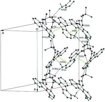
bonds between the amide H atoms and the two O atoms (N—H1N···O1 and N—H2N···O2) to form cyclic structures with
R44(24) graph-set motifs (Bernstein et al.., 1995). This bonding pattern results in a network of connected R44(24) rings
lying on pleated layers parallel to the ab plane (Table 1 and Fig. 4). The tert-butyl moieties are packed between the layers.
The linkage inside each layer is further supported by weak π—π stacking interactions among the central five-membered
ring and one of the attached six-membered rings of the fluorene moieties. Specifically, the C1′-C4′-C5′-C8′-C9 fulvene
ring at (x, y, z) and the C1′-C1-C2-C3-C4-C4′ aryl ring at (1/2+x, y, 3/2-z) are almost parallel forming between them a
dihedral angle of 6.5 (2)°. The centroid separation of the two rings is 3.583 (2) Å and the perpendicular distance of the
first centroid on the plane of the second ring is 3.421 (1) Å, corresponding to a centroid offset of 1.07 (2) Å.
S2. Experimental
A mixture of 4-amino-9H-fluoren-9-one (0.78 g, 4 mmol) and ylide BCMP (3.16 g, 8.4 mmol) in anhydrous DMF (4 ml)
was stirred at 100 oC for 4 days under an atmosphere of argon. The resulting solution was diluted with 30 ml e thylacetate
(EtOAc) and then washed with H2O (3 x 10 ml). The organic layer was dried (Na2SO4) and evaporated to dryness under
reduced pressure. The residue was subjected to flash column chromatography using as eluant the solvent system
PhMe/EtOAc (9.5:0.5).
The fractions with Rf 0.3 in the same solvent system were pooled and evaporated to leave 1.1 g (95% yield) of pure
compound in the form of a reddish oil. 1H NMR of the product revealed the presence of the two geometrical isomers in
the ratio 1:0.8 calculated on the basis of the integration of the two peaks at 8.839 and 8.329 p.p.m. where proton H-8
resonates for the isomers 5a and 5 b, respectively.The product was dissolved in the minimum volume of hot toluene. The
resulting solution was then left to attain ambient temperature and then cooled at 5 oC for 2 days. The crystalline
precipitate was collected, washed with ice-cold toluene and dried under reduced pressure. It weighed 60 mg. 1H NMR of
the crystals thus obtained showed them to be almost the pure isomer (5a:5 b=1:10) with H-8 resonating at 8.329 p.p.m..
The mother liquor from the crystallization was evaporated to dryness and the residue was crystallized also from toluene
to provide 70 mg of the almost pure alternative isomer with H-8 resonating at 8.839 p.p.m.. Recrystallization of the first
crystalline isomer finally gave reddish crystals suitable for X-ray analysis. This isomer had m.p. 386–387 K.
S3. Refinement
The amine H atoms and that attached to C14 were located in difference Fourier maps and their positions were refined
freely along with Uiso(H) equal to 1.5Ueq and 1.2Ueq of their parent atoms, respectively. The methyl H atoms were
constrained to an ideal geometry [C—H = 0.96 Å and Uiso(H) = 1.5Ueq(C)], but were allowed to rotate freely about the C
—C bonds. The remaining phenyl group H atoms were placed in geometrically idealized positions and constrained to ride
on their parent C atoms [C—H = 0.93 Å and Uiso(H) = 1.2Ueq(C)]. Five low-angle reflections were omitted from the final
cycles of refinement because their observed intensities were significantly lower than the calculated values, being
Figure 1
Figure 2
Figure 3
The molecular structure of the title compound (5 b) with the atom-labelling scheme. Displacement ellipsoids are drawn at
Figure 4
Part of the crystal structure of compound 5b, showing the network of the connected R44(24) cyclic structures parallel to
the ab plane and the π—π stacking interactions among the fluorene moieties (green dotted lines). For the sake of clarity,
only the H-atoms involved in the bonding pattern (dashed lines) are drawn. [Symmetry codes: (i) -x, y+1/2, -z+3/2, (ii)
x-1/2, y, -z+3/2, (iii) -x, -0.5+y, 1.5-z and (iv) 0.5+x, y, 1.5-z]
(Z)-tert-butyl 2-(4-amino-9H-fluoren-9-ylidene)acetate
Crystal data
C19H19NO2 Mr = 293.35
Orthorhombic, Pbca Hall symbol: -P 2ac 2ab a = 9.0820 (12) Å b = 13.7330 (17) Å c = 24.568 (3) Å V = 3064.2 (7) Å3 Z = 8
F(000) = 1248 Dx = 1.272 Mg m−3
Mo Kα radiation, λ = 0.71073 Å Cell parameters from 3945 reflections θ = 3.1–30.2°
µ = 0.08 mm−1 T = 100 K
Prism, light yellow 0.32 × 0.26 × 0.16 mm
Data collection
Oxford Diffraction Xcalibur-3 with Sapphire CCD
diffractometer
Radiation source: Enhance (Mo) X-ray source Graphite monochromator
ω and φ scans
Absorption correction: multi-scan
(CrysAlis RED; Oxford Diffraction, 2008) Tmin = 0.956, Tmax = 0.989
18331 measured reflections 2671 independent reflections
1671 reflections with I > 2σ(I) Rint = 0.119
θmax = 25.0°, θmin = 3.2° h = −8→10
k = −16→14 l = −27→29
Refinement
Refinement on F2 Least-squares matrix: full R[F2 > 2σ(F2)] = 0.063 wR(F2) = 0.143 S = 1.01 2671 reflections 211 parameters 0 restraints
Primary atom site location: structure-invariant direct methods
Secondary atom site location: difference Fourier map
Hydrogen site location: difference Fourier map H atoms treated by a mixture of independent
and constrained refinement w = 1/[σ2(F
o2) + (0.069P)2] where P = (Fo2 + 2Fc2)/3 (Δ/σ)max < 0.001
Δρmax = 0.30 e Å−3 Δρmin = −0.22 e Å−3
Special details
Experimental. IR (KBr, n, cm-1): 3404 and 3338 (w), 1700 (s), 1628 (m); 1H NMR (400 MHz, CDCl
3): d 8.329 (d, J = 7.6 Hz, 1H, H-8), 7.623 (d, J = 7.6 Hz, 1H, H-5), 7.528 (d, J= 7.6 Hz, 1H, H-1), 7.319 (t, J = 6.8 Hz, 1H, H-7), 7.152 (t, J= 6.8 Hz, 1H, H-6), 7.089 (t, J = 8 Hz, 1H, H-2), 6.707 (d, J= 7.6 Hz, 1H, H-3), 6.628 (s, 1H, H-10), 3.959 (s, 2H, NH2),1.541 (s, 9H, H-13) p.p.m.; EI—MS m/z: 293 (M+, 9), 237 (100), 220 (11), 193 (13), 180 (17), 165 (12). Geometry. All e.s.d.'s (except the e.s.d. in the dihedral angle between two l.s. planes) are estimated using the full covariance matrix. The cell e.s.d.'s are taken into account individually in the estimation of e.s.d.'s in distances, angles and torsion angles; correlations between e.s.d.'s in cell parameters are only used when they are defined by crystal symmetry. An approximate (isotropic) treatment of cell e.s.d.'s is used for estimating e.s.d.'s involving l.s. planes.
Refinement. Refinement of F2 against ALL reflections. The weighted R-factor wR and goodness of fit S are based on F2, conventional R-factors R are based on F, with F set to zero for negative F2. The threshold expression of F2 > σ(F2) is used only for calculating R-factors(gt) etc. and is not relevant to the choice of reflections for refinement. R-factors based on F2 are statistically about twice as large as those based on F, and R- factors based on ALL data will be even larger.
Fractional atomic coordinates and isotropic or equivalent isotropic displacement parameters (Å2)
x y z Uiso*/Ueq
C8 0.3365 (3) −0.1077 (2) 0.80637 (11) 0.0248 (7) H8 0.3956 −0.1492 0.7859 0.030* C8′ 0.2461 (3) −0.04057 (18) 0.78088 (11) 0.0216 (7) C9 0.2258 (3) −0.02063 (19) 0.72239 (12) 0.0224 (7) C10 0.2879 (3) −0.0755 (2) 0.68355 (12) 0.0241 (7) H10 0.350 (3) −0.129 (2) 0.6933 (11) 0.029* C11 0.2785 (3) −0.0635 (2) 0.62405 (12) 0.0258 (7) C12 0.2695 (3) −0.1621 (2) 0.54059 (11) 0.0268 (7) C13 0.2656 (4) −0.2720 (2) 0.53364 (13) 0.0450 (9) H13A 0.3497 −0.3003 0.5512 0.067* H13B 0.2675 −0.2878 0.4956 0.067* H13C 0.1772 −0.2974 0.5497 0.067* C14 0.4087 (4) −0.1212 (3) 0.51722 (13) 0.0445 (9) H14A 0.4062 −0.0514 0.5194 0.067* H14B 0.4175 −0.1406 0.4798 0.067* H14C 0.4915 −0.1453 0.5374 0.067* C15 0.1325 (4) −0.1164 (2) 0.51698 (13) 0.0421 (9) H15A 0.0472 −0.1425 0.5349 0.063* H15B 0.1272 −0.1304 0.4787 0.063* H15C 0.1358 −0.0472 0.5223 0.063* N −0.0702 (3) 0.18283 (18) 0.83630 (11) 0.0298 (6) H1N −0.147 (4) 0.218 (2) 0.8360 (13) 0.045* H2N −0.095 (3) 0.132 (2) 0.8587 (13) 0.045* O1 0.2699 (2) −0.15191 (12) 0.60048 (7) 0.0262 (5) O2 0.2815 (3) 0.01219 (14) 0.59924 (8) 0.0384 (6)
Atomic displacement parameters (Å2)
U11 U22 U33 U12 U13 U23
N 0.0346 (16) 0.0172 (14) 0.0374 (16) 0.0019 (12) 0.0056 (13) −0.0018 (12) O1 0.0446 (13) 0.0125 (10) 0.0214 (11) −0.0021 (9) 0.0032 (10) −0.0001 (8) O2 0.0723 (17) 0.0127 (10) 0.0303 (12) −0.0024 (10) −0.0025 (11) 0.0043 (9)
Geometric parameters (Å, º)
C1′—C1 1.391 (4) C8′—C9 1.475 (4) C1′—C4′ 1.409 (4) C9—C10 1.340 (4) C1′—C9 1.493 (4) C10—C11 1.474 (4) C1—C2 1.391 (4) C10—H10 0.96 (3) C1—H1 0.9300 C11—O2 1.205 (3) C2—C3 1.380 (4) C11—O1 1.347 (3) C2—H2 0.9300 C12—O1 1.478 (3) C3—C4 1.399 (4) C12—C14 1.498 (4) C3—H3 0.9300 C12—C15 1.510 (4) C4—N 1.398 (4) C12—C13 1.519 (4) C4—C4′ 1.400 (4) C13—H13A 0.9600 C4′—C5′ 1.478 (4) C13—H13B 0.9600 C5′—C5 1.377 (4) C13—H13C 0.9600 C5′—C8′ 1.412 (4) C14—H14A 0.9600 C5—C6 1.387 (4) C14—H14B 0.9600 C5—H5 0.9300 C14—H14C 0.9600 C6—C7 1.387 (4) C15—H15A 0.9600 C6—H6 0.9300 C15—H15B 0.9600 C7—C8 1.384 (4) C15—H15C 0.9600 C7—H7 0.9300 N—H1N 0.85 (3) C8—C8′ 1.384 (4) N—H2N 0.92 (3) C8—H8 0.9300
C5—C5′—C4′ 132.9 (3) H13B—C13—H13C 109.5 C8′—C5′—C4′ 106.9 (2) C12—C14—H14A 109.5 C5′—C5—C6 119.1 (3) C12—C14—H14B 109.5 C5′—C5—H5 120.4 H14A—C14—H14B 109.5 C6—C5—H5 120.4 C12—C14—H14C 109.5 C5—C6—C7 120.9 (3) H14A—C14—H14C 109.5 C5—C6—H6 119.6 H14B—C14—H14C 109.5 C7—C6—H6 119.6 C12—C15—H15A 109.5 C8—C7—C6 120.4 (3) C12—C15—H15B 109.5 C8—C7—H7 119.8 H15A—C15—H15B 109.5 C6—C7—H7 119.8 C12—C15—H15C 109.5 C8′—C8—C7 119.3 (3) H15A—C15—H15C 109.5 C8′—C8—H8 120.4 H15B—C15—H15C 109.5 C7—C8—H8 120.4 C4—N—H1N 111 (2) C8—C8′—C5′ 120.1 (3) C4—N—H2N 116 (2) C8—C8′—C9 129.7 (2) H1N—N—H2N 104 (3) C5′—C8′—C9 110.1 (2) C11—O1—C12 120.9 (2) C10—C9—C8′ 122.4 (3)
C4′—C1′—C1—C2 −0.3 (4) C7—C8—C8′—C5′ 0.0 (4) C9—C1′—C1—C2 179.3 (3) C7—C8—C8′—C9 179.4 (3) C1′—C1—C2—C3 −1.4 (4) C5—C5′—C8′—C8 0.8 (4) C1—C2—C3—C4 0.8 (4) C4′—C5′—C8′—C8 178.5 (2) C2—C3—C4—N 178.5 (3) C5—C5′—C8′—C9 −178.8 (2) C2—C3—C4—C4′ 1.4 (4) C4′—C5′—C8′—C9 −1.0 (3) N—C4—C4′—C1′ 179.9 (2) C8—C8′—C9—C10 6.5 (5) C3—C4—C4′—C1′ −3.1 (4) C5′—C8′—C9—C10 −174.0 (3) N—C4—C4′—C5′ 1.6 (4) C8—C8′—C9—C1′ −177.6 (3) C3—C4—C4′—C5′ 178.7 (3) C5′—C8′—C9—C1′ 1.9 (3) C1—C1′—C4′—C4 2.6 (4) C1—C1′—C9—C10 −6.4 (5) C9—C1′—C4′—C4 −177.1 (2) C4′—C1′—C9—C10 173.2 (3) C1—C1′—C4′—C5′ −178.8 (2) C1—C1′—C9—C8′ 178.3 (3) C9—C1′—C4′—C5′ 1.5 (3) C4′—C1′—C9—C8′ −2.0 (3) C4—C4′—C5′—C5 −4.5 (5) C8′—C9—C10—C11 −179.2 (3) C1′—C4′—C5′—C5 177.1 (3) C1′—C9—C10—C11 6.2 (5) C4—C4′—C5′—C8′ 178.1 (3) C9—C10—C11—O2 39.7 (5) C1′—C4′—C5′—C8′ −0.3 (3) C9—C10—C11—O1 −142.0 (3) C8′—C5′—C5—C6 −0.7 (4) O2—C11—O1—C12 2.5 (4) C4′—C5′—C5—C6 −177.8 (3) C10—C11—O1—C12 −175.8 (2) C5′—C5—C6—C7 −0.1 (4) C14—C12—O1—C11 60.3 (3) C5—C6—C7—C8 0.8 (5) C15—C12—O1—C11 −65.1 (3) C6—C7—C8—C8′ −0.7 (4) C13—C12—O1—C11 177.6 (2)
Hydrogen-bond geometry (Å, º)
D—H···A D—H H···A D···A D—H···A
N—H2N···O2ii 0.92 (3) 2.24 (3) 3.133 (3) 163 (3)