7,7
000,8,8
000-Tetramethoxy-4,4
000-dimethyl-3,3
000-bicoumarin
Hoong-Kun Fun,a*‡ Samuel Robinson Jebas,a§ Mehtab Parveen,bZakia Khanamb and Raza Murad Ghalibb
aX-ray Crystallography Unit, School of Physics, Universiti Sains Malaysia, 11800
Universiti Sains Malaysia, Penang, Malaysia, andbDepartment of Chemistry, Aligarh
Muslim University, Aligarh 202002 (UP), India Correspondence e-mail: [email protected]
Received 5 May 2009; accepted 8 May 2009
Key indicators: single-crystal X-ray study;T= 100 K; mean(C–C) = 0.002 A˚; Rfactor = 0.059;wRfactor = 0.155; data-to-parameter ratio = 18.7.
In the crystal structure, the title compound, C24H22O8, lies on a
twofold rotation axis and the asymmetric unit comprises one half-molecule. The dihedral angle formed by the coumarin unit with the symmetry-related part is 74.78 (14). One of the
methoxy groups attached to the coumarin unit is considerably twisted, making an angle of 87.17 (17) with respect to the
coumarin unit; the other is twisted by 0.66 (19). No classical
hydrogen bonds are found in the sturcture; only a weak C— H interaction and short intramolecular O O contacts [2.683 (2)–2.701 (2) A˚ ] are observed.
Related literature
For the biological activity of coumarins, see: El-Agrodyet al.
(2001); El-Farargy (1991); Emmanuel-Giota et al. (2001); Ghate et al.(2005); Laaksoet al. (1994); Nofalet al.(2000); Pratibha & Shreeya (1999); Shaker (1996); Yanget al.(2005). For the pharmaceutical properties of coumarin derivatives, see: Kennedy & Thornes (1997). For natural and synthetic coumarins, see: Carltonet al. (1996); Zhouet al.(2000). For related bond-length data, see: Allenet al.(1987). For stability of the temperature controller used in the data collection, see: Cosier & Glazer (1986).
Experimental
Crystal data
C24H22O8 Mr= 438.42 Monoclinic,C2=c a= 21.715 (9) A˚
b= 7.138 (3) A˚
c= 15.511 (6) A˚
= 121.801 (5)
V= 2043.3 (14) A˚3 Z= 4
MoKradiation
= 0.11 mm1 T= 100 K
0.280.190.06 mm
Data collection
Bruker SMART APEXII CCD area-detector diffractometer Absorption correction: multi-scan
(SADABS; Bruker, 2005)
Tmin= 0.971,Tmax= 0.994
27961 measured reflections 3527 independent reflections 2710 reflections withI> 2(I)
Rint= 0.065
Refinement
R[F2> 2(F2)] = 0.059 wR(F2) = 0.155 S= 1.08 3527 reflections
189 parameters
All H-atom parameters refined
max= 0.50 e A˚
3
min=0.21 e A˚
[image:1.610.93.249.616.687.2]3
Table 1
Hydrogen-bond geometry (A˚ ,).
D—H A D—H H A D A D—H A
C6—H6 Cg1i
0.96 (2) 2.86 (2) 3.676 (2) 143.5 (18)
Symmetry code: (i)x;y;z1
2.Cg1 is the centroid of the C4–C9 ring.
Data collection:APEX2(Bruker, 2005); cell refinement:SAINT (Bruker, 2005); data reduction:SAINT; program(s) used to solve structure: SHELXTL (Sheldrick, 2008); program(s) used to refine structure:SHELXTL; molecular graphics:SHELXTL; software used to prepare material for publication:SHELXTLandPLATON(Spek, 2009).
HKF and SRJ thank the Malaysian Government and Universiti Sains Malaysia for the Science Fund grant No. 305/ PFIZIK/613312. SRJ thanks Universiti Sains Malaysia for a post–doctoral research fellowship. HKF also thanks Universiti Sains Malaysia for the Research University Golden Goose grant No. 1001/PFIZIK/811012.
Supplementary data and figures for this paper are available from the IUCr electronic archives (Reference: IS2417).
References
Allen, F. H., Kennard, O., Watson, D. G., Brammer, L., Orpen, A. G. & Taylor, R. (1987).J. Chem. Soc. Perkin Trans. 2, pp. S1–S19.
Bruker (2005).APEX2,SAINTandSADABS. Bruker AXS Inc., Madison, Wisconsin, USA.
Carlton, B. D., Aubrun, J. C. & Simon, G. S. (1996).Fundam. Appl. Toxicol.30, 145–151.
Cosier, J. & Glazer, A. M. (1986).J. Appl. Cryst.19, 105–107.
El-Agrody, A. M., Abd El-Latif, M. S., El-Hady, N. A., Fakery, A. H. & Bedair, A. H. (2001).Molecules,6, 519–527.
El-Farargy, A. F. (1991).Egypt. J. Pharm. Sci,32, 625–625.
Emmanuel-Giota, A. A., Fylaktakidou, K. C., Hadjipavlou-Litina, D. J., Litinas, K. E. & Nicolaides, D. N. (2001).J. Heterocycl. Chem.38, 3, 717–722. Ghate, M., Kusanur, R. A. & Kulkarni, M. V. (2005).Eur. J. Med. Chem.40,
882–887.
organic compounds
o1294
Funet al. doi:10.1107/S1600536809017334 Acta Cryst.(2009). E65, o1294–o1295 Acta Crystallographica Section EStructure Reports
Online
ISSN 1600-5368
‡ Thomson Reuters Researcher ID: A-3561-2009.
and Mode of Action. New York: John Wiley & Sons.
Laakso, J. A., Narske, E. D., Gloer, J. B., Wicklow, D. T. & Dowd, P. F. (1994).J. Nat. Prod.,57, 128–133.
Nofal, Z. M., El-Zahar, M. & Abd El-Karim, S. (2000).Molecules,5, 99–113. Pratibha, S. & Shreeya, P. (1999).Indian J. Chem.38B, 1139–1142.
Sheldrick, G. M. (2008).Acta Cryst.A64, 112–122. Spek, A. L. (2009).Acta Cryst.D65, 148–155.
supporting information
sup-1
Acta Cryst. (2009). E65, o1294–o1295
supporting information
Acta Cryst. (2009). E65, o1294–o1295 [doi:10.1107/S1600536809017334]
7,7
′
,8,8
′
-Tetramethoxy-4,4
′
-dimethyl-3,3
′
-bicoumarin
Hoong-Kun Fun, Samuel Robinson Jebas, Mehtab Parveen, Zakia Khanam and Raza Murad
Ghalib
S1. Comment
Coumarins are a large group of naturally occurring oxygen heterocycles representing 2H-1-benzopyran-2-one derivative.
Many natural coumarins are reputed for their wide range of biological activites such as antibacterial (El-Agrody et al.,
2001; Pratibha & Shreeya, 1999), antifungal (Shaker, 1996; El-Farargy, 1991), antioxidant (Yang et al., 2005), analgesic
(Ghate et al., 2005), anti-inflammatory (Emmanuel-Giota et al., 2001) and antitumor (Nofal et al., 2000) properties. Bi
and tri-coumarins are comparatively new groups which are widely spread in nature and their biological properties are also
well known (Laakso et al., 1994). One of the characteristic pharmacological properties of coumarin derivatives is the
anticoagulant action (Kennedy & Thornes, 1997). A large number of natural and semisynthetic coumarin and bicoumarin
derivatives have been reported to demonstrate chemopreventive (Carlton et al., 1996) and anti-HIV (Zhou et al., 2000)
activities. Keeping in view of these biological importance of coumarins and their dimers, we have synthesized the title
compound (I) and report here its structure.
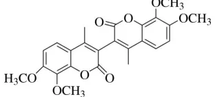
The asymmetric unit of (I) (Fig. 1), contains half of the 7,7′,8,8′-4,4′-dimethyl-3,3′-bicoumarin molecule. The other half
is symmetry generated [symmetry code: -x, y, -z + 1/2]. The coumarin unit is planar with the maximum deviation from
the mean plane of 0.0295 (15) Å for atom C2. One of the methyl group attached to the coumarin unit is twisted as
evidenced by the torsion angle of C10—O3—C8—C9 = 87.17 (17)°. The dihedral angle formed by the coumarin unit
(O1/C1—C9) with the symmetry related coumarin unit (O1A/C1A—C9A) is 74.78 (14)°, indicating that they are almost
perpendicular to each other. The bond lengths (Allen et al., 1987) and bond angles are normal.
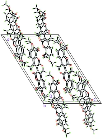
The crystal packing (Fig. 2) (Table 1) is stabilized by weak C—H···π interactions and intramolecular O···O = 2.683 (2)
to 2.701 (2) Å short contacts.
S2. Experimental
A mixture of 7,8-dimethoxy-4-methyl coumarin (2.20 g, 10 mmol) and manganese(III) acetate (0.774 g, 1 mmol) was
stirred at room temperature, then 70% perchloric acid (0.8 g, 6 mmol) was added. The reaction mixture was heated under
reflux at 114°C with stirring in the atmosphere of nitrogen for 3 h. The reaction mixture was cooled and diluted with 50
ml of benzene. The benzene solution was washed with water and aq. NaHCO3, dried over anhydrous Na2SO4 and left to
evaporate. The residue showed two major compounds which were separated by column chromatography followed by
preparative thin layer chromatography (Benzene: EtOAc, 9:1) into the title compound (I) (260 mg, 12%).
S3. Refinement
Figure 1
The molecular structure of (I), showing 50% probability displacement ellipsoids and the atom numbering scheme.
supporting information
sup-3
[image:5.610.141.472.71.519.2]Acta Cryst. (2009). E65, o1294–o1295 Figure 2
The crystal packing of (I). Molecules are stacked along the b axis.
(I)
Crystal data
C24H22O8
Mr = 438.42
Monoclinic, C2/c
Hall symbol: -C 2yc
a = 21.715 (9) Å
b = 7.138 (3) Å
c = 15.511 (6) Å
β = 121.801 (5)°
V = 2043.3 (14) Å3
Z = 4
F(000) = 920
Dx = 1.425 Mg m−3
Mo Kα radiation, λ = 0.71073 Å
Cell parameters from 6562 reflections
θ = 2.7–31.8°
µ = 0.11 mm−1
T = 100 K
Bruker SMART APEXII CCD area-detector diffractometer
Radiation source: fine-focus sealed tube Graphite monochromator
φ and ω scans
Absorption correction: multi-scan
(SADABS; Bruker, 2005)
Tmin = 0.971, Tmax = 0.994
27961 measured reflections 3527 independent reflections 2710 reflections with I > 2σ(I)
Rint = 0.065
θmax = 32.0°, θmin = 2.2°
h = −32→32
k = −10→10
l = −23→23
Refinement
Refinement on F2
Least-squares matrix: full R[F2 > 2σ(F2)] = 0.059
wR(F2) = 0.155
S = 1.08
3527 reflections 189 parameters 0 restraints
Primary atom site location: structure-invariant direct methods
Secondary atom site location: difference Fourier map
Hydrogen site location: inferred from neighbouring sites
All H-atom parameters refined w = 1/[σ2(F
o2) + (0.0752P)2 + 1.2567P]
where P = (Fo2 + 2Fc2)/3 (Δ/σ)max < 0.001
Δρmax = 0.50 e Å−3
Δρmin = −0.21 e Å−3
Special details
Experimental. The crystal was placed in the cold stream of an Oxford Cyrosystems Cobra open-flow nitrogen cryostat (Cosier & Glazer, 1986) operating at 100.0 (1) K.
Geometry. All e.s.d.'s (except the e.s.d. in the dihedral angle between two l.s. planes) are estimated using the full covariance matrix. The cell e.s.d.'s are taken into account individually in the estimation of e.s.d.'s in distances, angles and torsion angles; correlations between e.s.d.'s in cell parameters are only used when they are defined by crystal symmetry. An approximate (isotropic) treatment of cell e.s.d.'s is used for estimating e.s.d.'s involving l.s. planes.
Refinement. Refinement of F2 against ALL reflections. The weighted R-factor wR and goodness of fit S are based on F2,
conventional R-factors R are based on F, with F set to zero for negative F2. The threshold expression of F2 > σ(F2) is used
only for calculating R-factors(gt) etc. and is not relevant to the choice of reflections for refinement. R-factors based on F2
are statistically about twice as large as those based on F, and R- factors based on ALL data will be even larger.
Fractional atomic coordinates and isotropic or equivalent isotropic displacement parameters (Å2)
x y z Uiso*/Ueq
O1 0.15443 (5) 0.04445 (13) 0.38705 (7) 0.0178 (2)
O2 0.05473 (6) −0.06775 (15) 0.37300 (8) 0.0242 (2)
O3 0.30066 (5) 0.03278 (14) 0.48961 (7) 0.0212 (2)
O4 0.37511 (5) 0.26053 (15) 0.43926 (8) 0.0221 (2)
C1 0.08045 (7) 0.05014 (19) 0.34517 (10) 0.0175 (3)
C2 0.03949 (7) 0.19639 (19) 0.27048 (10) 0.0163 (3)
C3 0.07228 (7) 0.32194 (19) 0.24158 (10) 0.0168 (3)
C4 0.15015 (7) 0.31354 (18) 0.28870 (10) 0.0161 (3)
C5 0.19034 (8) 0.44106 (19) 0.26927 (10) 0.0185 (3)
C6 0.26498 (8) 0.42852 (19) 0.31768 (11) 0.0194 (3)
C7 0.30199 (7) 0.28719 (19) 0.38881 (10) 0.0174 (3)
C8 0.26401 (7) 0.15966 (18) 0.41265 (9) 0.0164 (3)
C9 0.18892 (7) 0.17416 (18) 0.36139 (10) 0.0154 (2)
supporting information
sup-5
Acta Cryst. (2009). E65, o1294–o1295
C11 0.41539 (8) 0.3869 (2) 0.41549 (13) 0.0267 (3)
C12 0.02974 (8) 0.4697 (2) 0.16315 (12) 0.0247 (3)
H5 0.1654 (10) 0.540 (3) 0.2226 (15) 0.024 (5)*
H6 0.2908 (11) 0.516 (3) 0.3016 (16) 0.033 (5)*
H10A 0.3396 (14) −0.139 (4) 0.429 (2) 0.063 (8)*
H10B 0.3324 (13) −0.224 (3) 0.514 (2) 0.046 (6)*
H10C 0.2573 (16) −0.202 (4) 0.410 (2) 0.067 (8)*
H11A 0.3985 (12) 0.382 (3) 0.3405 (19) 0.044 (6)*
H11B 0.4125 (11) 0.519 (3) 0.4338 (15) 0.028 (5)*
H11C 0.4642 (10) 0.340 (3) 0.4535 (14) 0.022 (4)*
H12A 0.0472 (11) 0.483 (3) 0.1149 (17) 0.035 (5)*
H12B 0.0359 (12) 0.591 (3) 0.1955 (18) 0.041 (6)*
H12C −0.0217 (12) 0.440 (3) 0.1264 (17) 0.036 (5)*
Atomic displacement parameters (Å2)
U11 U22 U33 U12 U13 U23
O1 0.0162 (5) 0.0193 (5) 0.0163 (4) 0.0013 (3) 0.0074 (4) 0.0050 (4)
O2 0.0213 (5) 0.0253 (5) 0.0244 (5) −0.0003 (4) 0.0111 (4) 0.0083 (4)
O3 0.0217 (5) 0.0219 (5) 0.0134 (4) 0.0047 (4) 0.0048 (4) 0.0029 (4)
O4 0.0144 (5) 0.0250 (5) 0.0230 (5) −0.0015 (4) 0.0071 (4) −0.0020 (4)
C1 0.0167 (6) 0.0199 (6) 0.0149 (6) 0.0003 (5) 0.0076 (5) 0.0008 (5)
C2 0.0155 (6) 0.0179 (6) 0.0140 (6) 0.0004 (4) 0.0067 (5) −0.0003 (4)
C3 0.0166 (6) 0.0176 (6) 0.0145 (6) 0.0013 (4) 0.0071 (5) 0.0018 (4)
C4 0.0166 (6) 0.0174 (6) 0.0133 (5) 0.0013 (4) 0.0072 (5) 0.0009 (4)
C5 0.0199 (6) 0.0185 (6) 0.0165 (6) 0.0007 (5) 0.0091 (5) 0.0027 (5)
C6 0.0202 (6) 0.0200 (6) 0.0186 (6) −0.0016 (5) 0.0106 (5) −0.0002 (5)
C7 0.0142 (6) 0.0211 (6) 0.0143 (6) −0.0007 (4) 0.0058 (5) −0.0037 (5)
C8 0.0168 (6) 0.0176 (6) 0.0108 (5) 0.0018 (4) 0.0045 (5) −0.0003 (4)
C9 0.0175 (6) 0.0155 (6) 0.0125 (5) −0.0012 (4) 0.0075 (5) −0.0006 (4)
C10 0.0564 (12) 0.0239 (8) 0.0302 (9) 0.0173 (8) 0.0241 (9) 0.0093 (7)
C11 0.0188 (7) 0.0267 (8) 0.0339 (8) −0.0059 (6) 0.0134 (6) −0.0047 (6)
C12 0.0183 (7) 0.0265 (7) 0.0254 (7) 0.0030 (5) 0.0089 (6) 0.0112 (6)
Geometric parameters (Å, º)
O1—C9 1.3750 (16) C5—H5 0.952 (19)
O1—C1 1.3801 (17) C6—C7 1.395 (2)
O2—C1 1.2085 (17) C6—H6 0.96 (2)
O3—C8 1.3701 (16) C7—C8 1.4025 (19)
O3—C10 1.428 (2) C8—C9 1.3912 (19)
O4—C7 1.3640 (17) C10—H10A 1.01 (3)
O4—C11 1.4334 (19) C10—H10B 0.92 (3)
C1—C2 1.4618 (19) C10—H10C 1.02 (3)
C2—C3 1.3592 (19) C11—H11A 1.02 (2)
C2—C2i 1.482 (3) C11—H11B 1.00 (2)
C3—C4 1.4472 (19) C11—H11C 0.963 (19)
C4—C9 1.4034 (18) C12—H12C 0.97 (2)
C5—C6 1.384 (2)
C9—O1—C1 121.36 (10) O3—C8—C9 121.00 (12)
C8—O3—C10 114.77 (12) O3—C8—C7 120.42 (12)
C7—O4—C11 116.63 (12) C9—C8—C7 118.42 (12)
O2—C1—O1 116.99 (12) O1—C9—C8 115.95 (11)
O2—C1—C2 125.28 (13) O1—C9—C4 121.49 (12)
O1—C1—C2 117.72 (11) C8—C9—C4 122.56 (12)
C3—C2—C1 121.77 (12) O3—C10—H10A 108.3 (16)
C3—C2—C2i 123.20 (11) O3—C10—H10B 108.3 (15)
C1—C2—C2i 115.03 (10) H10A—C10—H10B 106 (2)
C2—C3—C4 118.80 (12) O3—C10—H10C 108.6 (16)
C2—C3—C12 121.60 (13) H10A—C10—H10C 115 (2)
C4—C3—C12 119.59 (12) H10B—C10—H10C 110 (2)
C5—C4—C9 117.13 (12) O4—C11—H11A 111.7 (13)
C5—C4—C3 124.03 (12) O4—C11—H11B 112.6 (11)
C9—C4—C3 118.80 (12) H11A—C11—H11B 108.8 (17)
C6—C5—C4 121.74 (13) O4—C11—H11C 104.2 (11)
C6—C5—H5 119.7 (11) H11A—C11—H11C 107.7 (16)
C4—C5—H5 118.6 (11) H11B—C11—H11C 111.8 (16)
C5—C6—C7 119.83 (13) C3—C12—H12A 111.0 (12)
C5—C6—H6 119.7 (13) C3—C12—H12B 110.3 (14)
C7—C6—H6 120.5 (13) H12A—C12—H12B 107.2 (17)
O4—C7—C6 124.46 (12) C3—C12—H12C 110.3 (12)
O4—C7—C8 115.26 (12) H12A—C12—H12C 110.5 (18)
C6—C7—C8 120.28 (13) H12B—C12—H12C 107.4 (17)
C9—O1—C1—O2 −179.03 (12) C5—C6—C7—O4 178.75 (12)
C9—O1—C1—C2 1.45 (18) C5—C6—C7—C8 −1.0 (2)
O2—C1—C2—C3 −178.65 (14) C10—O3—C8—C9 87.17 (17)
O1—C1—C2—C3 0.83 (19) C10—O3—C8—C7 −97.47 (17)
O2—C1—C2—C2i 1.3 (2) O4—C7—C8—O3 6.82 (18)
O1—C1—C2—C2i −179.20 (11) C6—C7—C8—O3 −173.39 (12)
C1—C2—C3—C4 −2.0 (2) O4—C7—C8—C9 −177.70 (11)
C2i—C2—C3—C4 178.01 (13) C6—C7—C8—C9 2.09 (19)
C1—C2—C3—C12 178.72 (13) C1—O1—C9—C8 176.73 (11)
C2i—C2—C3—C12 −1.3 (2) C1—O1—C9—C4 −2.48 (18)
C2—C3—C4—C5 −176.43 (13) O3—C8—C9—O1 −5.18 (18)
C12—C3—C4—C5 2.8 (2) C7—C8—C9—O1 179.37 (11)
C2—C3—C4—C9 1.01 (19) O3—C8—C9—C4 174.02 (12)
C12—C3—C4—C9 −179.71 (13) C7—C8—C9—C4 −1.42 (19)
C9—C4—C5—C6 1.4 (2) C5—C4—C9—O1 178.84 (11)
C3—C4—C5—C6 178.94 (13) C3—C4—C9—O1 1.22 (19)
supporting information
sup-7
Acta Cryst. (2009). E65, o1294–o1295
C11—O4—C7—C6 −0.66 (19) C3—C4—C9—C8 −177.94 (12)
C11—O4—C7—C8 179.12 (12)
Symmetry code: (i) −x, y, −z+1/2.
Hydrogen-bond geometry (Å, º)
D—H···A D—H H···A D···A D—H···A
C6—H6···Cg1ii 0.96 (2) 2.86 (2) 3.676 (2) 143.5 (18)