1,2-Bis(1
H
-tetrazol-5-yl)benzene
dihydrate
Hai-Jun Xu,* Yi-Jie Pan and Li-Jing Cui
Ordered Matter Science Research Center, College of Chemistry and Chemical Engineering, Southeast University, Nanjing 210096, People’s Republic of China Correspondence e-mail: xuhj@seu.edu.cn
Received 11 May 2009; accepted 14 May 2009
Key indicators: single-crystal X-ray study;T= 294 K; mean(C–C) = 0.002 A˚;
Rfactor = 0.040;wRfactor = 0.098; data-to-parameter ratio = 12.6.
The asymmetric unit of the title compound, C8H6N82H2O, contains one half-molecule, with the benzene ring on a centre of symmetry, and two uncoordinated water molecules. The benzene ring is oriented at a dihedral angle of 34.43 (12)with respect to the tetrazole ring. Strong O—H N hydrogen bonds link the water molecules to the N atoms of the tetrazole ring. In the crystal structure, strong intermolecular O—H O and O—H N hydrogen bonds link the molecules into a network. One of the water H atoms is disordered over two positions and was refined with occupancies of 0.50.
Related literature
For general background, see: Luo et al. (2006). For related structures, see: Guzei & Bikzhanova (2002); Panet al.(2007).
Experimental
Crystal data
C8H6N82H2O
Mr= 286.27
Monoclinic,C2=c a= 14.510 (3) A˚
b= 12.427 (3) A˚
c= 7.2576 (15) A˚
= 96.29 (3) V= 1300.7 (5) A˚3
Z= 4
MoKradiation
= 0.12 mm1
T= 294 K
0.200.200.20 mm
Data collection
Rigaku SCXmini diffractometer Absorption correction: multi-scan
(Blessing, 1995)
Tmin= 0.971,Tmax= 0.979
5963 measured reflections 1276 independent reflections 1041 reflections withI> 2(I)
Rint= 0.044
Refinement
R[F2> 2(F2)] = 0.040
wR(F2) = 0.098
S= 1.06 1276 reflections 101 parameters
H atoms treated by a mixture of independent and constrained refinement
max= 0.20 e A˚
3
min=0.15 e A˚
3
Table 1
Hydrogen-bond geometry (A˚ ,).
D—H A D—H H A D A D—H A
N4—H4A O1W 0.91 (2) 1.78 (2) 2.682 (2) 171.9 (19) O1W—H1WA N2i
0.85 2.02 2.8658 (19) 173 O1W—H1WB O2Wii 0.85 1.98 2.813 (2) 168 O2W—H2WA N1 0.85 2.06 2.896 (2) 169 O2W—H2WB O2Wiii
0.85 1.97 2.813 (3) 170 O2W—H2WC O2Wiv
0.85 2.01 2.814 (3) 158
Symmetry codes: (i)xþ1 2;y
1 2;zþ
1 2; (ii)x
1 2;y
1 2;zþ
1
2; (iii)xþ1;y;z;
(iv)xþ1;y;z1 2.
Data collection: CrystalClear (Rigaku/MSC (2005); cell refine-ment:CrystalClear; data reduction:CrystalClear; program(s) used to solve structure: SHELXS97(Sheldrick, 2008); program(s) used to refine structure:SHELXL97(Sheldrick, 2008); molecular graphics: ORTEP-3 for Windows(Farrugia, 1997); software used to prepare material for publication:SHELXL97.
HJX acknowledges a start-up grant from Southeast University, China.
Supplementary data and figures for this paper are available from the IUCr electronic archives (Reference: HK2687).
References
Blessing, R. H. (1995).Acta Cryst.A51, 33–38. Farrugia, L. J. (1997).J. Appl. Cryst.30, 565.
Guzei, I. A. & Bikzhanova, G. A. (2002).Acta Cryst.E58, o937–o939. Luo, J., Zhang, X.-R., Cui, L.-L., Dai, W.-Q. & Liu, B.-S. (2006).Acta Cryst.
C62, m614–m616.
Pan, W.-L., Chen, X.-Y. & Hu, C.-W. (2007).Acta Cryst.E63, o1606–o1608. Rigaku/MSC (2005).CrystalClear. Rigaku/MSC, The Woodlands, Texas, USA. Sheldrick, G. M. (2008).Acta Cryst.A64, 112–122.
Acta Crystallographica Section E
Structure Reports Online
supporting information
Acta Cryst. (2009). E65, o1331 [doi:10.1107/S1600536809018224]
1,2-Bis(1H-tetrazol-5-yl)benzene dihydrate
Hai-Jun Xu, Yi-Jie Pan and Li-Jing Cui
S1. Comment
The tetrazole functional group has currently been received considerable attention mainly because of a wide range of
applications in coordination chemistry, medicinal chemistry and materials science (Luo et al., 2006). However, there are a
few crystal structure reports of organic tetrazolates compounds in the literature (Guzei & Bikzhanova, 2002). We reported
herein the synthesis and the crystal structure of the title compound.
The asymmetric unit of the title compound contains one-half molecule, with benzene ring on a centre of symmetry, and
two uncoordinated water molecules (Fig. 1). The bond lengths and angles are in accordance with the corresponding
values reported (Pan et al., 2007). The benzene ring is oriented with respect to the tetrazole ring at a dihedral angle of
34.43 (12)°. Strong intramolecular O-H···N hydrogen bonds (Table 1) link the water molecules to the nitrogens of the
tetrazole ring.
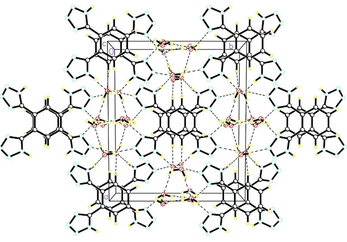
In the crystal structure, strong intermolecular O-H···O and O-H···N hydrogen bonds (Table 1) link the molecules into a
network (Fig. 2), in which they may be effective in the stabilization of the structure.
S2. Experimental
For the preparation of the title compound, phthalonitrile (1.28 g, 10 mmol), NH4Cl (1.38 g, 26 mmol) and NaN3 (1.69 g,
13 mmol) were dissolved in DMF (60 ml). The mixture was heated to 353 K, and stirred for 48 h. Then, it was cooled to
room temperature and poured into cold water and acidified to pH = 2 with concentrated hydrochloric acid. After 12 h at
277 K, the suspension was filtrated, and the residue was washed with H2O and H2O/EtOH (1/1), and then dried. Crystals
suitable for X-ray analysis were obtained from an EtOH solution.
S3. Refinement
One of the H atoms bonded to O2W was disordered. During the refinement process, the disordered H atom was refined
with occupancies of 0.50 and 0.50. H atom (for NH) was located in difference Fourier synthesis and refined isotropically.
The remaining H atoms were positioned geometrically with O-H = 0.85 Å (for H2O) [Uiso(H) = 0.060 (15)-0.086 (9) Å2]
Figure 1
The molecular structure of the title molecule, with the atom-numbering scheme. Displacement ellipsoids are drawn at the
30% probability level.
Figure 2
[image:3.610.131.481.427.671.2]1,2-Bis(1H-tetrazol-5-yl)benzene dihydrate
Crystal data
C8H6N8·2H2O Mr = 286.27
Monoclinic, C2/c Hall symbol: -C 2yc a = 14.510 (3) Å b = 12.427 (3) Å c = 7.2576 (15) Å β = 96.29 (3)° V = 1300.7 (5) Å3 Z = 4
F(000) = 600 Dx = 1.462 Mg m−3
Mo Kα radiation, λ = 0.71073 Å Cell parameters from 5656 reflections θ = 3.3–27.5°
µ = 0.12 mm−1 T = 294 K Prism, colorless 0.20 × 0.20 × 0.20 mm
Data collection
Rigaku SCXmini diffractometer
Radiation source: fine-focus sealed tube Graphite monochromator
Detector resolution: 13.6612 pixels mm-1 ω scans
Absorption correction: multi-scan (Blessing, 1995)
Tmin = 0.971, Tmax = 0.979
5963 measured reflections 1276 independent reflections 1041 reflections with I > 2σ(I) Rint = 0.044
θmax = 26.0°, θmin = 3.3° h = −17→17
k = −15→15 l = −8→8
Refinement
Refinement on F2 Least-squares matrix: full R[F2 > 2σ(F2)] = 0.040 wR(F2) = 0.098 S = 1.06 1276 reflections 101 parameters 0 restraints
Primary atom site location: structure-invariant direct methods
Secondary atom site location: difference Fourier map
Hydrogen site location: inferred from neighbouring sites
H atoms treated by a mixture of independent and constrained refinement
w = 1/[σ2(F
o2) + (0.0394P)2 + 0.846P] where P = (Fo2 + 2Fc2)/3
(Δ/σ)max < 0.001 Δρmax = 0.20 e Å−3 Δρmin = −0.15 e Å−3
Extinction correction: SHELXL97 (Sheldrick, 2008), Fc*=kFc[1+0.001xFc2λ3/sin(2θ)]-1/4 Extinction coefficient: 0.0220 (19)
Special details
Geometry. All e.s.d.'s (except the e.s.d. in the dihedral angle between two l.s. planes) are estimated using the full covariance matrix. The cell e.s.d.'s are taken into account individually in the estimation of e.s.d.'s in distances, angles and torsion angles; correlations between e.s.d.'s in cell parameters are only used when they are defined by crystal symmetry. An approximate (isotropic) treatment of cell e.s.d.'s is used for estimating e.s.d.'s involving l.s. planes.
Refinement. Refinement of F2 against ALL reflections. The weighted R-factor wR and goodness of fit S are based on F2, conventional R-factors R are based on F, with F set to zero for negative F2. The threshold expression of F2 > σ(F2) is used only for calculating R-factors(gt) etc. and is not relevant to the choice of reflections for refinement. R-factors based on F2 are statistically about twice as large as those based on F, and R- factors based on ALL data will be even larger.
Fractional atomic coordinates and isotropic or equivalent isotropic displacement parameters (Å2)
x y z Uiso*/Ueq Occ. (<1)
O1W 0.20523 (9) −0.46870 (10) 0.4630 (2) 0.0479 (4)
H1WB 0.1528 −0.4390 0.4684 0.084 (9)*
O2W 0.52104 (9) −0.10467 (11) −0.0561 (2) 0.0467 (4)
H2WA 0.4931 −0.1528 −0.0003 0.076 (8)*
H2WB 0.5036 −0.0452 −0.0135 0.060 (15)* 0.50
H2WC 0.4968 −0.1163 −0.1663 0.078 (19)* 0.50
N1 0.40777 (10) −0.24613 (11) 0.1426 (2) 0.0332 (4)
N2 0.33249 (10) −0.18460 (11) 0.1621 (2) 0.0397 (4)
N3 0.27393 (10) −0.23572 (12) 0.2522 (2) 0.0417 (4)
N4 0.31147 (10) −0.33246 (12) 0.2923 (2) 0.0353 (4)
H4A 0.2797 (14) −0.3834 (17) 0.350 (3) 0.053 (6)*
C1 0.45124 (10) −0.43665 (12) 0.2407 (2) 0.0264 (4)
C2 0.40490 (11) −0.53513 (12) 0.2324 (2) 0.0308 (4)
H2A 0.3404 −0.5358 0.2205 0.037*
C3 0.45222 (11) −0.63163 (13) 0.2414 (2) 0.0327 (4)
H3A 0.4198 −0.6963 0.2360 0.039*
C4 0.39372 (11) −0.33838 (12) 0.2258 (2) 0.0277 (4)
Atomic displacement parameters (Å2)
U11 U22 U33 U12 U13 U23
O1W 0.0353 (8) 0.0370 (8) 0.0737 (10) −0.0091 (6) 0.0168 (7) −0.0132 (7) O2W 0.0504 (9) 0.0396 (8) 0.0527 (9) −0.0046 (6) 0.0166 (7) 0.0005 (7)
N1 0.0323 (8) 0.0253 (7) 0.0421 (9) 0.0035 (6) 0.0052 (6) 0.0021 (6)
N2 0.0379 (9) 0.0283 (8) 0.0528 (10) 0.0071 (6) 0.0040 (7) 0.0015 (7) N3 0.0321 (9) 0.0304 (8) 0.0630 (11) 0.0082 (6) 0.0067 (8) −0.0009 (7) N4 0.0271 (8) 0.0262 (7) 0.0537 (10) 0.0020 (6) 0.0103 (7) 0.0017 (7) C1 0.0265 (8) 0.0235 (8) 0.0298 (9) 0.0009 (6) 0.0059 (6) 0.0010 (7) C2 0.0250 (9) 0.0280 (9) 0.0401 (10) −0.0024 (6) 0.0065 (7) −0.0014 (7) C3 0.0357 (9) 0.0225 (8) 0.0408 (10) −0.0050 (7) 0.0082 (8) −0.0005 (7) C4 0.0235 (8) 0.0255 (8) 0.0341 (9) −0.0011 (6) 0.0026 (7) −0.0017 (7)
Geometric parameters (Å, º)
O1W—H1WA 0.8500 C1—C2 1.394 (2)
O1W—H1WB 0.8501 C1—C1i 1.407 (3)
O2W—H2WA 0.8499 C1—C4 1.476 (2)
O2W—H2WB 0.8500 C2—C3 1.380 (2)
O2W—H2WC 0.8500 C2—H2A 0.9300
N1—N2 1.353 (2) C3—C3i 1.378 (3)
N2—N3 1.294 (2) C3—H3A 0.9300
N3—N4 1.339 (2) C4—N1 1.322 (2)
N4—H4A 0.91 (2) C4—N4 1.338 (2)
H1WA—O1W—H1WB 110.3 C2—C1—C4 117.17 (14)
H2WA—O2W—H2WB 105.2 C1i—C1—C4 124.17 (8)
H2WA—O2W—H2WC 99.2 C3—C2—C1 121.71 (15)
H2WB—O2W—H2WC 112.4 C3—C2—H2A 119.1
N3—N2—N1 110.96 (14) C3i—C3—C2 119.65 (9)
N2—N3—N4 106.06 (13) C3i—C3—H3A 120.2
C4—N4—N3 109.19 (14) C2—C3—H3A 120.2
C4—N4—H4A 130.1 (13) N1—C4—N4 107.80 (14)
N3—N4—H4A 120.6 (13) N1—C4—C1 129.54 (15)
C2—C1—C1i 118.65 (9) N4—C4—C1 122.58 (14)
Symmetry code: (i) −x+1, y, −z+1/2.
Hydrogen-bond geometry (Å, º)
D—H···A D—H H···A D···A D—H···A
N4—H4A···O1W 0.91 (2) 1.78 (2) 2.682 (2) 171.9 (19)
O1W—H1WA···N2ii 0.85 2.02 2.8658 (19) 173
O1W—H1WB···O2Wiii 0.85 1.98 2.813 (2) 168
O2W—H2WA···N1 0.85 2.06 2.896 (2) 169
O2W—H2WB···O2Wiv 0.85 1.97 2.813 (3) 170
O2W—H2WC···O2Wv 0.85 2.01 2.814 (3) 158