Original Article
microRNA-194 in the development of aortic
aneurysms and dissection
Jian Li1, Dayong Liu2
Departments of 1Cardiology, 2Cardiothoracic Surgery, Da Qing Long Nan Hospital, Daqing, Heilongjiang Province,
China
Received February 8, 2018; Accepted March 22, 2018; Epub May 15, 2018; Published May 30, 2018
Abstract: Objective: Our aim was to investigate the role and mechanism of microRNA-194 in aortic aneurysms and dissection. Methods: Fifteen patients with aortic aneurysms and dissection undergoing aortic valve replacement and thirteen normal donors without aortic lesions were selected to analyze expression levels of microRNA-194 in aortic tissue.The effect of microRNA-194 knockdown on expression of α-SMA, extracellular matrix secretory func -tion (collagen III and osteopontin), in human smooth muscle cells was also investigated. Addi-tionally, expression of Smad1 and Smad4, key molecules in TGF-β1 signaling pathway, was analyzed by qRT-PCR and Western blot. Results: Compared with normal aorta, microRNA-194 was significantly downregulated in aorta with aneurysms and dissection (P=0.019). Knockdown of microRNA-194 expression promoted contractile-to-synthetic conversion of human vascular smooth muscle cells (P=0.005), increased expression of collagen III and osteopontin (P<0.001, P=0.022), and promoted expression of Smad1 and Smad4 mRNA (P=0.016, P=0.001) and protein (P<0.001, P<0.001). Conclusion: Inhibition of microRNA-194 expression can activate TGF-β1 signaling pathways and affect the phenotype and function of vascular smooth muscle cells, possibly relating to development of aortic dissection.
Keywords: microRNA-194, aortic dissection, aortic aneurysm, vascular smooth muscle cells, transforming growth factor-β1 (TGF-β1)
Introduction
Aortic aneurysm is a local expansion of the blood vessel wall because of the patient’s aor-tic wall atherosclerosis, decreased elasaor-ticity, and inability to withstand hemodynamic impact [1]. In the case of aortic aneurysms and other underlying lesions, due to the impact of blood
flow, aortic dissection can lead to progressive
intimal tear and the formation of true and false lumens of arterial lumen with high risk of major bleeding. The majority of patients die because of failure to get timely surgical treatment. It is a common critical illness in cardiovascular dis-ease [2, 3]. Currently, the formation mecha-nism of aortic aneurysms and dissection is thought to be mainly related with changes of the structure and function of aortic tunica media, vascular smooth muscle cells, and extracellular matrix, the main components of tunica media [4]. Vascular smooth muscle cells include both contractile and synthetic forms. Contractile smooth muscle cells play a major
role in elasticity and tensile strength of the vas-cular wall. Synthetic smooth muscle cells play an important role in cell proliferation and
migra-tion. When the aortic vessel wall is damaged in
aortic aneurysms and dissection, smooth mus-cle cells can be converted from contractile to synthetic cells, resulting in promotion of devel-opment of aortic dissection [5].
microRNA, a highly conserved non-coding RNA containing about 22 nucleotides, can negative-ly regulate target gene expression through
spe-cific binding to the 3’untranslated region of
mRNA. It plays an important role in aortic
ath-erosclerosis, inflammation, cancer, and other
diseases. It has been reported in the literature that some microRNAs play a key role in the pathogenesis of aortic aneurysms and dissec-tion. For example, aorta microRNA-4787-5p
pathogenesis of aortic dissection by regulating signaling pathway of Polycystin-1 (PKD1) and
transforming growth factor-β1 (TGF-β1) [6]. In
addition, another study found that microRNA-15a and microRNA-23a have a clinical diagnos-tic value of aordiagnos-tic dissection by performing microRNA screening of 37 patients with acute aortic dissection, 26 patients with chronic aor-tic dissection, and 40 controls [7]. Therefore, study of the role and mechanism of microRNA in aortic dissection has important clinical value for aortic dissection diagnosis, treatment, and prognosis.
microRNA-194 is an important regulator of cell proliferation, differentiation, and migration and is widely involved in development of melanoma, degenerative disc disease, psoriasis, diabetes,
influenza, pneumonia, and other diseases
[8-13]. However, the role of microRNA-194 in aortic aneurysms and dissection remains unclear. Therefore, in this study, we analyzed expression of microRNA-194 in the aortic wall by selecting aortic dissection patients and
nor-mal control subjects as research objects. We
studied the function of microRNA-194 in cul-tured vascular smooth muscle cells.
Materials and methods
Aortic dissection tissue collection
This study was reviewed and approved by the Ethics Committee and informed consent was obtained. Aortic tissue samples were taken from 15 aortic dissection patients, undergoing aortic valve replacement in our Department of Thoracic Surgery, and 13 normal donors with-out aortic lesionsfrom May 2014 to December 2017. Inclusion criteria for aortic dissection patients was Stanford Type A thoracic aortic dissection with ascending aortic dissection determined by aortic arch digital subtraction angiography. Exclusion criteria included genetic factors (like familial aortic aneurysm), trauma, infections (such as syphilis), connective tissue disease (such as Marfan syndrome), and other causes of aortic lesions.
Construction of lentiviral vector for downregu-lation of microRNA-194
After the complementary sequence (TCCACAT-GGAGTTGCTGTTACA) and nonsense sequence
(TTCTCCGAACGTGTCACGT) of synthetic
microR-NA-194 were synthesized and annealed to
dou-ble-stranded sequence, it was inserted into a
lentiviral vector Plkd-CMV-G & PR (Shanghai Heyuan) by molecular biology methods such as restriction endonuclease, ligation, etc. [14, 15]. Then, the constructed lentiviral vectors were divided into two groups: lentiviral vector con-structed by microRNA-194 complementary
sequence as microRNA-194 intervention group
(Anti-miR194 group) and lentiviral vector
con-structed by nonsense sequence as microR -NA-194 control Group (Scramb-Anti-miR194 group).
Culture and treatment of human aortic smooth muscle cells
Human aortic wall smooth muscle cells were
purchased from ATCC cell bank. Cells were first
incubated with high glucose DMEM medium containing 10% FBS, 1% penicillin, and strepto-mycin at a 37°C cells incubator with 5% CO2. Cells were inoculated into 10 cm petri dishes and cultured until the logarithmic growth phase. The medium was discarded and 15 mL of Opti-MEM medium was added. Transfection solution
(25 μg of plasmids constructed above and 25 μL Obio transfection solution were respectively dissolved in 800 μL of Opti-MEM medium) was
also added. After culturing for 6 hours, the medium was discarded. Culturing continued for 48 hours with high glucose DMEM medium
containing 10% FBS. Subsequently, the cells were stimulated with TGF-β1 (PeproTech) at a final concentration of 10 ng/mL for 12 hours before collection for qRT-PCR and Western blot
[16].
Detection of mRNA expression by fluorescence quantitative PCR
Expression of microRNA-194 mRNA in aortic
tissue and expression of α-smooth muscle actin (α-SMA), collagen III, Osteopontin, Smad1
and Smad4 mRNA in human aortic smooth
muscle cells was detected. Specifically, RNA
extraction was performed using TRIzol one-step method. RNA was reverse transcribed into
cDNA using miScript RT Kit (QIAGEN) and fluo
-rescence quantitative PCR amplification was
Co., Ltd. [14, 17]. Specific sequences are pro -vided in Table 1.
Smad1 and Smad4 protein expression detect-ed by Western blotting
After collecting the cells, cell lysis and BCA
(bicinchoninic acid) protein quantification were performed sequentially [18]. Western blotting
was performed at the same total protein con-centration. Samples were subjected to electro-phoresis in a 10% polyacrylamide gel and then protein was electrotransferred to a
polyvinyli-dene difluoride membrane. 3% bovine serum
albumin was used for block at room tempera-ture for 1 hour. Anti-Smad1 (1:500), anti-Smad4 (1:500), and anti-GAPDH (1:1,000) (Abcam company) diluted with 3% bovine serum albu-min were added for overnight incubation at 4°C. After washing with PBST buffer three times, horseradish peroxidase-labeled goat anti-rabbit secondary antibody (BOSTER) was added for 1 hour at room temperature. After washing with PBST buffer three times,
mem-branes were covered with ECL liquid (Beyotime)
and finally photographed in the gel imaging
system.
Statistical analysis
All measurement data are expressed as mean ± standard deviation. SPSS 20.0 was used for data analysis. Measurement data were subject-ed to normal distribution test and comparsubject-ed using independent t-test. P<0.05 means the
difference is statistically significant.
Results
microRNA-194 mRNA is downregulated in the aorta with dissection
To analyze expression of microRNA-194 in
aor-tic dissection, we first performed microR
-NA-194 fluorescence quantitative PCR detec -tion on tissues from aortic dissec-tion patients and control participants. Results in Figure 1 show that microRNA-194 mRNA expression
[image:3.612.90.523.84.191.2]was significantly decreased in patients with
Table 1. Primer sequence of fluorescent quantitative PCR detection
Gene Upstream sequence Downstream sequence
microRNA-194 5’CATGATCAGCTGGGCCAAGATCCACATGGAGT3’ 5’TGTAACAGCAACTCCA3’
α-SMA 5’TGGCTACTCCTTCGTGACCA3’ 5’GCCGACTCCATACCGATGAA3’
Collagen III 5’AAGAGCGGAGAATACTGGG3’ 5’CAATGTCATAGGGTGCGATA3’
Osteopontin 5’TGATGCTACAGACGAGGAC3’ 5’ACTATCAATCACATCGGAAT3’
Smad1 5’ACAGTCTGTGAACCATGGATTTGA3’ 5’TGAGGTGAACCCATTTGAGTAAGAA3’
Smad4 5’GCGACGAAGGTCATCAACAC3’ 5’TCATCGACAGATGCAGCAGC3’
GAPDH 5’CCTCTGACTTCAACAGCGACA3’ 5’TGGTCCAGGGGTCTTACTCC3’
[image:3.612.91.287.227.367.2]Note: α-SMA, α-smooth muscle actin.
Figure 1. Expression of microRNA-194 mRNA in aor-tic dissection. AD group compared with the normal group, *P<0.05.
[image:3.612.325.522.230.356.2]aortic dissection (P=0.019), suggesting that microRNA-194 may play a role in the pathogen-esis of aortic dissection.
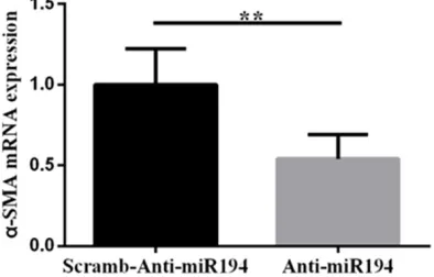
microRNA-194 knockdown promotes phe-notypic transformation of human vascular smooth muscle cells
In order to further study the mechanism of decreased expression of microRNA-194 in aortic dissection, we constructed a lentiviral vector which could downregulate expression of microRNA-194 and transfected it into human vascular smooth muscle cells to observe the effects of microRNA-194 on human vascular smooth muscle cells.Results in Figure 2 show
that expression of α-SMA, a specific molecule
of contracted vascular smooth muscle cells
[19], decreased significantly by downregulation
of microRNA-194 expression in human vascu-lar smooth muscle cells (P=0.005).
microRNA-194 knockdown promotes expres-sion of collagen III and osteopontin
[image:4.612.325.520.72.205.2]To further investigate whether intervention of microRNA-194 can further affect the function of smooth muscle cells to promote progression of aortic dissection, we selected collagen III and osteopontin as the indicator [20-22]. Results in Figures 3 and 4 show that downregu-lating expression of microRNA-194 can pro-mote expression of collagen III and osteopontin (P<0.001, P=0.022). Collagen III and osteopon-tin are components of the vascular wall but do not have the function of maintaining elasticity of the vessel wall. Excessive levels of collagen III and osteopontin can cause stiffness and lack of elasticity of the vessel wall, which is associated with remodeling of the vessel wall [17, 23].
[image:4.612.90.287.73.204.2]These results imply that downregulation of microRNA-194 expression can promote the Figure 3. Downregulating expression of
microR-NA-194 can promote expression of collagen III mRNA. Anti-miR194 group compared with the Scramb-Anti-miR194 group, **P<0.01.
[image:4.612.324.520.279.407.2]Figure 4. Downregulating expression of microR-NA-194 can promote expression of osteopontin mRNA. Anti-miR194 group compared with the Scramb-Anti-miR194 group, *P<0.05.
Figure 5. Downregulating expression of microR-NA-194 can promote expression of Smad1 mRNA. Anti-miR194 group compared with the Scramb-Anti-miR194 group, *P<0.05.
[image:4.612.93.285.281.414.2]transformation of human vascular smooth mu- scle cells from contractile to synthetic. There- fore, downregulating expression of microR-NA-194 can further affect function of smooth muscle cells and play an important role in the pathological progress of aortic dissection. microRNA-194 downregulation is associated with TGF-β1 signaling activation
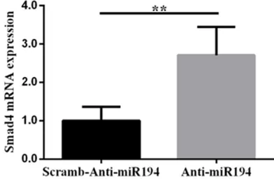
In order to further explore specific mechanisms
of phenotypic transformation and functional changes by microRNA-194 in smooth muscle
cells, we analyzed the TGF-β1 signaling path -way. Figures 5 and 6 showthat downregulation of microRNA-194 could promote expression of Smad1 and Smad4 mRNA (P=0.016, P=0.001).
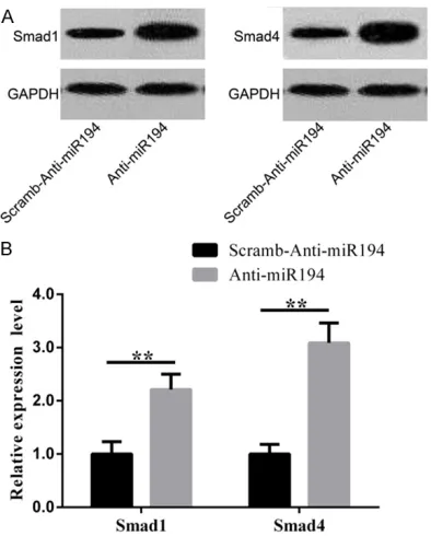
We also conducted Western blot to detect pro -tein levels. Results in Figure 7A show that
Smad1 and Smad4 proteins were significantly
increased when microRNA-194 was downregu-lated. Figure 7B shows that Smad1 and Sm-
ad4 proteins were significantly increased after
microRNA-194 knockdown (P<0.001, P<0.001).
As key molecules in TGF-β1 signaling pathway, significant increase in mRNA and protein
ex-pression of Smad1 and Smad4 indicate the
activation of TGF-β1 signaling pathway.
Discussion
Aortic dissection is a vascular system disease with high mortality and high disability. The main pathological changes are changes of extracel-lular matrix such as apoptosis, degeneration,
phenotypic transformation, and elastic fibers and collagen fibers in the aortic smooth muscle cells but the specific pathogenesis remains
unclear and there remains a lack of corre-sponding prevention and curative measures [4]. Aortic dissection is often accompanied by changes in expression of microRNAs. microR-NAs play an important role in the pathological progression of aortic dissection by regulating key cell and signaling pathways [24, 25]. microRNA-194 is widely involved in regulation of biological processes such as cell prolifera-tion, differentiaprolifera-tion, and apoptosis. It has been found that microRNA-194 plays an important regulatory role in cancer, infectious diseases, and metabolic diseases. However, little re- search has been done on aortic dissection. Therefore, we investigated expression levels of microRNA-194 in aortic dissection and studied
the specific mechanism of microRNA-194 in vitro using vascular smooth muscle cells.
We first performed microRNA-194 fluorescence quantitative PCR on aortic dissection and nor
-mal aorta tissues. We found that expression of microRNA-194 mRNA was significantly
decreased in patients with aortic dissection, suggesting that microRNA-194 may play a role in the pathogenesis of aortic dissection (P<0.05). Then, by constructing a lentiviral vec-tor to interfere with expression of microR-NA-194 in human vascular smooth muscle cells, we found that downregulation of microR-NA-194 can promote transformation of vascu-lar smooth muscle cells from contractile to syn-thetic and increase expression of collagen III and osteopontin. Previous studies have found that microRNA-194 could regulate differentia-tion of hepatocytes, osteoblasts, neurons, epi-thelial cells, and mesenchymal stem cells [26-28]. Overexpression of microRNA-194 can pro-mote differentiation of mesenchymal stem cells into osteoblasts [29]. However, there have not been any reports on the role of microR-NA-194 in vascular smooth muscle cells. Here,
we report for the first time that downregulation
[image:5.612.91.288.69.314.2]of microRNA-194 plays an important role in development of aortic dissection by promoting transformation from contractile to synthetic phenotypes of vascular smooth muscle cells. Figure 7. A: Downregulation of microRNA-194
Vascular smooth muscle cells are important cells that maintain the structure and function
of blood vessels. When they undergo apopto -sis, denaturation, and phenotypic transforma-tion by external stimuli, they can cause struc-tural remodeling of blood vessels and reduction of elasticity [30]. In a resting state, smooth muscle cells show contractile type. After stimu-lation, they can be converted to synthetic type, which can enhance cell proliferation with
elas-ticity significantly reduced, causing pathologi -cal changes such as vascular remodeling [31-33]. Pei et al. found that expression of collagen
III and osteopontin in the aortic wall was signifi -cantly increased in patients with aortic
aneu-rysm. Moreover, contractile-specific α-SMA of vascular smooth muscle cells was significantly
decreased in vitro cell experiments, while syn-thesis of collagen III and osteopontin was
sig-nificantly increased. Their results indicated that
phenotypic transformation of vascular smooth muscle cells is involved in development of aor-tic aneurysms pathogenesis [17]. Therefore,
expression of α-SMA, collagen III, and osteo -pontin in human vascular smooth muscle cells was also detected in our study. It was found that downregulation of microRNA-194 could
significantly reduce α-SMA, whereas osteopon -tin, a marker of synthetic vascular smooth
mus-cle cells, was significantly increased [19, 20].
These results suggest that microRNA-194 is involved in phenotypic modulation of vascu- lar smooth muscle cells. Furthermore, elastic
fibers and collagen fibers constitute the main
matrix components of blood vessels and their imbalance in proportion is the structural basis
for reduction of blood vessel elasticity. Wang et
al. found that connective tissue growth factor can promote expression of collagen III in the aortic wall and play a key role in the
pathogen-esis of aortic dissection [23]. We also found
that downregulation of microRNA-194 can
sig-nificantly increase expression of collagen III,
suggesting that microRNA-194 plays an impor-tant role in synthesis and regulation of extracel-lular matrix.
TGF-β1 can promote transformation of vascular
smooth muscle cells from contraction to syn-thetic, with biological functions of promoting cell migration and secretion, playing a key role in vascular remodeling. According to the
litera-ture, expression of TGF-β1 in aortic dissection is significantly increased, with the most promi -nent in the intima and media of vessels [34].
Therefore, in order to further explore the mech-anism by which microRNA-194 induces vascu-lar smooth muscle cell phenotype transforma-tion and extracellular matrix regulatransforma-tion, we detected Smad1 and Smad4, key molecules of
TGF-β1 signaling pathway. We found that Smad1 and Smad4 were significantly increased
at mRNA and protein levels. These results sug-gest that inhibition of microRNA-194 may play a role in the pathological progress of aortic
dis-section by activating the TGF-β1 signaling path -way to promote vascular smooth muscle cell phenotype transformation and extracellular matrix regulation.
We found that inhibition of microRNA-194 expression can activate TGF-β1 signaling path -way and affect the phenotype and function of vascular smooth muscle cells, thereby promot-ing development of aortic dissection. There were still many shortcomings: 1) The effect of microRNA-194 overexpression on the pheno-type and function of vascular smooth muscle cells was not studied; 2) Vascular endothelial cells are also important cells in the aortic wall, whether microRNA-194 can participate in development of aortic dissection by regulating function of vascular endothelial cells was not determined; and 3) Most of the experimental results were obtained by cell experiments in vitro and were not verified in animal experi -ments. Therefore, we plan to further carry out microRNA-194 overexpression in human vascu-lar smooth muscle cells and cultivate vascuvascu-lar endothelial cells in vitro to investigate whether
intervention of microRNA-194 influences the
biological function of vascular endothelial cells.
We plan to construct an animal model of aortic
dissection and conduct in vivo validation of the roles of microRNA-194.
In this study, we analyzed expression of microR-NA-194 in the aortic walls of 15 aortic dissec-tion patients undergoing aortic valve replace-ment and 13 normal donors without aortic
lesion. We found that inhibition of microR -NA-194 expression may be involved in the
pathogenesis of aortic dissection.
osteo-pontin. In addition, activation of TGF-β1 signal -ing pathway plays an important role. Therefore, inhibition of microRNA-194 expression can
activate TGF-β1 signaling pathways and affect
the phenotype and function of vascular smooth muscle cells, resulting in development of aortic dissection.
Disclosure of conflict of interest
None.
Address correspondence to: Dayong Liu, Depart- ment of Cardiothoracic Surgery, Da Qing Long Nan Hospital, No.35 Aiguo Road, Ranghulu District, Daqing 163001, Heilongjiang Province, China. Tel: +86-0459-5910211; E-mail: liudayong96@163. com
References
[1] Strayer RJ. Thoracic aortic syndromes. Emerg Med Clin North Am 2017; 35: 713-725. [2] Siddiqi HK, Bossone E, Pyeritz RE and Eagle
KA. Chronobiology of acute aortic syndromes. Heart Fail Clin 2017; 13: 697-701.
[3] Kuo EC and Han SM. Treatment of complex thoracoabdominal aortic disease. Cardiol Clin 2017; 35: 411-429.
[4] Nienaber CA, Clough RE, Sakalihasan N, Suzuki T, Gibbs R, Mussa F, Jenkins MP, Thompson MM, Evangelista A, Yeh JS, Cheshire N, Rosendahl U and Pepper J. Aortic dissec-tion. Nat Rev Dis Primers 2016; 2: 16071. [5] Peterss S, Mansour AM, Ross JA, Vaitkeviciute
I, Charilaou P, Dumfarth J, Fang H, Ziganshin BA, Rizzo JA, Adeniran AJ and Elefteriades JA. Changing pathology of the thoracic aorta from acute to chronic dissection: literature review and insights. J Am Coll Cardiol 2016; 68: 1054-1065.
[6] Wang L, Zhang S, Xu Z, Zhang J, Li L and Zhao G. The diagnostic value of microRNA-4787-5p and microRNA-4306 in patients with acute aortic dissection. Am J Transl Res 2017; 9: 5138-5149.
[7] Dong J, Bao J, Feng R, Zhao Z, Lu Q, Wang G, Li H, Su D, Zhou J, Jing Q and Jing Z. Circulating microRNAs: a novel potential biomarker for diagnosing acute aortic dissection. Sci Rep 2017; 7: 12784.
[8] Bai M, Zhang M, Long F, Yu N, Zeng A and Zhao R. Circulating microRNA-194 regulates human melanoma cells via PI3K/AKT/FoxO3a and p53/p21 signaling pathway. Oncol Rep 2017; 37: 2702-2710.
[9] Hu B, Xu C, Tian Y, Shi C, Zhang Y, Deng L, Zhou H, Cao P, Chen H and Yuan W. Inflammatory
microRNA-194 and -515 attenuate the bios- ynthesis of chondroitin sulfate during human intervertebral disc degeneration. Oncotarget 2017; 8: 49303-49317.
[10] Yu X, An J, Hua Y, Li Z, Yan N, Fan W and Su C. MicroRNA-194 regulates keratinocyte proli- feration and differentiation by targeting Grainyhead-like 2 in psoriasis. Pathol Res Pract 2017; 213: 89-97.
[11] Latouche C, Natoli A, Reddy-Luthmoodoo M, Heywood SE, Armitage JA and Kingwell BA. MicroRNA-194 modulates glucose metabolism and its skeletal muscle expression is reduced in diabetes. PLoS One 2016; 11: e0155108. [12] Wang K, Lai C, Gu H, Zhao L, Xia M, Yang P and
Wang X. miR-194 Inhibits innate antiviral im -munity by targeting FGF2 in influenza H1N1 virus infection. Front Microbiol 2017; 8: 2187. [13] Xie F, Yang L, Han L and Yue B. MicroRNA-194
regulates lipopolysaccharide-induced cell via-bility by inactivation of nuclear factor-kappa B pathway. Cell Physiol Biochem 2017; 43: 2470-2478.
[14] Couto LB and High KA. Viral vector-mediated RNA interference. Curr Opin Pharmacol 2010; 10: 534-542.
[15] Han K, Zhao T, Chen X, Bian N, Yang T, Ma Q, Cai C, Fan Q, Zhou Y and Ma B. microRNA-194 suppresses osteosarcoma cell proliferation and metastasis in vitro and in vivo by targeting CDH2 and IGF1R. Int J Oncol 2014; 45: 1437-1449.
[16] Shi N and Chen SY. Mechanisms simultane-ously regulate smooth muscle proliferation and differentiation. J Biomed Res 2014; 28: 40-46.
[17] Pei H, Tian C, Sun X, Qian X, Liu P, Liu W and Chang Q. Overexpression of MicroRNA-145 promotes ascending aortic aneurysm media remodeling through TGF-beta1. Eur J Vasc Endovasc Surg 2015; 49: 52-59.
[18] Li JL, Liu N, Chen XH, Sun M and Wang CB. Inhibition of UVA-induced apoptotic signaling pathway by polypeptide from Chlamys farreri in human HaCaT keratinocytes. Radiat Environ Biophys 2007; 46: 263-268.
[19] Mack CP. Signaling mechanisms that regulate smooth muscle cell differentiation. Arterioscler Thromb Vasc Biol 2011; 31: 1495-1505. [20] Yuan SM, Wang J, Huang HR and Jing H.
Osteopontin expression and its possible func-tions in the aortic disorders and coronary ar-tery disease. Rev Bras Cir Cardiovasc 2011; 26: 173-182.
[22] Parrish AR and Ramos KS. Differential pro-cessing of osteopontin characterizes the prolif-erative vascular smooth muscle cell pheno-type induced by allylamine. J Cell Biochem 1997; 65: 267-275.
[23] Wang X, LeMaire SA, Chen L, Shen YH, Gan Y, Bartsch H, Carter SA, Utama B, Ou H, Coselli JS and Wang XL. Increased collagen deposition and elevated expression of connective tissue growth factor in human thoracic aortic dissec-tion. Circulation 2006; 114: I200-205.
[24] Leeper NJ and Maegdefessel L. Non-coding RNAs: key regulators of smooth muscle cell fate in vascular disease. Cardiovasc Res 2018; 114: 611-621.
[25] Wang XJ, Huang B, Yang YM, Zhang L, Su WJ, Tian L, Lu TY, Zhang S, Fan XH and Hui RT. Differential expression of microRNAs in aortic tissue and plasma in patients with acute aortic dissection. J Geriatr Cardiol 2015; 12: 655-661.
[26] Zhuang H, Zhang R, Zhang S, Shu Q, Zhang D and Xu G. Altered expression of microRNAs in the neuronal differentiation of human Wharton’s Jelly mesenchymal stem cells. Neurosci Lett 2015; 600: 69-74.
[27] Jung KH, McCarthy RL, Zhou C, Uprety N, Barton MC and Beretta L. MicroRNA regulates hepatocytic differentiation of progenitor cells by targeting YAP1. Stem Cells 2016; 34: 1284-1296.
[28] Hino K, Tsuchiya K, Fukao T, Kiga K, Okamoto R, Kanai T and Watanabe M. Inducible expres -sion of microRNA-194 is regulated by HNF-1alpha during intestinal epithelial cell differen-tiation. Rna 2008; 14: 1433-1442.
[29] Li J, He X, Wei W and Zhou X. MicroRNA-194 promotes osteoblast differentiation via do- wnregulating STAT1. Biochem Biophys Res Commun 2015; 460: 482-488.
[30] Al-Huseini I, Ashida N and Kimura T. Deletion of IkappaB-Kinase beta in smooth muscle cells Induces vascular calcification through beta-catenin-runt-related transcription factor 2 sig-naling. J Am Heart Assoc 2018; 7.
[31] Leeper NJ, Raiesdana A, Kojima Y, Chun HJ, Azuma J, Maegdefessel L, Kundu RK, Quertermous T, Tsao PS and Spin JM. MicroRNA-26a is a novel regulator of vascular smooth muscle cell function. J Cell Physiol 2011; 226: 1035-1043.
[32] Maegdefessel L, Azuma J, Toh R, Merk DR, Deng A, Chin JT, Raaz U, Schoelmerich AM, Raiesdana A, Leeper NJ, McConnell MV, Dalman RL, Spin JM and Tsao PS. Inhibition of microRNA-29b reduces murine abdominal aor-tic aneurysm development. J Clin Invest 2012; 122: 497-506.
[33] Yu Y, Shi E, Gu T, Tang R, Gao S, Wang Y and Liu H. Overexpression of microRNA-30a contrib-utes to the development of aortic dissection by targeting lysyl oxidase. J Thorac Cardiovasc Surg 2017; 154: 1862-1869.