Original Article
miR-9-5p prompts malignancies of acute myeloid
leukemia cells mainly by targeting p27
Huizhe Zheng1, Yuanbo Sun2, Tao Zhan4, Kaidong Zeng3, Canzhang Liu3, Jingli Zhang2
1Pathological Diagnosis Center, Hongqi Hospital, Mudanjiang Medical University, Mudanjiang, P.R. China; 2
Depart-ment of Hematology, Hongqi Hospital, Mudanjiang Medical University, Mudanjiang City, Heilongjiang Province, P.R. China; 3Department Four of Circulation, Zhongshan Hospital Affiliated to Dalian University, Dalian, P.R. China; 4Department of Pathology, School of Basic Medical Sciences, Mudanjiang Medical University, Mudanjiang, P.R.
China
Received July 6, 2017; Accepted December 17, 2017; Epub March 15, 2018; Published March 30, 2018
Abstract: MicroRNAs play key roles in the initiation and progression of acute myeloid leukemia (AML). The abnormal expression of miR-9-5p has been widely reported in various tumors. However, the expression pattern of miR-9-5 in AML has never been explored. The current study aims to explore the role of miR-9-5p in the progression of AML. First, real-time quantitative PCR was used to determine the level of miR-9-5p in the bone marrow and whole blood of AML patients. Then, a dual-luciferase reporter assay was performed to explore the possible target genes of miR-9-5p in HL-60 cells. According to our study, the level of miR-miR-9-5p was significantly increased in the bone marrow and peripheral blood of AML patients compared with that of healthy controls. Furthermore, a close correlation was identified between the miR-9-5p level and clinicopathological parameters of AML, including FAB classification and cytogenetics. Additionally, the dual-luciferase reporter assay demonstrated that the cyclin-dependent kinase (Cdk) inhibitor p27Kip1 (p27) was a target of miR-9-5p. Transfection with miR-9-5p inhibitors into HL-60 cells resulted in decreased proliferation and enhanced cell apoptosis. In summary, these results indicated that miR-9-5p functions as an oncogenic regulator mainly by targeting p27 in HL-60 cells.
Keywords: MiR-9-5p, acute myeloid leukemia, cell cycle, p27
Introduction
As a key component of hematopoiesis, mye- loid development is characterized by sequen-tial regulator expression [1]. The dysregulation of those regulators may lead to the failed dif-ferentiation of myeloid development, which in-duces the initiation of myeloid leukemia. My- eloid leukemia can be divided into acute my- eloid leukemia (AML) and chronic myeloid leu-kemia (CML), both of which arise from abnor-mal white blood cells. Among them, acute my- eloid leukemia is a malignant disorder, which is characterized by the arrest of maturation and rapid proliferation of immature precurs- ors [2]. Due to substantial genetic abnormali-ties, the clinicopathological features and pro- gnosis of AML patients are widely variable. It has been reported that the early and timely diagnosis of acute myeloid leukemia (AML) is effective in reducing morbidity and mortality [3].
and enhanced cell apoptosis [9, 10]. Thus, it is important to explore the possible mechani- sm by which the expression of p27 is regulat- ed in AML.
microRNAs (miRNAs) are a class of small, non-coding RNAs with up to 22 nucleotides, which are stable in the blood and exhibit tissue-spe-cific expression patterns [11]. Therefore, miR-NAs may be utilized as non-invasive biomark- ers of specific cancers. In the current study, we mainly focused on miR-9-5p, which has been widely explored in various tumors. For example, in cervical cancer cells, miR-9-5p was reported to act as a tumor suppressor mainly by targeting astrocyte-elevated gene-1 [12]. Furthermore, miR-9-5p was also found to be upregulated in the bone marrow of AML patients carrying NPM1 mutations, which are present in approximately 35% of patients with
The median leukocyte count at diagnosis was 20,606/μL (range 420-352, 906/μL). The cli- nical characteristics of 200 pediatric AML pa- tients are summarized in Table 1. The healthy control group consisted of 30 healthy volun-teers with no clinical symptoms of cancer or other diseases.
Leukemic blasts from AML samples and mono-nucleated fractions from normal BM specim- ens were isolated by Ficoll-Hypaque (Nygaard) centrifugation and then cryopreserved. After thawing, cells were washed with HBSS and processed for RNA isolation.
RNA extraction
[image:2.612.91.310.110.462.2]Total RNA from the bone marrow (2 mg) and whole blood samples (5 mL) collected in tubes containing EDTA was extracted with RNAVzol
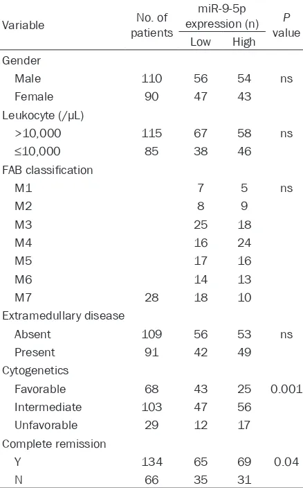
Table 1. Correlation of peripheral blood miR-9-5p level with clinical characteristics of 200 AML patients
Variable patientsNo. of
miR-9-5p expression (n) P
value Low High Gender
Male 110 56 54 ns
Female 90 47 43
Leukocyte (/µL)
>10,000 115 67 58 ns
≤10,000 85 38 46
FAB classification
M1 7 5 ns
M2 8 9
M3 25 18
M4 16 24
M5 17 16
M6 14 13
M7 28 18 10
Extramedullary disease
Absent 109 56 53 ns
Present 91 42 49
Cytogenetics
Favorable 68 43 25 0.001 Intermediate 103 47 56
Unfavorable 29 12 17 Complete remission
Y 134 65 69 0.04
N 66 35 31
AML [13, 14]. However, few studies have focused on the expression of miR-9-5p in pediatric AML patients. Here, we evaluated the role of miR-9-5p in AML, which has ne- ver been explored. Therefore, the present st- udy aims to evaluate the expression of miR-9-5p in the peripheral whole blood of AML patients and healthy controls, thereby inv- estigating the potential of blood miR-9-5p level as a non-invasive AML diagnostic bio- marker.
Materials and methods
Patient samples
Bone marrow (BM) specimens were isolat- ed from 200 pediatric AML patients and 30 healthy volunteers before any interventio- nal measures between February 2014 and October 2015 in Hongqi Hospital. All peri- pheral whole blood samples of patients and specimens were handled anonymously according to ethical and legal standards. The study was approved by the hospital’s Ethical Committee with informed consent from the patients.
LS (Vigorous Biotechnology, Beijing) in strict accordance with the manufacturer’s instruc-tions. The concentration and purity of the RNA samples was determined by OD260/OD280.
Cell culture
The human AML cell line HL-60 was derived at the Institute of Haematology & Blood Dis- eases Hospital, Chinese Academy of Medical Sciences & Peking Union Medical College (Tianjin, China). HL-60 was cultured in IMDM (HyClone, Logan, UT, USA) supplemented wi- th 20% fetal bovine serum (Gibco, Life Tech- nologies, USA).
Transient transfection
Cells were seeded at 106 cells/well in 6-well plates. Meanwhile, miR-9-5p mimic, inhibitor, or miR negative control (GenePharma) was mixed with HiPerFect transfection reagent (Qiagen) and incubated at room temperature for 10 min. Then, the complex was added to the culture medium for 48 h.
Reverse transcription (RT) and quantitative (q) polymerase chain reaction (PCR)
For the synthesis of the cDNA of the specific miR, a TaqMan MicroRNA Reverse Transcrip- tion Kit (Applied Biosystems) was used. To qu- antify miR-9-5p, a quantitative real-time PCR assay was performed using SYBR Green Su- permix (Bio-Rad) on a Bio-Rad iCycler iQ real-time PCR detection system according to the manufacturer’s instructions. The thermal cy- cling conditions included a hot start step at 95°C for 10 min, followed by 40 cycles at 95°C for 15 s and 60°C for 1 min. The 2-del- ta delta Ct analysis method was used to de- termine the relative quantity of miR-9-5p. U6 was used as an internal control. The primers were listed as follows: RT-miR-9-5p: CGTAT- CCAGTGCAGGGTCCGAGGTATTCGCACTGGATAC- GACGCCAAT; RT-U6: TCGTATCCAGTGCAGGGT- CCGAGGTATTCGCACTGGATACGACAAATATG; miR-9-5p-F: GCTCTTAGCAGCACAGAAAT; U6-F: GCGTCGTGAAGCGTTC; Universe reverse prim-er: GTGCAGGGTCCGAGGT.
Protein extraction and Western blot analysis
Protein samples were extracted in RIPA buffer (1% TritonX-100, 15 mmol/L NaCl, 5 mmol/L EDTA, and 10 mmol/L Tris-HCl (pH 7.0) (Solar- bio, China)) supplemented with a protease and
phosphatase inhibitor cocktail (Sigma) and then separated by 10% SDS-PAGE, followed by electrophoretic transfer to PVDF membr- anes. After incubation with 8% milk in PBST (pH 7.5) for 2 h at room temperature, the me- mbranes were incubated with the following primary antibodies: p27, p-AKT, anti-AKT, anti-Bcl-2, anti-Bax and anti-β-actin (Cell Signalling). Immunodetection was performed by an enhanced chemiluminescence detection system (Millipore) according to the manufac-turer’s instructions. The housekeeping gene GAPDH was used as the internal control.
Luciferase target assay
The TargetScan program (http://www.targets- can.org/vert_71/) was used to identify the possible target genes of miR-9-5p. The 3’un- translated region (UTR) of p27 containing the predicted binding site was cloned into the pmirGLO (Promega) luciferase reporter vector. The PCR procedures were as follows: a hot st- art step at 95°C for 10 min, 40 cycles at 95°C for 15 s and 55°C for 45 s, and 72°C for 30 s. To construct the mutant vector, the Fast Mutagenesis System was used (TransGen Biotech, Beijing, China).
For luciferase reporter assays, cells were seed-ed at 5×10⁴ cells/well in 24-well plates in a 500-μL volume for 18 h. Then, the modified firefly luciferase vector (500 ng/μL) was mix- ed with the VigoFect transfection reagent ac- cording to the manufacturer’s instructions. Af- ter transfection for 48 h, the dual-luciferase reporter assay system (Promega) was used to determine the changes in relative luciferase units (RLU). Renilla luciferase activity was us- ed as the internal control.
Apoptosis assay
TUNEL
Nuclear fragmentation was detected by TUNEL staining with an apoptosis detection kit (R & D Systems) according to the manufacturer’s protocol.
Statistical analysis
The data are presented as the mean ± SD. Differences were analyzed with Student’s t test. ROC curves were used to assess miR-9- 5p as a biomarker, and the AUC was reported.
eral blood of AML patients (6.47 ± 10.22) co- mpared with that of healthy controls (1.0 ± 0.58).
Correlation between miR-9-5p expression in the peripheral blood with clinical parameters of AML
[image:4.612.94.371.74.292.2]Next, the correlation between miR-9-5p expre- ssion in the peripheral blood and clinical par- ameters of AML was analyzed. According to
Table 1, no correlation was identified between miR-9-5p and gender, age, leukocytes, extra-medullary disease and complete remission. St- rikingly, a significant correlation between the peripheral blood miR-9-5p level and FAB clas-sification (p=0.0023) and cytogenetics (p= 0.001) was found in AML patients (Table 1). In addition, the ROC curve was applied to eva- luate whether miR-9-5p expression can be us- ed as a potential diagnostic marker for AML. It was shown that the level of miR-9-5p expres-sion could be available as a potential diagnos-tic biomarker for screening AML from controls with an AUC of 0.976 (95% CI=0.931-1.000; P
< 0.0001; Figure 2).
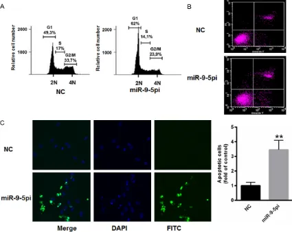
Decreased miR-9-5p induced cell cycle arrest and enhanced apoptosis
Next, we explored the specific role of miR-9-5p in AML cells. As shown in Figure 3A, the
inhibi-Figure 1. Increased miR-9-5p level in the bone marrow and peripheral blood of AML patients. Real-time PCR analysis of miR-9-5p expression in the bone marrow (A) or peripheral blood (B) of AML patients compared with that of healthy controls. **P < 0.01, ***P < 0.001 vs. control.
P < 0.05 was used to define a statistically significant diffe- rence.
Results
Increased miR-9-5p in the bone marrow and peripheral blood of AML patients
First, the expression level of miR-9-5p was explored in leu-kemia cells isolated from the bone marrow and peripheral blood of AML patients and he- althy controls. As shown in
Figure 1A, the level of miR-9-5p was markedly enhanced in the leukemic cells of bone ma- rrow from AML patients (5.55 ± 13.75) compared with that of healthy controls (1.0 ± 1.56). We also found that miR-9-5p was increased in the leukemic cells isolated from the
[image:4.612.93.287.366.514.2]tion of miR-9-5p in HL-60 cells markedly in- duced cell cycle arrest. Furthermore, TUNEL staining demonstrated that suppressed miR- 9-5p significantly enhanced apoptosis in HL- 60 cells (Figure 3B). In addition, apoptosis was increased by up to 3.45-fold in HL-60 ce- lls transfected with miR-9-5p inhibitors com-pared with those transfected with the nega- tive control (Figure 3C). These data indicated an oncogenic role of miR-9-5p in AML cells.
p27 is the target gene of miR-9-5p
We further analyzed the target gene of miR-9-5p, and TargetScan prediction demonstrated that there was a conserved binding site of miR-9-5p in the 3’UTR of p27 (Figure 4A). Then, the 3’UTR of p27 was cloned into the pmirGLO plasmid, and further study demonstrated that
miR-9-5p suppressed the relative luciferase reporter activity of pmirGLO-p27-3’UTR com-pared with that of the blank vector (Figure 4B). Next, the overexpression of miR-9-5p signifi-cantly suppressed the protein levels of p27 and bcl-2, while it markedly enhanced the ex- pression of bax (Figure 4C). In contrast, the inhibition of miR-9-5p markedly increased the expression of p27 and bcl-2, while it inhibited the level of bax in HL-60 cells (Figure 4D). Together, these data showed that miR-9-5p inhibited the apoptosis of HL-60 cells by tar- geting p27.
Discussion
[image:5.612.93.518.73.411.2]Among older patients, AML is a different dis-ease due to the aging of hematopoietic stem cells (HSCs) caused by DNA damage, telomere
Figure 4. p27 was the target gene of miR-9-5p. A. Schematic analysis of the binding site for miR-9-5p in the 3’UTR of p27. B. Dual-luciferase reporter assay demonstrated that miR-9-5p suppressed the relative luciferase re-porter activity of pmirGLO-p27-3’UTR compared with that of the blank vec-tor. C. The overexpression of miR-9-5p significantly suppressed the protein levels of p27 and bcl-2, while it markedly enhanced the expression of bax. D. The inhibition of miR-9-5p markedly increased the expression of p27 and bax, while it inhibited the level of bax in HL-60 cells. *P < 0.05, **P < 0.01 vs. control.
shortening, and oxidative stress [15]. It has been reported that the 5-year survival rate is approximately 50% among pediatric AML pa- tients, while the mortality is even higher among older AML patients [16, 17]. miRNAs have be- en widely reported to be dysregulated in dif- ferent tumors and any disruption of miRNAs may result in oncogenesis and metastasis [18]. For example, the upregulation of miR-155 is correlated with initial progression and poor outcomes in Chinese pediatric AML patients [19]. In addition, miR-22-mediated MECOM de- gradation has been shown to play a key role in normal hematopoiesis and AML
develop-ment, indicating its potential value in AML diagnosis and th- erapy [20]. Therefore, investi-gations on miRNAs may shed light on the understanding of AML progression.
Previous studies have shown the enhanced expression of miR-9-5p in the bone marrow of AML patients carrying NP- M1 mutations [13, 14]. In ad- dition, miR-9-5pb was also fo- und to be overexpressed in mixed lineage leukemia (MLL) -associated AML in the bead-based miRNA profiling of 57 samples [21]. Here, we first ex- plored the expression of miR-9-5p in the bone marrow and peripheral blood of AML pati- ents and healthy controls. We first demonstrated that miR-9-5p was markedly increased in the bone marrow and periph-eral blood of AML patients compared with that of healthy controls. Further studies dem-onstrated the significant corre-lation of peripheral blood miR-9-5p levels with FAB classifi- cation (p=0.0023) and cyto- genetics (p=0.001). ROC anal-ysis indicated that peripheral blood miR-9-5p could be us- ed to distinguish AML patients from healthy controls. Together, these data indicated miR-9-5p as a potential biomarker for AML diagnosis.
p27Kip1 is an inhibitor of cell cycle progres-sion, which is encoded by the CDKN1B gene and suppresses the activation of cyclin E-CD- K2 and cyclin D-CDK4 complexes, thereby blocking the cell cycle at G1 phase [22]. Studi- es have indicated that p27 plays a key role in carcinogenesis and may also be utilized as a prognostic factor in different tumors [23, 24]. In the present study, we found that miR-9-5p markedly suppressed the relative luciferase re- porter activity of p27, and Western blot show- ed that the protein level of p27 was markedly inhibited by p27, suggesting that p27 is a target gene of miR-9-5p. These findings demonstrate an oncogenic role of miR-9-5p mediated by p27 in the progression of AML.
Acknowledgements
This work was supported by Doctoral Project Startup Grants from the Mudanjiang Medical University [MDJ-2015-45].
Disclosure of conflict of interest
None.
Address correspondence to: Dr. Jingli Zhang, De- partment of Hematology, Hongqi Hospital, Mudan- jiang Medical University, 5 Township Road, Aimin District, Mudanjiang 157011, Heilongjiang Provin- ce, P.R. China. Tel: +86-453-6582800; E-mail: zhangjingli666@yeah.net
References
[1] Bissels U, Bosio A and Wagner W. MicroRNAs are shaping the hematopoietic landscape. Haematologica 2012; 97: 160-167.
[2] Chen J, Odenike O and Rowley JD. Leukaemo-genesis: more than mutant genes. Nat Rev Cancer 2010; 10: 23-36.
[3] Aydin M, Flenaugh EL and Nichols M. Hemopty-sis, anemia and respiratory failure: a rare ini-tial presentation of acute leukemia. J Natl Med Assoc 2005; 97: 1550-1552.
[4] Xia Q, Cai Y, Peng R, Wu G, Shi Y and Jiang W. The CDK1 inhibitor RO3306 improves the re-sponse of BRCA-pro fi cient breast cancer cells to PARP inhibition. Int J Oncol 2014; 44: 735-744.
[5] Macaluso M, Montanari M, Cinti C and Giorda-no A. Modulation of cell cycle components by epigenetic and genetic events. Semin Oncol 2005; 32: 452-457.
[6] Chu IM, Hengst L and Slingerland JM. The Cdk inhibitor p27 in human cancer: prognostic
po-tential and relevance to anticancer therapy. Nat Rev Cancer 2008; 8: 253-267.
[7] Porter PL, Barlow WE, Yeh IT, Lin MG, Yuan XP, Donato E, Sledge GW, Shapiro CL, Ingle JN, Haskell CM, Albain KS, Roberts JM, Livingston RB and Hayes DF. p27(Kip1) and cyclin E ex-pression and breast cancer survival after treat-ment with adjuvant chemotherapy. J Natl Can-cer Inst 2006; 98: 1723-1731.
[8] Pohl G, Rudas M, Dietze O, Lax S, Markis E, Pirker R, Zielinski CC, Hausmaninger H, Kubis-ta E, Samonigg H, Jakesz R and Filipits M. High p27Kip1 expression predicts superior relapse-free and overall survival for premenopausal women with early-stage breast cancer receiv-ing adjuvant treatment with tamoxifen plus goserelin. J Clin Oncol 2003; 21: 3594-3600. [9] Diaz-Rodriguez E, Hernandez-Garcia S, Sanz E,
and Pandiella A. Antitumoral effect of Ocoxin on acute myeloid leukemia. Oncotarget 2016; 7: 6231-6242.
[10] Zhang F, Zhu FB, Li JJ, Zhang PP and Zhu JF. Hyperoside enhances the suppressive effects of arsenic trioxide on acute myeloid leukemia cells. Int J Clin Exp Med 2015; 8: 15290-15295.
[11] Zhi F, Cao X, Xie X, Wang B, Dong W, Gu W, Ling Y, Wang R, Yang Y and Liu Y. Identification of circulating microRNAs as potential biomarkers for detecting acute myeloid leukemia. PLoS One 2013; 8: e56718.
[12] Zhang X, Cai D, Meng L and Wang B. MicroR-NA-124 inhibits proliferation, invasion, migra-tion and epithelial-mesenchymal transimigra-tion of cervical carcinoma cells by targeting astrocyte-elevated gene-1. Oncol Rep 2016; 36: 2321-2328.
[13] Jongen-Lavrencic M, Sun SM, Dijkstra MK, Valk PJ and Lowenberg B. MicroRNA expres-sion profiling in relation to the genetic hetero-geneity of acute myeloid leukemia. Blood 2008; 111: 5078-5085.
[14] Schotte D, Pieters R and Den Boer ML. MicroR-NAs in acute leukemia: from biological players to clinical contributors. Leukemia 2012; 26: 1-12.
[15] Liang Y, Van Zant G and Szilvassy SJ. Effects of aging on the homing and engraftment of mu-rine hematopoietic stem and progenitor cells. Blood 2005; 106: 1479-1487.
[16] Thol F and Ganser A. Molecular pathogenesis of acute myeloid leukemia: a diverse disease with new perspectives. Front Med China 2010; 4: 356-362.
treat-ment of acute myeloid leukemia. Semin Oncol 2011; 38: 215-224.
[18] Bouyssou JM, Manier S, Huynh D, Issa S, Roc-caro AM and Ghobrial IM. Regulation of mi-croRNAs in cancer metastasis. Biochim Bio-phys Acta 2014; 1845: 255-265.
[19] Xu LH, Guo Y, Cen JN, Yan WY, He HL, Niu YN, Lin YX, Chen CS and Hu SY. Overexpressed miR-155 is associated with initial presentation and poor outcome in Chinese pediatric acute myeloid leukemia. Eur Rev Med Pharmacol Sci 2015; 19: 4841-4850.
[20] Shen C, Chen MT, Zhang XH, Yin XL, Ning HM, Su R, Lin HS, Song L, Wang F, Ma YN, Zhao HL, Yu J and Zhang JW. The PU.1-modulated mi-croRNA-22 is a regulator of monocyte/macro-phage differentiation and acute myeloid leuke-mia. PLoS Genet 2016; 12: e1006259. [21] Li Z, Huang H, Chen P, He M, Li Y, Arnovitz S,
Jiang X, He C, Hyjek E, Zhang J, Zhang Z, Elkahloun A, Cao D, Shen C, Wunderlich M, Wang Y, Neilly MB, Jin J, Wei M, Lu J, Valk PJ, Delwel R, Lowenberg B, Le Beau MM, Vard-iman J, Mulloy JC, Zeleznik-Le NJ, Liu PP, Zhang J and Chen J. miR-196b directly targets both HOXA9/MEIS1 oncogenes and FAS tumour suppressor in MLL-rearranged leukaemia. Nat Commun 2012; 3: 688.
[22] Sherr CJ and Roberts JM. CDK inhibitors: posi-tive and negaposi-tive regulators of G1-phase pro-gression. Genes Dev 1999; 13: 1501-1512. [23] Hernandez-Garcia S, Gonzalez V, Sanz E, and
Pandiella A. Effect of oncoxin oral solution in HER2-overexpressing breast cancer. Nutr Can-cer 2015; 67: 1159-1169.