ISSN Online: 2327-509X ISSN Print: 2327-5081
DOI: 10.4236/jbm.2019.710007 Oct. 22, 2019 72 Journal of Biosciences and Medicines
Depolymerization of α- & β-Chitosan by e-Beam
Irradiation
Sueng Hwan Jo
1, Changyong Choi
2, Soo-Kyung Choi
31School of Medicine, Chosun University, Gwangju, Republic of Korea
2Petrochemical Process Engineering Department, Hanyeong University, Yeosu, Jeollanam-do, Republic of Korea
3Technical Research Center, LCGen Co., Ltd., Gwangju, Republic of Korea
Abstract
α- and β-chitosan with molecular weight of 190,000 and 800,000 respectively, were depolymerized by e-beam irradiation with various doses. The radiation yield of scission (Gs) and degradation rate of the chitosans were identified.
The synergistic chemical degradation in the presence of hydrogen peroxide is more effective at lower doses. Mw of β-chitosan was dramatically decreased
from 800,000 to 21,030 at the irradiation dose 5 kGy, on the other hand, that of α-chitosan was decreased much more gradually from 190,000 to 36,000. The values of Gs at 10 kGy in the solution without H2O2 and with H2O2 were
respectively 6.09 × 10⁻⁵ mol/cal and 30.6 × 10⁻⁵ mol/cal for α-Chitosan, and
8.18 × 10⁻⁵ mol/cal and 43.8 × 10⁻⁵ mol/cal for β-chitosan. It was obviously
effective on depolymerization by using the combination of e-beam and
H2O2. α-Chitosan molecules are likely to adopt a diffuse conformation in
the solution and make the different morphologies depending on the con-centration.
Keywords
α- and β-Chitosan, Conformation, Depolymerization, e-Beam Irradiation, Hydrogen Peroxide
1. Introduction
Chitosan which is composed of a copolymer of N-acetylglucosamine and gluco-samine is a linear polysaccharide composed of randomly distributed β-(1→4)- linkage, deacetylated unit and acetylated unit. The amine group mainly exists in the form of NH3+, making it a charged polycation in the chitonsan molecule,
which was reported to have either a rigid rod-type structure. The different mo-lecular conformations of α- and β-chitosan influence their antibacterial
mechan-How to cite this paper: Jo, S.H., Choi, C.Y. and Choi, S.-K. (2019) Depolymerization of α- & β-Chitosan by e-Beam Irradiation. Journal of Biosciences and Medicines, 7, 72-83.
https://doi.org/10.4236/jbm.2019.710007
Received: August 30, 2019 Accepted: October 19, 2019 Published: October 22, 2019 Copyright © 2019 by author(s) and Scientific Research Publishing Inc. This work is licensed under the Creative Commons Attribution International License (CC BY 4.0).
http://creativecommons.org/licenses/by/4.0/
DOI: 10.4236/jbm.2019.710007 73 Journal of Biosciences and Medicines
isms, such as the interactions between the protonated amino groups of chitosan and the negatively charged bacterial cell membranes [1] [2] [3].
The low solubility of chitosan exerts its limitation in use, especially in medi-cine and food science. To overcome the shortcoming, several researchers have attempted to reduce the molecular weight of chitosan with various chemicals such as enzymes, acids, and hydrogen peroxide [4]-[8]. Furthermore, depolyme-rization by high energy beam radiation has been recently investigated, since this technique provides a useful tool to separate oligo-chitosans without the need of additional processing [4] [5]. However, less attention has been paid to study on electron beam (EB) irradiation [6] [7] [8]. An EB can provide faster processing than a gamma ray [7] from 60Co to irradiate the same radiation doses. It is also
not needed to concern the radioactive waste disposal. As we previously described
[8], hydrogen peroxide was found to have a synergistic effect in the process of EB-depolymerization of polysaccharides. The combined H2O2/EB process for
depolymerization of polysaccharide is based on the formation of a reactive hy-droxyl radical due to the dissociation of hydrogen peroxide in the presence of
e-beam. Generally, chitosan is a charged polycationic molecule and which is re-ported to have either a rigid rod-type structure. Thus, it can be expected that chitosan can be applied to specific areas as long as the conformation of the mo-lecule is correctly identified. Characteristics of chitosan in solubility and struc-ture of the molecular chain vary according to the factors such as collection ori-gin, processing method, degree of deoxidation and amine distribution [9] [10] [11] [12].
Physical characteristics, such as inherent viscosity and Mark-Houwink (MH) coefficients, are closely related to polymer chain conformation. The dependence of intrinsic viscosity on molecular weight provides information on the confor- mation and extension of polymers. The simple MH Equation (1) expressing this fact is given by:
[ ]
[ ]
ln η =ln K +alnMv (1)
where K and a are empirical constants that are valid for a specific polymer-solvent pair within one to two orders of magnitude of molecular weight [13] [14] and Mv
is the viscosity average of the molecular weight. The so-called Mark-Houwink constants K and a depend upon the kind of polymer, solvent and the tempera-ture used during the viscosity measurement. The plot of the ln[η] vs. ln[M] typ-ically gives a straight line with slope a and intercept ln[K]. The slope, a, can vary from 0 (compact sphere) over 0.65 - 0.85 (random coil) to 2.0 (very rigid rod chain) revealing information about the polymer conformation in solution. Conformation of polymers in solution influences strongly macroscopic polymer effects like viscosity.
proper-DOI: 10.4236/jbm.2019.710007 74 Journal of Biosciences and Medicines
ties, etc. The different intra- and inter-molecular behaviors between α- and
β-chitosan could alter the chitosan conformations in the solution, hence, the physical properties of α-chitosan are different from those of β-chitosan in solu-bility, reactivity, and swelling ability.
In this paper, the depolymerization of chitosan by means of high energy ir-radiation with various doses in H2O2 solution was conducted using an electron
beam as an irradiation source. We will discuss the conformation of α- and
β-chitosan depoloymerized by EB irradiation with/without hydrogen peroxide using a recent advancement in the analysis of the molar mass and size depen-dencies of radiation yield of scission and the MH coefficients [15].
2. Experimental
To prepare the 1 wt% of α-chitosan (av. Mw; 190,000 g/mol, ChitoLife Co. Ltd.,
Korea) and β-chitosan (800,000 g/mol, ChitoLife Co. Ltd., Korea) solution, 1% lactic acid aqueous solution was used as a solvent. The α- and β-chitosan solu-tion were irradiated with electron beam in sealed state with or without hydrogen peroxide. The radiation yield of scission Gs (mol/J) of depolymerized chitosan
was calculated by irradiating with 0, 5, 10, 20, 30, 40 kGy at 2.5 MeV energy us-ing E-beam process system (EB Tech, Korea). The electron beam irradiated chi-tosan solution was precipitated in ethanol, filtered or centrifuged, washed with ethanol, and then dried for analysis. The molecular weight of depolymerized chitosan was determined by GPC-MALLS (WYATT Technology Corporation, Detector-MALLS: DAWN EOS-RI: OPTILAB DSP). Intrinsic viscosities of de-polymerized chitosan were measured by using Ubbelohde viscometer at 35˚C and the inherent viscosity (ηinh) was measured at concentrations of 1.0, 0.5, and
0.25 mg/ml. The morphology of depolymerized chitosan was investigated by TEM with FE-SEM (JEOL, JSM840A), and particle size and zeta potential (ELSZ-2000) were measured.
The radiation yield of scission of chitosan (Gs) was determined in connection
with concentrations of chitosan solution. Polymer degradation can be expressed in terms of molecular weight reduction due to polymer chain scission and its ef-ficiency can be estimated by radiation yield of scission Gs (mol/J) [16]. The Gs of
chitosan by e-beam irradiation was calculated by the following Equation (2).
0
2 1 1
s
w
c G
D d M M
= −
⋅ (2)
where Gs is radiation yield of scission (mol/J), D; absorbed dose (Gy), d; solution
density (kg/dm3), c; polymer concentration (g/dm3), M
w, Mo; weight-average
molecular weight of polymer after and before irradiation respectively. The in-trinsic viscosities were determined as the average of extrapolating both ln (ηsp/c)
DOI: 10.4236/jbm.2019.710007 75 Journal of Biosciences and Medicines
the aggregation of chitosan depolymerized. For MH plot to avoid any accidentally occurrence of aggregated material we started to filter the sample solutions care-fully. The morphological examination of samples was conducted using SEM and BIO-TEM (Tecnai G2 spirit Biotwin, FEI, Portland, USA) using an acceleration tension of 100 kV. The diluted samples were placed on carbon coated copper grid for TEM observation.
3. Results and Discussion
The chitosans in different conformation were depolymerized by e-beam irradia-tion in hydrogen peroxide aqueous soluirradia-tion. Table 1 & Table 2 and Figure 1
display the plots of MH equations for α- (Figure 1(a) and Figure 1(c)) and
β-chitosan (Figure 1(b) and Figure 1(d)). As illustrated, the molecular weights of the chitosans were decreased rapidly with increasing irradiation dose up to 20 kGy in both the α- and β-chitosan solutions under the condition without H2O2
(Figure 2(a) and Figure 2(b)), and then gradually leveled off as the dose
[image:4.595.212.539.395.538.2]in-creased. This pattern may be attributed to the fact that the drastic tion occurs in amorphous region in the beginning, and the slow depolymeriza-tion at higher doses occurs in crystalline regions. Similar results were also re-ported at other irradiated polysaccharides [4] [17].
Table 1. Molecular weight (Mw) and intrinsic viscosity of depolymerised Chitosan.
Dose (kGy)
α-Chitosan β-Chitosan
*
w
M ln[η]a *
w
M ln[η]a
0 190,000 5.220 800,000 6.892 5 36,000 5.030 21,030 4.854 10 28,470 4.879 23,910 5.204 20 4950 3.172 7470 - 30 5530 - 8270 4.323 40 5520 3.539 7020 -
*Measured by GPC-MALLS, athe intrinsic viscosity was evaluated by Equation (1).
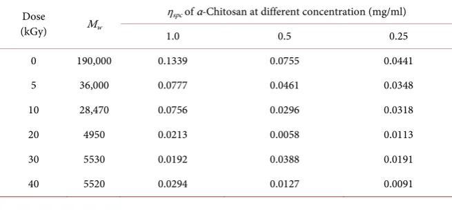
Table 2.Specific viscosities (ηspc) of depolymerised α-Chitosan at different concentration.
Dose
(kGy) Mw
ηspc of α-Chitosan at different concentration (mg/ml)
[image:4.595.210.538.583.735.2]DOI: 10.4236/jbm.2019.710007 76 Journal of Biosciences and Medicines Figure 1. Intrinsic viscosity of (a) α- and (b) β-Chitosan and MHS plots of (c) α- and (d) β-Chitosan.
Figure 2. Molecular weight vs. dose of (a) α- and (b) β-Chitosan and Gsof (c) α- and (d) β-Chitosan.
Concentration(g/ml)
0.0000 0.0002 0.0004 0.0006 0.0008 0.0010 0.0012
ηspc
/C 0 50 100 150 200 250 185.1804 152.8519 131.4299 23.8638 83.0837 34.4446 Mw;190,000 Mw; 36,000 Mw;28,470 Mw;5,530 (a) Concentration(g/ml)
0.0000 0.0002 0.0004 0.0006 0.0008 0.0010 0.0012
ηspc /C 0 200 1000 1200 984.3730 128.2068 182.0003 75.4330 Mw; 800000 Mw; 23910 Mw;21030 Mw; 8270 (b)
ln Mw
0 2 4 6 8 10 12 14
ln [ η ] 0 1 2 3 4 5 6
α−Chitosan MHS
a=0.2358
lnK=2.4316 K=11.377
a=3.3481 Mw=7400
(c) β−Chitosan MHS
ln Mw
0 2 4 6 8 10 12 14 16
ln [ η ] 0 2 4 6 8 a=0.5479 lnK=-0.5237, K=0.5923 (d) α−chitosan Dose(kGy)
0 10 20 30 40
Mw (g /mo l)X 10 4 0 5 10 15 20
without H2O2
with H2O2 (a)
Dose(kGy)
0 10 20 30 40
Mw (g /mo l)X 10 5 0 2 4 6 8 10
with H2O2 without H2O2 β-chitosan
(b)
α−chitosan
Dose(kGy)
0 10 20 30 40
G s( m o/ J) X 10 5 0 20 40 60 80 100 without H2O2 With H2O2 (c) Dose(kGy)
0 10 20 30 40
G s( m ol /J )X 10 5 0 20 40 60 80 100
without H2O2
[image:5.595.215.534.395.692.2]DOI: 10.4236/jbm.2019.710007 77 Journal of Biosciences and Medicines
ln[η] was plotted against ln[Mw] and the MH exponent, “a” values were
eva-luated for the α-chitosan in various concentrations (Figure 1(c)). ln[η] of the
α-chitosans whose molecular weights are 7400 or higher increased slightly with increasing molecular weight, it gave 0.236 in MH value, a. Whereas ln[η] of
α-chitosan whose molecular weights were lower than 7400 showed a pronounced increase of 1.854 in slope. It means that compact sphere changes to very rigid rod chain in the solution at around Mw 7400. This indicates that molecular
weight-induced conformational transition occurred in this Mw range. On the
other hand, the a for β-chitosan was 0.548 which means random coil in its con-formation.
Several reports suggested that irradiation-induced scissions of glycosidic bonds of chitosan caused an inconsistent reduction in molecular weight of the poly-mers in different conformation [18] [19]. As shown in Table 1, the molecular weight of β-chitosan was dramatically decreased from 800,000 to 21,030 at the irradiation dose 5 kGy, on the other hand, that of α-chitosan was decreased much more gradually from 190,000 to 36,000. That means that synergistic de-gradation in the presence of hydrogen peroxide is rather effective at low doses such as around 5 kGy. It is well known that chitin has two forms, named α and β
chitin, in which α-chitin is very stable with intra-chain and inter-sheet hydrogen bonds from the antiparallel sheets along with c-axis in orthorhombic cell, while
β-chitin has no hydrogen bonds between two inter-sheets owing to their parallel directions [18] [19] [20].
Kurita reported [21] that α-chitin is more rigid and more crystalline and can be less susceptible to deacetylation compared to β-chitin. Therefore, the poly-meric structures of chitosan deacetylated from different forms of chitin may not be identical and β-chitosan can have higher solubility with less crystallinity, thus providing much higher radiation susceptibility than α-chitosan. In order to es-timate the radiation sensitivity of the α- and β-chitosan molecules in the pres-ence or abspres-ence of H2O2, the radiation yield of scission Gs were calculated (Table
3 and Figure 2(c) and Figure 2(d)) in the range from 0 to 40 kGy. The results
showed that the depolymerization of chitosan by e-beam irradiation was much faster in the solution with H2O2, than in that without H2O2 up to 20 kGy (Figure
3). It was obvious that the rate of chitosan molecular weight decreased by the combination of e-beam and H2O2 was much higher than that by e-beam
irradia-tion alone. This result showed that the synergistic effect for degradairradia-tion of chi-tosan with e-beam and H2O2 gained the most effective outcome, especially at the
lowest absorbed dose studied of 5 kGy. In both cases, Gs increased dramatically
at 5 kGy and then decreased gradually. The values of Gs at 10 kGy in the solution
without H2O2 and with H2O2 were 6.09 × 10⁻⁵ mol/cal and 30.6 × 10⁻⁵ mol/cal
respectively for α-Chitosan, and 8.18 × 10⁻⁵ mol/cal and 43.8 × 10⁻⁵ mol/cal for β-chitosan. It means that it was obvious that the rate of chitosan molecular weight decreased by the combination of e-beam and H2O2 was much higher than
solu-DOI: 10.4236/jbm.2019.710007 78 Journal of Biosciences and Medicines
tions. One Gs value obtained by other authors for chitosan irradiated in aqueous
solution with molecular weight of 100 kDa was 0.382 × 10⁻⁷ mol/J [22]. The
[image:7.595.209.540.457.585.2]rad-iation depolymerization yield in chitosan solution is strongly influenced by the presence of hydrogen peroxide, initial molecular arrangement and irradiation conditions such as crystallinity, moisture content, radiation dose [7] [23] [24]. The observation from electron microscope (SEM and TEM) revealed informa-tion on the shape and size of the particles. The irradiainforma-tions of the α-chitosan gave the individual spherical shape. On the other hand, it was difficult to identify the irradiated β-chitosan as a spherical shape. The α and β-chitosan particles in these samples were found in variable shapes (Figure 4 & Figure 5). To investi-gate the effect of radiation dose and concentration of chitosan during irradiation on the size and size distribution of aggregates, the aggregates from SEM and TEM images were randomly collected and determined for all irradiation condi-tions. The result indicated that both factors influenced not only aggregate shape but also aggregate size (Figure 4) as well as size distribution (Figure 6). The size distribution plots in Figure 6 clearly show that the e-beam irradiation gave chi-tosan particles with very narrow size distribution when compared to the original one. The present experimental plots prove that aggregate size distribution is possibly controlled by using e-beam irradiation. Radiation seems to influence the particle size over an extended radiation dose period as observed on the left-shift in the distribution plot. The relationship between radiation dose and aggregate size as shown in Table 4 and Figure 6 confirmed that the higher the radiation dose, the bigger the aggregate size. This is inconsistent with nanopar-ticles formed from chitosan by high energy irradiation [4] [5] [6] [24].
Table 3.Gs of depolymerised Chitosan with/without hydrogen peroxide.
Dose (kGy)
α-Chitosan β-Chitosan
Without H2O2 With H2O2 Without H2O2 With H2O2
Gs (×105) Gs (×105) Gs (×105) Gs (×105)
0 0.0000 0.0000 0.0000 0.0000 5 9.0959 65.0168 18.7084 69.7559 10 6.0320 30.6579 8.1958 43.7562 20 19.8848 20.0597 13.4017 27.1489 30 11.8282 - 8.0548 - 40 8.8828 - 7.1255 -
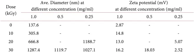
Table 4. Average diameters and zeta potential of α-Chitosan at different concentration.
Dose (kGy)
Ave. Diameter (nm) at
different concentration (mg/ml) at different concentration (mg/ml) Zeta potential (mV) 1.0 0.5 0.25 1.0 0.5 0.25 0 137.6 - - 2.87 - - 10 305.8 - - 14.8 - - 20 666.8 - 1188.7 13.0 - 5.07 30 1287.4 1119.7 1027.1 16.2 18.03 2.52
[image:7.595.209.539.617.709.2]DOI: 10.4236/jbm.2019.710007 79 Journal of Biosciences and Medicines Figure 3. Molecular weight and specific viscosity of depolymerized
[image:8.595.220.528.284.519.2]α-Chitosan with different dose.
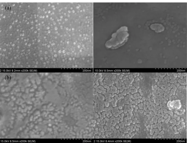
Figure 4. SEM images of depolymerised α-Chitosan (30 kGy) at (a) 1.0 mg/ml (b) 0.5 mg/ml concentration.
Figure 5. TEM images of depolymerised α-Chitosan (30 kGy) at (a) 1.0 mg/ml (b) 0.5 mg/ml concentration (×100 K magnification).
Dose(kGy)
ηsp
c
0.00 0.05 0.10 0.15 0.20
1.0mg/ml 0.50mg/ml 0.25mg/ml
0 10 20 30 40
ln
Mw
0 2 4 6 8 10 12 ln Mw
(a)
(b)
[image:8.595.221.532.567.693.2]DOI: 10.4236/jbm.2019.710007 80 Journal of Biosciences and Medicines Figure 6. Particle size of raw α-Chitosan (black), depolymerised α-Chitosan at 10 kGy (Blue), 20 kGy (Yellow), and 30 kGy (Red) at 0.5 mg/ml concentration.
This pattern obtained from α-chitosan may be due to the formation of ionic clusters based on ion-ion interaction which could give much bigger aggregates. Additionally, the irradiation of the chitosan seems to bring the aggregate size bigger. The particle sizes are up to more than 100 nm, when using e-beam dose as low as 5 kGy.
The particle still formed significant circle-like shape as expected, especially in spherical form. TEM images implied that α-chitosan irradiated at low concen-tration (0.5 mg/ml) gave a smaller particle size with individual spherical shape than that from high concentration (1.0 mg/ml). SEM images have shown the morphological properties and surface appearance of aggregates. The aggregates have nearly spherical shape, smooth surface and size range of about 20 - 80 nm (Figure 4).
SEM imaging of the two solutions are consistent with the size profiles, mono-dispersed nanoparticles and agglomerates of several hundred nanometers were visible in the solution. In the present study, the results obtained by Zetasizer re-vealed that the zeta potential of the α-chitosan nanoparticles can greatly influ-ence their stability in suspension by means of electrostatic repulsion between the particles. Our results demonstrated respective zeta potentials of α-chitosan na-noparticles of 2.5 and 28.7 mV. It is supposed that the α-chitosan molecules de-polymerized by e-beam radiation are not uniform. Chitosan molecules are likely to adopt a diffuse conformation in the solution because of electrostatic repulsion force existing between amine groups along the molecular chain [8] [10] [23]. The carboxyl groups on the surface of a large protein molecule may form hy-drogen bonds with amine groups at certain sites along the chitosan chain [23].
DOI: 10.4236/jbm.2019.710007 81 Journal of Biosciences and Medicines
chitosan nanoparticles with different sizes via electrostatic interaction of chito-san and methacrylic acid [25]. The mechanism for the synergistic effect was pre-sented by Kang, Dai, Zhang, and Chen (2007) [5]. Particularly, the degradation of chitosan by the gamma-ray irradiation alone can be ascribed to the direct ac-tion of radiaac-tion on the chitosan chains. In the present study the effect of differ-ent chitosan concdiffer-entrations (0.25, 0.5, 1.0 mg/mL) on molecular weight and viscosity was evaluated (Table 2) and measured the size and zeta potential by Zetasizer (Table 4, Figure 6). Our results showed that by increasing the chitosan concentration from 0.25 to 1.0 mg/mL in the absence of hydrogen peroxide, the size of nanoparticles increased (Table 1). Table 2 also shows that specific vis-cosity of chitosan decreased with decreasing chitosan molecular weight in solu-tions. For chitosan of the same molecular weight, specific viscosity decreased with increasing solution concentration. Zetasizer revealed that the size of the aggregates of α-chitosan was increased from 138 nm to 1287 nm with increasing radiation dose from 0 to 30 kGy, possibly due to Zeta potential of chitosan par-ticles can greatly influence their stability in suspension by means of electrostatic repulsion between the particles [26]. The results indicate that the chain flexibili-ties of higher molecular weight chitosans were higher than those of lower mole-cular weight ones.
However, strong pre-peaks in the detector responses (especially in the light scattering detector) and an increase or curvature of the ln[Mw] distribution over
the retention volume indicate the presence of aggregates. These effects were completely absent for the samples measured. Yanagisawa et al.[27] showed that the absence of aggregates is an important prerequisite for a reliable conforma-tional analysis. For MH plot to avoid any occurrence of aggregates we filtered the sample solutions carefully. Chitosan molecules are likely to adopt a diffuse conformation in the solution because of electrostatic repulsion force existing between amine groups along the molecular chain.
4. Conclusion
The chitosan with two different molecular weights in different conformation was depolymerized by e-beam irradiation in aqueous solution. Radiation sensitivity was evaluated by the determination of radiation yield of scission Gs and
degra-dation behavior. The e-beam irradiation, used as a method to reduce the mole-cular weight of chitosan, showed even greater synergy when used with H2O2 and
its synergistic degradation is more effective at lower irradiation doses. The zeta potentials of the chitosans demonstrated the molecules depolymerized by e-beam are not uniform
Acknowledgements
DOI: 10.4236/jbm.2019.710007 82 Journal of Biosciences and Medicines
Conflicts of Interest
The authors declare no conflicts of interest regarding the publication of this pa-per.
References
[1] Rinaudo, M. (2006) Chitin and Chitosan: Properties and Applications. Progress in Polymer Science, 31, 603-632.https://doi.org/10.1016/j.progpolymsci.2006.06.001
[2] Paul, W. and Sharma, C.P. (2000) Chitosan, a Drug Carrier for the 21st Century: A Review. STP Pharma Sciences, 10, 5-22.
[3] Brugnerotto, J.D.J., Heux, L., Mazeau, K. and Rinaudo, M. (2001) Overview on Structural Characterization of Chitosan Molecules in Relation with Their Behavior in Solution. Macromolecular Symposia, 168, 1-20.
https://doi.org/10.1002/1521-3900(200103)168:1<1::AID-MASY1>3.0.CO;2-W
[4] Ahn, K.-J., Choi, W.-S., Lee, D.-W., Byun, M.-W. and Park, H.-J. (2002) Prepara-tion of Chitosan Oligomers by IrradiaPrepara-tion. Polymer Degradation and Stability, 78, 533-538.https://doi.org/10.1016/S0141-3910(02)00226-4
[5] Kang, Y.D.D.B., Zhang, H.Q. and Chen, D. (2007) Synergetic Degradation of Chi-tosan with Gamma Radiation and Hydrogen Peroxide. Polymer Degradation and Stability, 92, 359-362.https://doi.org/10.1016/j.polymdegradstab.2006.12.006
[6] Kim, H. (2013) Molecular Weight Control of Chitosan Using Gamma Ray and Electron Beam Irradiation. Journal of Radiation Industry, 7, 51-54.
https://www.ksri.kr/0304/view/page/25/id/293
[7] Tahtat, D., Mahlous, M., Benamer, S., Khodja, A.N. and Youcef, S.L. (2012) Effect of Molecular Weight on Radiation Chemical Degradation Yield of Chain Scission of γ-Irradiated Chitosan in Solid State and in Aqueous Solution. Radiation Physics and Chemistry, 81, 659-665.https://doi.org/10.1016/j.radphyschem.2012.02.036
[8] Jo, B.W. and Choi, S.-K. (2014) Degradation of Fucoidans from Sargassum Fulvel-lum and Their Biological Activities, Carbohydrate Polymers. Carbohydrate Poly-mers, 111, 822-829. https://doi.org/10.1016/j.carbpol.2014.05.049
[9] Ulanski, P. and Rosiak, J.M. (1992) Preliminary Studies on the Radiation-Induced Changes in Chitosan. Radiation Physics and Chemistry, 39, 53-57.
https://doi.org/10.1016/1359-0197(92)90171-B
[10] Nagasawa, H.M.N., Yoshii, F. and Kume, T. (2000) Radiation-Induced Degradation of Sodium Alginate. Polymer Degradation and Stability, 69, 279-285.
https://doi.org/10.1016/S0141-3910(00)00070-7
[11] Wu, W.J., Huang, K.S., Chen, J.B. and Lian, H.S. (2008) Application of Low-Mole- cular-Weight Chitosan in Durable Press Finishing. Carbohydate Polymers, 73, 254-260.https://doi.org/10.1016/j.carbpol.2007.11.023
[12] Diep, T.B., Hai, L., Nagasawa, N., Yoshii, F. and Kume, T. (2003) Radiation Depo-lymerization of Chitosan to Prepare Oligomers. Nuclear Instruments and Methods in Physics Research B, 208, 466-470.
https://doi.org/10.1016/S0168-583X(03)01181-9
[13] Bohdanecky, M. and Kovar, J. (1982) Viscosity of Polymer Solutions. Elsevier Scien-tific Publishing Company, Amsterdam.
DOI: 10.4236/jbm.2019.710007 83 Journal of Biosciences and Medicines
https://doi.org/10.1016/0144-8617(93)90140-Y
[15] Wang, W., Bo, S., Li, S. and Qin, W. (1991) Determination of the Mark-Houwink Equation for Chitosans with Different Degrees of Deacetylation. International Journal of Biological Macromolecules, 13, 281-285.
https://doi.org/10.1016/0141-8130(91)90027-R
[16] Chemat, F., Teunissen, P.G.M., Chemat, S. and Bartels, P.V. (2001) Sono-Oxidation Treatment of Humic Substances in Drinking Water. Ultrasonics Sonochemistry, 8, 247-250.https://doi.org/10.1016/S1350-4177(01)00084-0
[17] Aliste, A.J., Vieira, F.F. and DelMastro, N.L. (2000) Radiation Effects on Agar, Al-ginates and Carrageenan to Be Used as Food Additives. Radiation Physics and Chemistry, 57, 305-308.https://doi.org/10.1016/S0969-806X(99)00471-5
[18] Lamarque, G., Cretenet, M., Viton, C. and Domard, A. (2005) New Route of Deace-tylation of α- and β-Chitins by Means of Freeze-Pump Out-Thaw Cycles. Bioma-cromolecules, 78, 1380-1388.https://doi.org/10.1021/bm049322b
[19] Dweltz, N.E. (1961) The Structure of β-Chitin. Biochimica et Biophysica Acta, 51, 283-294.https://doi.org/10.1016/0006-3002(61)90169-X
[20] Minke, R. and Blackwell, J. (1978) The Structure of α-Chitin. Journal of Molecular Biology, 120, 167-181.https://doi.org/10.1016/0022-2836(78)90063-3
[21] Kurita, K., Yoshino, H., Nishimura, S.-I. and Ishii, S. (1993) Preparation and Bio-degradability of Chitin Derivatives Having Mercapto Groups. Carbohydrate Poly-mers, 20, 239-245.https://doi.org/10.1016/0144-8617(93)90095-L
[22] Hien, N.Q., Phu, D.V., Duy, N.N. and Lan, N.T.K. (2012) Degradation of Chitosan in Solution by Gamma Irradiation in the Presence of Hydrogen Peroxide. Carbohy-drate Polymers, 87, 935-938.https://doi.org/10.1016/j.carbpol.2011.08.018
[23] Pasanphan, W., Choofong, S., Rimdusit, P. and Piroonpan, T. (2010) Systematic Fabrication of Chitosan Nanoparticle by Gamma Irradiation. Radiation Physics and Chemistry, 79, 1095-1102.https://doi.org/10.1016/j.radphyschem.2010.04.003
[24] Pillai, C.K.S., Paul, W. and Sharma, C.P. (2009) Chitin and Chitosan Polymers: Chemistry, Solubility and Fiber Formation. Progress in Polymer Science, 34, 641-678.
https://doi.org/10.1016/j.progpolymsci.2009.04.001
[25] De Moura, M.R., Aouada, F.A. and Mattoso, L.H.C. (2008) Preparation of Chitosan Nanoparticles Using Methacrylic Acid. Journal of Colloid and Interface Science, 321, 477-483.https://doi.org/10.1016/j.jcis.2008.02.006
[26] Wu, Y., Yang, W., Wang, C., Hu, J. and Fu, S. (2005) Chitosan Nanoparticles as a Novel Delivery System for Ammonium Glycyrrhizinate. International Journal of Pharmaceutics, 295, 235-245.https://doi.org/10.1016/j.ijpharm.2005.01.042