Themed Section: Molecular Pharmacology of GPCRs
REVIEW
The evolving small-molecule
fluorescent-conjugate
toolbox for Class A GPCRs
Andrea J Vernall
1, Stephen J Hill
2and Barrie Kellam
11School of Pharmacy, Centre for Biomolecular Sciences,University of Nottingham,Nottingham,
UK, and2Institute of Cell Signalling, School of Biomedical Science, Queen’s Medical Centre, University of Nottingham,Nottingham, UK
Correspondence
Dr Barrie Kellam, School of Pharmacy, Centre for
Biomolecular Sciences, University of Nottingham, Nottingham, NG7 2RD, UK. E-mail:
---Keywords
G-protein coupled receptor; fluorescent ligand conjugate; binding; screening; fluorescence microscopy; imaging
---Received
9 April 2013
Revised
9 May 2013
Accepted
17 May 2013
The past decade has witnessed fluorescently tagged drug molecules gaining significant attraction in their use as
pharmacological tools with which to visualize and interrogate receptor targets at the single-cell level. Additionally, one can generate detailed pharmacological information, such as affinity measurements, down to almost single-molecule detection limits. The now accepted utilization of fluorescence-based readouts in high-throughput/high-content screening provides further evidence that fluorescent molecules offer a safer and more adaptable substitute to radioligands in molecular
pharmacology and drug discovery. One such drug-target family that has received considerable attention are the GPCRs; this review therefore summarizes the most recent developments in the area of fluorescent ligand design for this important drug target. We assess recently reported fluorescent conjugates by adopting a receptor-family-based approach, highlighting some of the strengths and weaknesses of the individual molecules and their subsequent use. This review adds further strength to the arguments that fluorescent ligand design and synthesis requires careful planning and execution; providing examples illustrating that selection of the correct fluorescent dye, linker length/composition and geographic attachment point to the drug scaffold can all influence the ultimate selectivity and potency of the final conjugate when compared with its unlabelled precursor. When optimized appropriately, the resultant fluorescent conjugates have been successfully employed in an array of assay formats, including flow cytometry, fluorescence microscopy, FRET and scanning confocal microscopy. It is clear that fluorescently labelled GPCR ligands remain a developing and dynamic research arena.
LINKED ARTICLES
This article is part of a themed section on Molecular Pharmacology of GPCRs. To view the other articles in this section visit http://dx.doi.org/10.1111/bph.2014.171.issue-5
Introduction
Recent years have witnessed a rapid expansion in the use of fluorescence-based techniques with which to interrogate bio-logical processes and receptors of physiobio-logical and pharma-cological importance. There are a number of methods by which a fluorescent probe can be generated, including genetic manipulation to label a protein with a fluorophore
(Dedeckeret al., 2013), measurement of the inherent
fluores-cence of a compound (Beltran et al., 2011; Burchak et al.,
2011) or by using synthetic chemistry to covalently link a biologically active compound to a fluorophore of choice, creating a fluorescent ligand conjugate (Daly and McGrath, 2003). Fluorescent ligands can be designed to interact with different entities, for example, as reaction-based probes, which offer a powerful technique for detecting and studying small molecules and/or metal ions of interest in living
systems (Chanet al., 2012). However, the most prevalent use
of fluorescent ligands has been the study of protein–protein
interactions (Kaleet al., 2012) or ligand–receptor interactions
(Leopoldoet al., 2009) in biological systems.
One of the most important human receptor families, from a drug discovery and development perspective, is the GPCRs
(Alexander et al., 2013). GPCRs are 7-transmembrane
span-ning receptors, which account for nearly 4% of the
protein-encoding human genome (Bjarnadóttiret al., 2006) and are
the target of approximately 30% of all marketed drugs
(Overingtonet al., 2006). GPCRs have been classified into five
different classes (http://www.gpcr.org/7tm/proteinfamily), of which Class A is the largest and generally regarded as the most understood. GPCRs are signalling powerhouses and can regulate various intracellular biological cascades via the binding of extracellular endogenous ligands, such as pep-tides, hormones and neurotransmitters. There is significant interest surrounding the development of fluorescent ligands with which to study GPCRs, and research reports of fluores-cent GPCR ligands have been previously reviewed (Middleton and Kellam, 2005; Kuder and Kiec´-Kononowicz, 2008; Böhme
British Journal of Pharmacology (2014)1711073–1084 1073 © 2013 The Authors. British Journal of Pharmacology published by John Wiley &
molecule (non-peptide) fluorescent conjugates for Class A GPCRs that have been reported subsequent to these afore-mentioned reviews.
Fluorescent ligands are powerful tools to study GPCRs as they can be employed in many varied experiments to reveal insight into receptor structure and function in native, live
cells (Briddonet al., 2011). High-affinity fluorescent
antago-nists can be used to label the target GPCR, and fluorescently tagged agonists can provide a means to monitor dynamic processes such as receptor internalization and trafficking (cf. examples within this review). A fluorescent ligand can be used as the competing probe in a competition-based binding
assay (Cottetet al., 2011; Sextonet al., 2011; Stoddartet al.,
2012) instead of a radiolabelled ligand, thereby avoiding the inherent safety risks, legal issues and disposal costs associated with the latter. In addition to measuring the direct displace-ment of a competing fluorescent ligand from a GPCR orthos-teric site, fluorescent ligands have enormous potential for revealing elaborate and intricate details about receptor oli-gomerization through the use of FRET and BRET assays
(Albizuet al., 2010; Cottetet al., 2011; 2012). Kinetic
meas-urements of the fluorescent ligand–receptor interaction can
reveal insight into receptor allosterism (Hillet al., 2014, this
issue) and receptor dimerization (May et al., 2011), while
ligand–receptor diffusion times measured using techniques such as fluorescence correlation spectroscopy (FCS) can be used to distinguish different receptor complexes (Briddon
and Hill, 2007; Jakobset al., 2012). This brief precis highlights
just some of the possible applications of fluorescent ligands in what is becoming a rapidly expanding field.
Fluorescent ligands for GPCRs
The design of a small-molecule-based fluorescent probe begins with selecting an amenable parent pharmacophore, surveying where to append the linker, determining what linker to use and, lastly, deciding what fluorophore to cova-lently tether (Jacobson, 2009). The linker position on the parent ligand must be tolerant to chemical change, which is often driven by existing structure activity relationship (SAR) data where available. There is an increasingly diverse range of commercially available fluorophores, and often the commer-cial fluorophore can be purchased as the pre-activated
N-hydroxysuccinimidyl (NHS) ester primed for coupling to
an amine on the pharmacophore-linker congener. Properties to consider when selecting the appropriate fluorophore to append to a congener include the absorption and emission profile of the fluorophore, lipophilicity (which can influence the conjugate’s ability to diffuse across the cell membrane) and whether the fluorophore is quenched in certain environ-ments. From a GPCR imaging perspective, the fluorescent ligand will ideally not enter the cells (unless bound to the internalized receptor), show very low levels of non-specific membrane binding, be quenched when not bound to the receptor and/or cell membrane and, for most applications, be displaceable using higher concentrations of a known non-fluorescent ligand that targets the same receptor. Once assembled, the fluorescent conjugate must be rigorously pharmacologically characterized, as its profile in terms of
the parent ligand. In the following sections, we review the small-molecule-based fluorescent conjugates that have been developed for Class A receptor families since publication of earlier reviews of this subject area (Middleton and Kellam, 2005; Kuder and Kiec´-Kononowicz, 2008; Böhme and Beck-Sickinger, 2009).
Adenosine receptor
The use of fluorescent probes for studying the adenosine receptor has recently been comprehensively reviewed by
Kozmaet al. (2013b). The fluorescent ligand toolbox for the
adenosine receptor family is relatively advanced compared with other Class A GPCRs, with many reports of both antago-nist and agoantago-nist-based probes built around different
pharma-cophores (predominately for the A1- and A3-adenosine
receptor subtypes) by the research groups of Jacobson and
Hill/Kellam (refer to references within Kozmaet al., 2013b).
Use of fluorescent antagonists for the adenosine A1- and
A3-receptors is now at a stage where they can be used in place
of radioligand-binding studies for screening purposes. A good example of this is a recent report from our laboratories of high-content screening of a fragment library to identify new
synthetic scaffolds for the human A1- and A3-adenosine
recep-tor family subtypes (Stoddartet al., 2012). Since the review by
Kozma and colleagues, there has been one additional account comparing the pharmacology and imaging properties of three
new agonist-based fluorescent adenosine A3-receptor probes
(Kozmaet al., 2013a) to five alternatives that had been
pre-viously reported (Tosh et al., 2009). The new compounds
included an IR dye 700 DX conjugate (1) linked through the C2 position of the adenine nucleoside ring, and two
N6-linked Alexa Fluor 488 probes (2) and (3) synthesized by
click-coupling between an azide and alkyne. The three novel conjugates unfortunately displayed a weaker adenosine
A3-receptor potency when compared with the originally
reported fluorescent ligands, and therefore, the authors pro-ceeded with imaging studies using the previously reported
Cy5-containing MRS5218 (Tosh et al., 2009) as their
first-choice fluorescent probe to visualize and study both the
human and the mouse A3-receptor.
Adrenoceptor
Martikkala et al. (2009) constructed three
europium(III)-labelled probes for theβ2-adrenoceptor by coupling amino
pindolol derivatives containing different linker lengths to isothiocyanate-activated europium chelates [the chemical moiety(s) of these chelates were not disclosed]. The com-pounds with the shortest (4) and longest (5) linker-length were employed in a competitive time-resolved fluorescence emission-binding assay using the beta-blocker propranolol as
the model drug. IC50values of 60 and 37 nM for the human
β2-adrenoceptor were obtained for propranolol using (4) and
(5), respectively, compared to a value of 33 nM calculated
from a [3H]dihydroalprenolol radioligand displacement assay.
It was interesting to note that an intermediate linker-length
europium conjugate (5minus one of the heptanamide units)
could not displace propranolol from the β2-adrenoceptor.
point, in any one direction. This demonstrates that a fluores-cent conjugate can possess a complex, unique and often unpredictable pharmacological profile compared with the parent pharmacophore.
In a comprehensive study from our laboratories, a series
of red-fluorescent β-adrenoceptor ligands were synthesized
based on three different orthostericβ-antagonist head groups;
namely propranolol, alprenolol and pindolol (Baker et al.,
2011). Using alkyl- or polyether-based linker extensions, the resultant propranolol (6) and alprenolol-based (7)
fluo-rescent β-blockers displayed high affinity for the human
β2-adrenoceptor. This study provided a further example of
how subtle changes in the structural nature of the linker can exert a significant impact on the final conjugate’s
pharma-cology. The 8-carbon linker analogue of6, where the
‘PEG-like’ linker was replaced with a hydrocarbon chain, showed
a 10-fold lower affinity for the β2-adrenoceptor compared
to6. In contrast, when the linker of6was replaced with a shorter 4-carbon linker, the conjugate’s affinity for the
β2-adrenoceptor was comparable to6. Conjugate6was used
to visualize ligand–receptor binding in CHO-β2cells
express-ing the human form of theβ2-adrenoceptor using confocal
microscopy, and displayed clear labelling of the membrane-bound receptors at 3 nM. This specific binding could be attenuated by incubation with various concentrations
(1–100 nM) of theβ2-selective antagonist ICI 118551. In this
study, it was also of interest to note that the pindolol-based fluorescent conjugates showed significant loss of affinity when compared with the native drug molecule. Even with three orthosteric ligands acting upon the equivalent receptor-binding pocket, one cannot therefore assume that installa-tion of a fluorophore onto the analogous posiinstalla-tion of a congener will afford similar pharmacological outcomes with regard to the final conjugate.
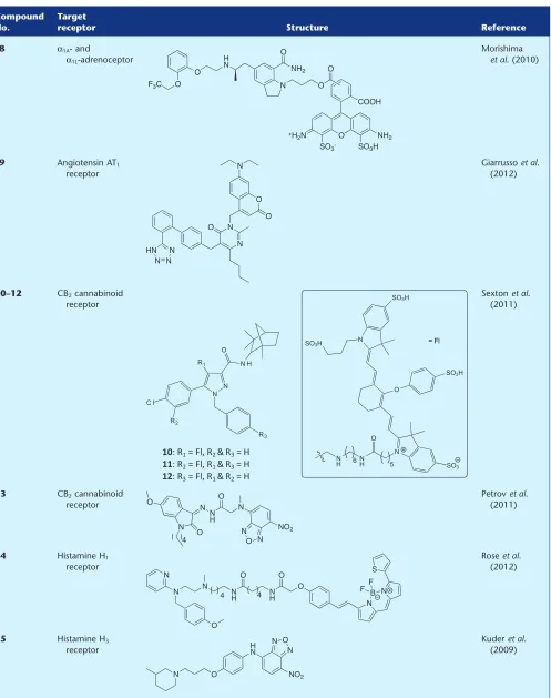
Morishimaet al. (2010) developed a high-affinity
fluores-cent probe (8) selective for the α1A-adrenoceptor and α1L
-adrenoceptor (thought to be anα1A-adrenoceptor phenotype)
over the α1B-adrenoceptor and α1D-adrenoceptor subtypes.
Theα1A-adrenoceptor subtype selective antagonist silodosin,
which is used to treat bladder outlet obstruction, was labelled with an Alexa Fluor 488 fluorophore. The authors did not disclose which isomer of the fluorophore was used, and
there-fore the Alexa Fluor 488 mixture of 5′and 6′isomers has been
depicted in Table 1. While fluorescent probe 8 displayed a
10-fold reduction in binding affinity across the human adrenoceptor receptor subtypes as compared with silodosin,
it retained anα1A-adrenoceptor selectivity profile (100- and
15-fold selective overα1B-adrenoceptor andα1D-adrenoceptor
respectively). Fluorescent confocal microscopy demonstrated
that8localized to the membrane of CHO cells overexpressing
theα1A-adrenoceptor, and this binding could be significantly
reduced using the high-affinity selective antagonist prazosin.
Building on this promising result,8was used to visualize the
α1L-adrenoceptor, an α1A-adrenoceptor phenotype, localized
to the muscle layer of the human prostate.
Angiotensin receptor
A fluorescent angiotensin II AT1 receptor (AT1R) ligand has
been reported, derived from a sartan-based pharmacophore
(Giarrussoet al., 2012). In place of the thiophene carboxylate
moiety of the antagonist milfasartan, Giarrussoet al. (2012)
instead installed various polyaromatic hydrocarbons or a
cou-marin fluorophore (e.g.9) by reacting an alkyl halide with
the pyrimidinone pharmacophore core. The authors remark that polyaromatic conjugates, such as those containing a
naphthalene moiety, were clearly unsuitable forin vivouse;
therefore, coumarin-conjugate 9was further evaluated as a
potential visual AT1R probe. Functional analysis using CHO
cells expressing the rat AT1aR revealed that9was an
antago-nist of the AT1R, with an estimated pKbvalue similar to the
native drug. Although the authors state that9was a selective
ligand for the AT1R, no pharmacological data were provided
for other angiotensin receptors to confirm this statement.
The ability of9to label the AT1R was evaluated, but
unfor-tunately, significant accumulation of fluorescence in the cell
cytoplasm was observed even with non-AT1R transfected
CHO cells. Development of less lipophilic fluorescent ligands, to eliminate this intracellular localization, is an ongoing work in the authors’ laboratory.
Cannabinoid receptor
In a recent report by Sextonet al. (2011), two newly designed
CB2cannabinoid receptor (CB2R) fluorescent probes (10) and
(11) were compared with the previously reported fluorescent
antagonist NIRmbc94 (12) (Baiet al., 2008). The purpose of
these new conjugates was to examine the influence of linker location around the core of the antagonist. The parent
phar-macophore, CB2R selective antagonist SR144528, lacked an
intrinsic biological handle such as an amine or carboxylic acid. Therefore, a 6-(aminohexyl)aminomethyl tether was incorporated in additional positions to that previously
reported for12, and then coupled to the near-infrared IRDye
800CW-NHS ester. The two new compounds did not demon-strate measurable binding to mouse delayed brain tumour
cells that heterologously express the mouse CB2R;
conse-quently, the fluorescent conjugate of choice remained the previously reported NIRmbc94 (12). This study reinforces the importance of identifying a tolerant location on the pharma-cophore for linker attachment and, as anticipated, demon-strates that different linker positions can have dramatic effects on final conjugate pharmacology. NIRmbc94 (12) was subsequently used as the competing probe in a
competition-binding assay (Sextonet al., 2011), and this methodology was
further elaborated by screening a small compound library to
reliably identify known CB2R binders. The authors then went
on to demonstrate that NIRmcb94 can identify endogenously
expressed CB2R in a mouse microglia cell line, BV-2.
Instead of the more common approach of conjugating a known, discrete orthosteric ligand via a linker to a fluoro-phore, the fluorescent moiety can instead be designed as part of the primary ‘pharmacophore’ scaffold with rational receptor–ligand interactions in mind. This approach was
employed by Petrov et al. (2011), who constructed isatin
acylhydrazone-based antagonist13that demonstrated
selec-tivity for the human CB2R (over human CB1R). A
methoxyi-satin derivative was linked to a 7-nitro-2,1,3-benzoxadiazole
(NBD) fluorophore to afford13, which, although displaying
slightly reduced affinity to the comparable non-fluorescent
compound fragment, retained the desired CB2R selectivity
profile. Using fluorescent confocal microscopy, the
associa-tion of13with T-cells could be visualized, and this
Recently reported small-molecule fluorescent conjugates with application to GPCRs
Compound No.
Target
receptor Structure Reference
1 Adenosine
receptor
Kozmaet al. (2013a)
2
3
4 β2-Adrenoceptor Martikkala
et al. (2009)
5
6 β2-Adrenoceptor Bakeret al.
(2011)
Table 1
Continued
Compound No.
Target
receptor Structure Reference
8 α1A- and
α1L-adrenoceptor
Morishima
et al. (2010)
9 Angiotensin AT1
receptor
Giarrussoet al. (2012)
10–12 CB2cannabinoid
receptor
N N R1
O N H
R3
C l
R2
O N
N SO3H
SO3H
SO3H
SO3
O
N
H 5
N H 6
= Fl
10: R1= Fl, R2 & R3= H
11: R2= Fl, R1 & R3= H
12: R3= Fl, R1 & R2= H
Sextonet al. (2011)
13 CB2cannabinoid
receptor
Petrovet al. (2011)
14 Histamine H1
receptor
Roseet al. (2012)
15 Histamine H3
receptor
Continued
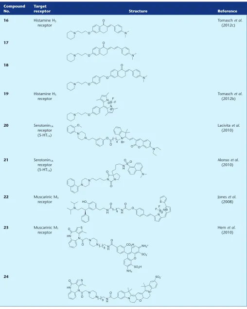
Compound No.
Target
receptor Structure Reference
16 Histamine H3
receptor
Tomaschet al. (2012c)
17
18
19 Histamine H3
receptor
Tomaschet al. (2012b)
20 Serotonin1A
receptor (5-HT1A)
Lacivitaet al. (2010)
21 Serotonin1A
receptor (5-HT1A)
Alonsoet al. (2010)
22 Muscarinic M3
receptor
Joneset al. (2008)
23 Muscarinic M1
receptor
Hernet al. (2010)
[image:6.594.44.552.98.732.2]Table 1
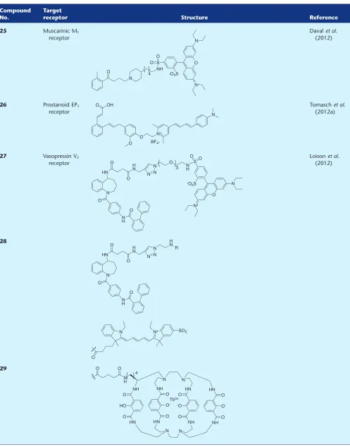
Continued
Compound No.
Target
receptor Structure Reference
25 Muscarinic M1
receptor
Davalet al. (2012)
26 Prostanoid EP3
receptor
Tomaschet al. (2012a)
27 Vasopressin V2
receptor
Loisonet al. (2012)
28
antagonist. Using flow cytometric analysis, the authors
also revealed that 13 is associated with the CB2R in B
lymphocytes.
Histamine receptor
A high-affinity fluorescent antagonist (14) for the human
histamine H1receptor (H1R) has been reported by Roseet al.
(2012) consisting of the high-affinity and H1R-selective
antagonist mepyramine linked to the BODIPY 630/650
fluo-rophore. Although fluorescent ligand14was purchased from
a commercial supplier and not synthesized by the authors in this publication, it represents the first disclosure of the
chemical structure of14. Conjugate14displayed comparable
affinity for the H1R compared with the parent
pharmacoph-Continued
Compound No.
Target
receptor Structure Reference
30 Nuclear
oestrogen receptor
Céspedes-Guiraoet al. (2011)
31 Nuclear
oestrogen receptor
H O
O H
H H
H N
N
RO2C
(CH2)4SO3
O3S
R =
O H O
O H O H H O
H3N
[image:8.594.41.563.98.624.2]ore mepyramine. Confocal microscopy revealed specific and
displaceable binding of14 to the H1R localized to the cell
membrane, and despite significant non-specific intracellular uptake, this probe proved very useful for studying the
recep-tor in single living cells. The diffusion coefficient of14-H1R
complexes was quantified using FCS, and these values were
different for CHO-K1 cell lines transiently expressing the H1R
with and without the yellow fluorescent protein receptor tag. The authors then developed this further, by showing that FCS
experiments using 14 can detect endogenously expressed
H1Rs in HeLa cells.
Kuder et al. (2009) have reported 15 as a selective
H3R fluorescent antagonist, which consists of a
piperidine-containing pharmacophore (related to the
known H3R selective antagonist pitolisant) linked to a
nitrobenzoxadiazole-based fluorophore. The only difference
between15and a fluorescent probe previously developed by
Amonet al. (2007) is the presence of a 3-methyl group on the
piperidine ring of15. Conjugate15was twofold less potent
for the human H3R than the previously reported
non-methylated derivative (Amon et al., 2007), and the authors
did not evaluate the selectivity or imaging properties of15.
In another report of an H3R fluorescent ligand, but with a
goal of making red-shifted probes, Tomasch et al., (2012c)
have tethered selective H3R antagonists, again based on a
piperidine moiety, to substituted chalcones. A series of pharmacophore–fluorophore combinations containing dif-ferent linker positions and lengths were synthesized, and all
exhibited nanomolar affinity for the human H3R and
selec-tivity over the H1R (one log unit) and H4R (two log units). The
authors then examined the ability of three conjugates (16– 18) to visualize the H3R in hH3-HEK-293 cells. Confocal
microscopy revealed enrichment of the fluorescent signal to the cell membrane, and the authors conclude that this was
specific binding to the H3R as when HEK-293 cells that do not
transiently express the H3R were treated with the fluorescent
ligands, no enriched membrane fluorescence was observed.
From the same laboratory, Tomasch et al. (2012b) have
used the same piperidine-based pharmacophore but now with the boron-dipyrromethene scaffold as the fluorophore (19). Synthesis of the fluorophore moiety was completed by reaction with boron trifluoroetherate as the final reaction in a stepwise synthesis, rather than the more common convergent approach of coupling a pre-activated (and often commercially available as the NHS ester) fluorophore
to a complementary pharmacophore/linker. Conjugate 19,
named Bodilisant by the authors, displayed a low nanomolar
affinity for the human H3R that was 10 times more potent
that the previous generation (16–18) of conjugates (Tomasch
et al., 2012c). Again, H3R subtype selectivity was maintained.
Fluorescence microscopy was used to visualize the human
H3R in H3-HEK-293 cells and showed that19predominately
localized to the cell membrane. In these experiments, the
authors concluded that fluorescent probe19was not
inter-nalized, as it did not overlap with the nuclear stain DAPI.
5-Hydroxytryptamine (serotonin) receptor
Lacivitaet al. (2010) designed and synthesized a fluorescent
probe (20) for the serotonin1Areceptor (5-HT1AR) in an effort
to improve on previous ligands from the same laboratory that showed high levels of non-specific binding. A
chromenone-containing fluorophore was synthesized in-house and coupled to a 1-arylpiperizine-based antagonist, affording a
conjugate with nanomolar affinity for the human 5-HT1AR;
approximately 10-fold less potent than the parent piperizine
pharmacophore. The authors then evaluated the ability of20
to visualize the 5-HT1AR, and using a high concentration of
20, showed fluorescent labelling of CHO-5-HT1Acells that was
reduced by application of serotonin. Along with the lead ligand (20), a conjugate containing a near-infrared fluoro-phore was also synthesized, and despite a similar affinity for
the 5-HT1AR compared to20, the authors commented that
due to the loss of fluorescent properties as measured in aqueous buffer, it was not useful as an imaging probe.
However, given the location of the 5-HT1AR, as with all Class
A GPCRs, in the cell membrane, it would be interesting to study the properties of this ligand when bound to the recep-tor. It can be advantageous to have a fluorescent probe that is
quenched in an aqueous environment (Bakeret al., 2010) but
fluoresces when associated with the receptor in a lipophilic membrane environment. For example, in competition-binding assays, this property can eliminate the need for thor-ough washing steps prior to analysing membrane-localized fluorescence.
Fluorescent 5-HT1AR probes have also been developed by
Alonsoet al. (2010) based on an arylpiperazine agonist
pre-viously reported from the same research group. A series of
compounds were synthesized using the 7a-position of the
bicyclohydantoin moiety to tether a dansyl fluorophore.
Several conjugates showed an affinity for the human 5-HT1AR
that were comparable to the starting arylpiperazine scaffold.
Conjugate 21 was identified as the lead fluorescent ligand
due to a high fluorescent intensity emission value. Fixed
CHO-5-HT1AR cells could be labelled with21and this could
be blocked using a reference non-fluorescent ligand. Despite being based on a known agonist, only radioligand competi-tion binding assays were carried out to determine the affinity
of21for the 5-HT1AR – no information was provided
regard-ing how linkage to the dansyl fluorophore influenced ligand efficacy. The authors did not comment on localized mem-brane fluorescence or the potential of the fluorescent conju-gate (if indeed an agonist) to internalize with the receptor. However, the timescale for the visualization experiments was only 10 min pre-incubation followed by a wash, fix and mount process, which may therefore have precluded this from occurring.
Muscarinic receptors
A fluorescent antagonist (22) of the muscarinic M3receptor
(M3R) has been reported by Joneset al. (2008), by linking the
non-subtype selective M3R antagonist tolterodine to the
com-mercially available fluorophore BODIPY 630/650-NHS ester.
Conjugate 22 displayed a threefold loss in affinity for the
human M3R compared with tolterodine, and also
approxi-mately the same fold-loss in affinity across the other human muscarinic receptor subtypes. The authors did not examine
the use of22as an imaging tool, but indicated that this was
an ongoing work in their laboratory.
Hern et al. (2010) have reported 23 as an M1R probe,
synthesized by the reaction of a telenzepine amino congener
to an Alexa Fluor 488 fluorophore. Conjugate 23 had a
monitor receptor–ligand complexes in living cells. Alongside
the previously reported high-affinity M1R ligand 24(Harris
et al., 2003), total reflection fluorescence microscopy was used to track the position of fluorescent ligand–receptor
com-plexes in live CHO cells expressing the human M1R.
Informa-tion about M1R mobility, clustering and, in particular,
dimerization could be obtained by simultaneously using
probes23and24that have different fluorescence emission
wavelengths.
Davalet al. (2012) synthesized fluorescent M1R ligand25
by coupling an agonist (based on AC-42) with high
func-tional selectivity for the M1R to the fluorophore lissamine
rhodamine B sulfonyl chloride. The emphasis in this study was on investigating how the fluorescent conjugate binds to
the M1R, and teasing out possible ‘non-orthosteric’ binding
mode(s) and receptor–ligand interactions. The parent agonist
pharmacophore and conjugate25displayed a similar affinity
towards the human M1R receptor, but interestingly,25could
no longer elicit a typical agonist-induced calcium response in
a functional assay. Instead,25fully reduced the functional
response to a known agonist, thereby classifying 25 as an
antagonist. In a series of very comprehensive and interesting experiments involving assays with reference allosteric ligands, receptor truncation, molecular modelling and even
application of 25 itself as a FRET tracer, the authors
con-cluded that binding of 25 to the M1R showed a bitopic
(Valantet al., 2012) nature. This work further demonstrates
the importance of treating the ligand-linker–fluorophore conjugate as a new chemical entity, which can have subtle or quite profound pharmacological differences compared with the starting drug molecule. These differences are often not captured in a single competition-based affinity assay, and there is a fascinating road ahead in terms of rationalizing
ligand–receptor interactions of fluorescent conjugates
beyond the confines of the orthosteric ligand-binding pocket.
Prostanoid receptor
From the same research laboratory as fluorescent H3R ligands
16–18 (Tomasch et al., 2012b; 2012c) was the report of a
fluorescent prostanoid EP3 receptor (EP3R) antagonist (26)
(Tomasch et al., 2012a). Based on an ortho-substituted
cin-namic acid antagonist, a series of fluorescent conjugates were synthesized containing different fluorophores, with
pyrylium-containing 26 showing the most promise as an
imaging tool. Although with approximately threefold
reduced affinity for the human EP3R compared with the
parent drug molecule, 26 maintained selectivity over the
EP1R, EP2, and EP4R subtypes. The authors then proceeded to
demonstrate that the EP3R receptor could be visualized using
26in murine kidney and human brain tissue.
Vasopressin receptor
Loisonet al. (2012) reported the first examples of selective,
fluorescent, non-peptidic ligands for the vasopressin V2
recep-tor (V2R) based on a tetrahydro-1H-benzo[b]azepine
antago-nist. A series of conjugates containing different linker lengths and fluorophores were synthesized, and from these, three lead compounds (27–29) were identified. Interestingly,
although compounds 27–29 displayed a slight decrease in
these fluorescent conjugates were comparatively more
selec-tive for the V2R over the V1AR and oxytocin receptor. Cyanine
probe28and terbium-containing29(the nature of the linker
housed within the fluorophore is not represented, it has been presumed this is the commercially available Lumi4-Tb-NHS ester as stated in the publication) were then used to develop
an acceptor/donor V2R TR-FRET-based assay and utilized as a
tool to study V2R-V1AR dimerization in association with a
previously reported V1AR probe (Albizuet al., 2010).
Other fluorescent Class A GPCR ligands
In addition to fluorescent probes specifically designed to target Class A GPCRs there have been recent reports of fluo-rescent ligands for alternative receptors, but which one would predict might also bind to Class A GPCRs. There are tworecent reports (Céspedes-Guiraoet al., 2011; Joseet al., 2011)
outlining the synthesis and application of fluorescent oestradiol-based probes (30) and (31) to target the nuclear hormone oestrogen receptor (ER). Although the ER is not classified as a GPCR, the other type of oestrogen receptor, GPER (or GPR30), is classified as a Class A GPCR. Oestradiol is a high-affinity ligand for both the ER and the GPER; there-fore, oestradiol-based conjugates developed for the ER may well be useful tools for studying GPER. In addition to the GPCR families covered in this review, there continues to be an interest in developing small-molecule fluorescent probes for the dopamine receptor, another Class A GPCR, although there have been no new reports published of novel ligands since the preceding review articles (Kuder and Kiec´-Kononowicz, 2008; Böhme and Beck-Sickinger, 2009). Another Class A GPCR for which developing fluorescent ligands is an exciting prospect is the opioid receptor, but since the last GPCR fluorescent ligand reviews, there have only been reports of peptide, or peptide-based ligands
conju-gated to a fluorophore (e.g. Josanet al., 2009), and is therefore
outside the scope of this review article. In addition to the novel fluorescent conjugates reported via peer-reviewed pub-lications that have been discussed in this review, there are a growing number of fluorescent conjugates available commer-cially, with and without the chemical structure disclosed. In an excellent example that showcased the power of using several commercially available fluorescent probes that emit at
different wavelengths, Dalyet al. (2010) investigated the
dis-tribution of adrenoceptors and ‘cannabinoid-like’ receptors in different cell types.
Conclusion
within this review have helped to crystallize many of the key factors for consideration when appending a fluorophore to a relatively small orthosteric drug molecule. The linker that connects the fluorescent moiety to the drug of interest must be attached to a position that is relatively insensitive to structural modification and that can potentially tolerate bulky substituents. In order to attenuate non-specific binding of the fluorescent-drug conjugate, one must also be mindful of the final physicochemical properties of the molecule. Finally, when using the generated probes for cellular-based imaging studies or certain types of assay, it has become clearer that judicious choice of the correct fluorophore is also of paramount importance. The work summarized in this and previous reviews unequivocally substantiates that if all these considerations are taken into account, the resulting fluores-cent conjugates are extremely powerful pharmacological tools that can be utilized in numerous cutting-edge assay formats. An interesting road lies ahead for all researchers in this area in attempting to rationalize the ligand–receptor interactions of fluorescent conjugates beyond the confines of the orthosteric ligand-binding pocket. In so-doing though, this will undoubtedly help cement the use of these fluores-cent reagents as versatile and extremely useful tools for modern-day molecular pharmacology and drug discovery.
Acknowledgements
Work in Barrie Kellam’s and Steve Hill’s laboratories related to this review was supported by the Medical Research Council (Grant No. G0800006), the BBSRC (Grant No. BB/0521581/1) and the University of Nottingham.
Conflicts of interest
The authors declare the following competing financial inter-est(s): B.K and S.J.H. are founding directors of the University of Nottingham spin-off company CellAura Technologies Ltd.
References
Albizu L, Cottet M, Kralikova M, Stoev S, Seyer R, Brabet Iet al. (2010). Time-resolved FRET between GPCR ligands reveals oligomers in native tissues. Nat Chem Biol 6: 587–594.
Alexander SPH, Benson HE, Faccenda E, Pawson AJ, Sharman JL, Catterall WA, Spedding M, Peters JA, Harmar AJ and CGTP Collaborators (2013). The Concise Guide to PHARMACOLOGY 2013/14: G Protein-Coupled Receptors. Br J Pharmacol 170: 1459–1581.
Alonso D, Vázquez-Villa H, Gamo AM, Martínez-Esperón MF, Tortosa M, Viso Aet al. (2010). Development of fluorescent ligands for the human 5-HT1Areceptor. ACS Med Chem Lett 1: 249–253.
Amon M, Ligneau X, Camelin JC, Berrebi Bertrand I, Schwartz JC, Stark H (2007). Highly potent fluorescence – tagged nonimidazole histamine H3 receptor ligands. ChemMedChem 2: 708–716.
Bai M, Sexton M, Stella N, Bornhop DJ (2008). MBC94, a
conjugable ligand for cannabinoid CB2 receptor imaging. Bioconjug Chem 19: 988–992.
Baker JG, Middleton R, Adams L, May LT, Briddon SJ, Kellam B et al. (2010). Influence of fluorophore and linker composition on the pharmacology of fluorescent adenosine A1receptor ligands. Br J
Pharmacol 159: 772–786.
Baker JG, Adams LA, Salchow K, Mistry SN, Middleton RJ, Hill SJ et al. (2011). Synthesis and characterization of high-affinity 4,4-difluoro-4-bora-3a,4a-diaza-s-indacene-labeled fluorescent ligands for humanβ-adrenoceptors. J Med Chem 54: 6874–6887.
Beltran B, Carrillo R, Martin T, Martin VS, Machado JD, Borges R (2011). Fluorescentβ-blockers as tools to study presynaptic mechanisms of neurosecretion. Pharmaceuticals 4: 713–725.
Bjarnadóttir TK, Gloriam DE, Hellstrand SH, Kristiansson H, Fredriksson R, Schiöth HB (2006). Comprehensive repertoire and phylogenetic analysis of the G protein-coupled receptors in human and mouse. Genomics 88: 263–273.
Böhme I, Beck-Sickinger AG (2009). Illuminating the life of GPCRs. Cell Commun Signal 7: 16–38.
Briddon SJ, Hill SJ (2007). Pharmacology under the microscope: the use of fluorescence correlation spectroscopy to determine the properties of ligand-receptor complexes. Trends Pharmacol Sci 28: 637–645.
Briddon SJ, Kellam B, Hill SJ (2011). Design and use of fluorescent ligands to study ligand–receptor interactions in single living cells. Methods Mol Biol 746: 211–236.
Burchak ON, Mugherli L, Ostuni M, Lacapère JJ, Balakirev MY (2011). Combinatorial discovery of fluorescent pharmacophores by multicomponent reactions in droplet arrays. J Am Chem Soc 133: 10058–10061.
Céspedes-Guirao FJ, Ropero AB, Font-Sanchis E, Nadal Á, Fernández-Lázaro F, Sastre-Santos Á (2011). A water-soluble perylene dye functionalised with a 17β-estradiol: a new fluorescent tool for steroid hormones. Chem Commun 47: 8307–8309.
Chan J, Dodani SC, Chang CJ (2012). Reaction-based
small-molecule fluorescent probes for chemoselective bioimaging. Nat Chem 4: 973–984.
Cottet M, Faklaris O, Zwier JM, Trinquet E, Pin JP, Durroux T (2011). Original fluorescent ligand-based assays open new perspectives in G protein-coupled receptor drug screening. Pharmaceuticals 4: 202–214.
Cottet M, Faklaris O, Maurel D, Scholler P, Doumazane E, Trinquet Eet al. (2012). BRET and Time-resolved FRET strategy to study GPCR oligomerization: from cell lines toward native tissues. Front Endocrinol (Lausanne) 3: article 92.
Daly CJ, McGrath JC (2003). Fluorescent ligands, antibodies, and proteins for the study of receptors. Pharmacol Ther 100: 101–118.
Daly CJ, Ross RA, Whyte J, Henstridge CM, Irving AJ, McGrath JC (2010). Fluorescent ligand binding reveals heterogeneous distribution of adrenoceptors and ‘cannabinoid-like’ receptors in small arteries. Br J Pharmacol 159: 787–796.
Daval SB, Valant C, Bonnet D, Kellenberger E, Hibert M, Galzi J-L et al. (2012). Fluorescent derivatives of AC-42 to probe bitopic orthosteric/allosteric binding mechanisms on muscarinic M1 receptors. J Med Chem 55: 2125–2143.
Schiesser CH (2012). Fluorescent angiotensin AT1receptor
antagonists. Asian J Org Chem 1: 274–279.
Harris A, Cox S, Burns D, Norey C (2003). Miniturization of fluorescence polarization receptor-binding assays using CyDye-labeled ligands. J Biomol Screen 8: 410–420.
Hern JA, Baig AH, Mashanov GI, Birdsall B, Corrie JET, Lazareno S et al. (2010). Formation and dissociation of M1muscarinic receptor
dimers seen by total internal reflection fluorescence imaging of single molecules. Proc Natl Acad Sci U S A 107: 2693–2698.
Hill SJ, May LT, Kellam B, Woolard J (2014). Allosteric interactions at adenosine A1and A3receptors: new insights into the role of
small molecules and receptor dimerization. Br J Pharmacol 171: 1102–1113.
Jacobson KA (2009). Functionalized congener approach to the design of ligands for G protein-coupled receptors (GPCRs). Bioconjug Chem 20: 1816–1835.
Jakobs D, Sorkalla T, Häberlein H (2012). Ligands for fluorescence correlation spectroscopy on G protein-coupled receptors. Curr Med Chem 19: 4722–4730.
Jones LH, Randall A, Napier C, Trevethick M, Sreckovic S, Watson J (2008). Design and synthesis of a fluorescent muscarinic antagonist. Bioorg Med Chem Lett 18: 825–827.
Josan JS, Morse DL, Xu L, Trissal M, Baggett B, Davis Pet al. (2009). Solid-phase synthetic strategy and bioevaluation of a labeled δ-opioid receptor ligand Dmt-Tic-Lys for in vivo imaging. Org Lett 11: 2479–2482.
Jose I, Deodhar KD, Desai UB, Bhattacharjee S (2011). Early detection of breast cancer: synthesis and characterization of novel target specific NIR-fluorescent estrogen conjugate for molecular optical imaging. J Fluoresc 21: 1171–1177.
Kale J, Liu Q, Leber B, Andrews DW (2012). Shedding light on apoptosis at subcellular membranes. Cell 151: 1179–1184.
Kozma E, Gizewski ET, Tosh DK, Squarcialupi L, Auchampach JA, Jacobson KA (2013a). Characterization by flow cytometry of fluorescent, selective agonist probes of the A3 adenosine receptor. Biochem Pharmacol 85: 1171–1181.
Kozma E, Suresh Jayasekara P, Squarcialupi L, Paoletta S, Moro S, Federico Set al. (2013b). Fluorescent ligands for adenosine receptors. Bioorg Med Chem Lett 23: 26–36.
Kuder K, Kiec´-Kononowicz K (2008). Fluorescent GPCR ligands as new tools in pharmacology. Curr Med Chem 15: 2132–2143.
Kuder KJ, Kottke T, Stark H, Ligneau X, Camelin JC, Seifert Ret al. (2009). Search for novel, high affinity histamine H3receptor ligands
with fluorescent properties. Inflamm Res 59: 247–248.
Lacivita E, Masotti AC, Jafurulla M, Saxena R, Rangaraj N, Chattopadhyay Aet al. (2010). Identification of a red-emitting fluorescent ligand for in vitro visualization of human serotonin 5-HT1Areceptors. Bioorg Med Chem Lett 20: 6628–6632.
Leopoldo M, Lacivita E, Berardi F, Perrone R (2009). Developments in fluorescent probes for receptor research. Drug Discov Today 14: 706–712.
et al. (2012). Selective fluorescent nonpeptidic antagonists for vasopressin V2GPCR: application to ligand screening and
oligomerization assays. J Med Chem 55: 8588–8602.
Martikkala E, Lehmusto M, Lilja M, Rozwandowicz-Jansen A, Lunden J, Tomohiro Tet al. (2009). Cell-basedβ2-adrenergic
receptor–ligand binding assay using synthesized europium-labeled ligands and time-resolved fluorescence. Anal Biochem 392: 103–109.
May LT, Bridge LJ, Stoddart LA, Briddon SJ, Hill SJ (2011). Allosteric interactions across native adenosine-A3receptor homodimers:
quantification using single-cell ligand-binding kinetics. FASEB J 25: 3465–3476.
Middleton RJ, Kellam B (2005). Fluorophore-tagged GPCR ligands. Curr Opin Chem Biol 9: 517–525.
Morishima S, Suzuki F, Nishimune A, Yoshiki H, Akino H, Yokoyama Oet al. (2010). Visualization and tissue distribution of α1L-adrenoceptor in human prostate by the fluorescently labeled ligand Alexa-488-Silodosin. J Urology 183: 812–819.
Overington JP, Al-Lazikani B, Hopkins AL (2006). How many drug targets are there? Nat Rev Drug Discov 5: 993–996.
Petrov RR, Ferrini ME, Jaffar Z, Thompson CM, Roberts K, Diaz P (2011). Design and evaluation of a novel fluorescent CB2ligand as
probe for receptor visualization in immune cells. Bioorg Med Chem Lett 21: 5859–5862.
Rose RH, Briddon SJ, Hill SJ (2012). A novel fluorescent histamine H1receptor antagonist demonstrates the advantage of using
fluorescence correlation spectroscopy to study the binding of lipophilic ligands. Br J Pharmacol 165: 1789–1800.
Sexton M, Woodruff G, Horne EA, Lin YH, Muccioli GG, Bai M et al. (2011). NIR-mbc94, a fluorescent ligand that binds to endogenous CB2receptors and is amenable to high-throughput
screening. Chem Biol 18: 563–568.
Stoddart LA, Vernall AJ, Denman JL, Briddon SJ, Kellam B, Hill SJ (2012). Fragment screening at adenosine-A3receptors in living cells
using a fluorescence-based binding assay. Chem Biol 19: 1105–1115.
Tomasch M, Schwed JS, Kuczka K, Meyer dos Santos S, Harder S, Nüsing RMet al. (2012a). Fluorescent human EP3receptor
antagonists. ACS Med Chem Lett 3: 774–779.
Tomasch M, Schwed JS, Paulke A, Stark H (2012b). Bodilisant – a novel fluorescent, highly affine histamine H3receptor ligand. ACS
Med Chem Lett 4: 269–273.
Tomasch M, Schwed JS, Weizel L, Stark H (2012c). Novel
chalcone-based fluorescent human histamine H3receptor ligands as
pharmacological tools. Front Syst Neurosci 6: article 14, 1–16.
Tosh DK, Chinn M, Ivanov AA, Klutz AM, Gao Z-G, Jacobson KA (2009). Functionalized congeners of A3adenosine receptor-selective
nucleosides containing a bicyclo[3.1.0]hexane ring system. J Med Chem 52: 7580–7592.
Valant C, Lane JR, Sexton PM, Christopoulos A (2012). The best of both worlds? Bitopic orthosteric/allosteric ligands of G