Original citation:
Lee, Yun, Sooranna, Suren R., Terzidou, Vasso, Christian, Mark, Brosens, Jan J.,
Huhtinen, Kaisa, Poutanen, Matti, Barton, Geraint, Johnson, Mark R. and Bennett, Phillip
R.. (2012) Interactions between inflammatory signals and the progesterone receptor in
regulating gene expression in pregnant human uterine myocytes. Journal of Cellular and
Molecular Medicine, 16 (10). pp. 2487-2503. jcmm0016-2487
Permanent WRAP url:
http://wrap.warwick.ac.uk/70729
Copyright and reuse:
The Warwick Research Archive Portal (WRAP) makes this work of researchers of the
University of Warwick available open access under the following conditions.
This article is made available under the Creative Commons Attribution-NonCommercial
2.5 (CC BY-NC 2.5) license and may be reused according to the conditions of the
license. For more details see:
http://creativecommons.org/licenses/by-nc/2.5/
A note on versions:
The version presented in WRAP is the published version, or, version of record, and may
be cited as it appears here.
Interactions between inflammatory signals and the progesterone
receptor in regulating gene expression in pregnant human
uterine myocytes
Yun Lee
a, Suren R. Sooranna
a, Vasso Terzidou
a, Mark Christian
a, Jan Brosens
a,
Kaisa Huhtinen
b, Matti Poutanen
b, c, Geraint Barton
d, Mark R. Johnson
a, Phillip R. Bennett
a,*
a
Parturition Research Group, Imperial College London, Institute of Reproductive and Developmental Biology, London, UK
b
Department of Physiology, Institute of Biomedicine, University of Turku, Turku, Finland
cTurku Center for Disease Modeling, University of Turku, Turku, Finland
d
Department of Bioinformatics, Imperial College London, London, UK
Received: October 31, 2011; Accepted: February 20, 2012
Abstract
The absence of a fall in circulating progesterone levels has led to the concept that human labour is associated with ‘functional progesterone
withdrawal’ caused through changes in the expression or function of progesterone receptor (PR). At the time of labour, the human uterus is
heavily infiltrated with inflammatory cells, which release cytokines to create a ‘myometrial inflammation’
via
NF-
j
B activation. The negative
interaction between NF-
j
B and PR, may represent a mechanism to account for ‘functional progesterone withdrawal’ at term. Conversely, PR
may act to inhibit NF-
j
B function and so play a role in inhibition of myometrial inflammation during pregnancy. To model this inter-relationship,
we have used small interfering (si) RNA-mediated knock-down of PR in human pregnant myocytes and whole genome microarray analysis to
identify genes regulated through PR. We then activated myometrial inflammation using IL-1
b
stimulation to determine the role of PR in
myome-trial inflammation regulation. Through PR-knock-down, we found that PR regulates gene networks involved in myomemyome-trial quiescence and
extra-cellular matrix integrity. Activation of myometrial inflammation was found to antagonize PR-induced gene expression, of genes normally
upregulated
via
PR. We found that PR does not play a role in repression of pro-inflammatory gene networks induced by IL-1
b
and that only
MMP10 was significantly regulated in opposite directions by IL-1
b
and PR. We conclude that progesterone acting through PR does not
gener-ally inhibit myometrial inflammation. Activation of myometrial inflammation does cause ‘functional progesterone withdrawal’ but only in the
context of genes normally upregulated
via
PR.
Keywords:
labour
parturition
progesterone
PR
inflammation
NF-kappaB.
Introduction
Progesterone is considered to play a major role in the maintenance of
pregnancy. In 1956 Csapo proposed that the essential role of
proges-terone in pregnancy is to ‘block’ myometrial contractility and that the
onset of labour therefore requires withdrawal of this progesterone
block. In the majority of mammals labour is preceded by a decline in
circulating progesterone concentrations, however, in the human,
there is no dramatic fall in progesterone concentrations prior to
labour, yet parturition can be induced using progesterone
antago-nists. This has led to the concept that, in the human, labour is
associ-ated with ‘functional progesterone withdrawal’ caused through
changes in the expression or function of PR rather than through
changes in circulating concentrations of progesterone [1, 2].
The genomic effects of progesterone upon target tissues are
gen-erally mediated through nuclear PR. The major PR isoforms, PR-A
and PR-B, are encoded from a single gene by differential promoter
usage. PR-A, a 94 kD protein, lacks the first N-terminal 164 amino
acids of PR-B, which is a 116 kD protein with an additional activation
*Correspondence to: Phillip BENNETT,Parturition Research Group, Imperial College London, Institute of Reproductive and Developmental Biology, Du Cane Road, London W12, ONN, UK.
function (AF-3) [1, 2]. In myometrial cells, PR-A has limited
transacti-vation properties at classical progesterone response elements and
has been shown to transrepress PR-B and other class I nuclear
recep-tor family members [3, 4]. The PR gene also contains additional
puta-tive downstream translational start sites predicted to encode proteins
of approximately 60
–
70 and 39 kD respectively. A 60 kD PR variant
found in T47D cells has been termed PR-C. This contains a
ligand-binding domain, but lacks a DNA ligand-binding domain and has been
reported to be cytosolic rather than nuclear [5].
A current theory for the mechanism of ‘functional progesterone
withdrawal’ in human myometrium involves a change in the
abun-dance of PR isoforms, leading to a decrease in the ratio of PR-B to
PR-A and/or PR-C [1, 2, 4]. Other proposed mechanisms include a
decline in the myometrial levels of PR coactivators at term [6], and a
decrease in the circulating concentration of bioactive progesterone
metabolites associated with decreased steroid 5 beta-reductase
expression in the uterus [7].
A further potential mechanism for ‘functional progesterone
with-drawal’ in the human involves the transcriptional antagonism between
the activated PR and inflammatory signal intermediates, especially
the inflammation-associated transcription factor NF-kappaB (NF-
j
B).
A central role for NF-
j
B in murine parturition has been demonstrated
[8]. Nuclear translocation of the p50 and p65 subunits of NF-
j
B
increases in the pregnant mouse uterus towards term and
intra-amni-otic injection of the NF-
j
B inhibitor peptide SN50 delays in the onset
of labour. Surfactant protein-A (SP-A), secreted by the maturing
foe-tus lung in increasing amounts towards term, was shown to enhance
p65 nuclear levels and to induce labour. It has been proposed that
SP-A triggers the onset of labour at term by inducing the migration of
macrophages to the maternal uterus, where local inflammatory
sig-nals activate NF-
j
B signalling in myocytes, resulting in the
stimula-tion of uterine contractility. Direct protein-protein interacstimula-tion between
p65 and PR, resulting in reciprocal functional antagonism, has been
demonstrated in breast cancer cells [9], and may also operate in
uter-ine cells [10, 11].
Activation of myometrial inflammation, or more specificially of the
NF-
j
B pathway in the myometrium appears to be an attractive
mecha-nism to account for ‘functional progesterone withdrawal’ at term.
However, the functional consequences of the interaction between PR
and NF-
j
B on myometrial gene expression in pregnancy has not yet
been determined. We have therefore combined small interfering (si)
RNA-mediated knock-down of PR in human term pregnant myocytes
with whole genome microarray analysis to identify those genes
regu-lated by activated PR. We then used IL-1
b
stimulation, which activates
NF-
j
B and induces myometrial inflammation, to determine the role of
PR in the transcriptional regulation of myometrial inflammation.
Materials and methods
Tissue collection and cell culture
Tissue was collected with local ethics committee approval and informed consent from patients at Queen Charlotte’s and Chelsea
Hospital, London. Myometrial tissue was obtained from the upper edge of lower uterine incision, made at the location of the bladder fold at the time of Caesarean section. Elective caesarean section was per-formed in each case at or after 39 weeks in uncomplicated pregnan-cies. Indications were breech presentation and previous caesarean section. Myometrial tissue was minced and digested for 45 min. in DMEM with 1 mg/ml collagenase type IA and IX (Sigma-Aldrich Com-pany Ltd, Poole, UK). Cells were centrifuged at 4009gfor 10 min. and grown in DMEM with 10% foetal calf serum,L-glutamine and pen-icillin-streptomycin (37°C and 5% CO2).Cells (passage number 3 or 4)
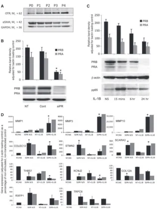
were incubated with 100nM medroxyprogesterone acetate (MPA) in 2% reduced serum for 24 hrs before IL-1b (R&D Systems, Europe Ltd., Abingdon, UK) was added to a final concentration of 1 ng/ml for 6 hrs. To confirm that the cells established in our cultures are myo-cytes and not fibroblasts or epithelial cells, we undertook Western analysis for alpha-smooth-muscle actin, and oxytocin receptor and found no significant changes in expression between passages 0–4 (Fig. 1A).
Transfection of siRNA
ON-TARGETplus SMART pool human RNA (Dharmacon, Lafayette, CO, USA) was used to knock-down PR. SiGLO (Dharmacon) was used as a positive control, giving a high transfection efficiency of approximately 90%, and ON-TARGETplus Non-Targeting Pool (Dharmacon) was used as a negative control. The siRNAs were transfected using Dharmafect 2 (Dharmacon) transfection reagent at a final concentration of 20lM according to manufacturer’s instructions.
Protein extraction
Myocytes were scraped into buffer A [10 mM HEPES pH 7.4, 10 mM KCl, 0.1 mM EDTA, 0.1 mM EGTA, 2 mM DTT, complete protease inhibitor tablets (Roche, Welwyn Garden City, UK) and 2 M KOH to adjust pH to 7.4]. The resulting suspension was collected and incubated on ice for 20 min. To the incubated mixture, NP-40 (Nonidet P-40, Sigma-Aldrich Company Ltd) was added to give a final concentration of 1% and carefully mixed. The cytosolic protein fraction was extracted by centrifuging at 13,0009gfor 30 sec. The pellet was then resuspended in buffer B (10 mM HEPES, 10 mM KCl, 0.1 mM EDTA, 0.1 mM EGTA, 2 mM DTT, 400 mM NaCl, 1% NP-40) and complete protease inhibitor tablets (Roche) to lyse the nuclear membrane. Samples were incubated on a shaking platform for 15 min. Cell suspensions were centrifuged at 13,0009g for 5 min. and supernatant (nuclear protein fraction) was collected, snap-frozen and stored at 80°C for later use.
Western blot analysis
Biotechnology, Heidelberg, Germany) was used with ECL Plus (Amer-sham Biosciences) chemiluminescent reagents for signal detection. Anti-bodies used were PR Novocastra NCL-L-PGR-312 (Novocastra Laboratories Ltd, Newcastle upon Tyne, UK) OTR Santa Cruz SC-8102,
alpha smooth muscle actin Sigma A2547, GAPDH Millipore MAB374, Millipore, Watford, UK, beta actin Abcam ab8226 (Abcam, Cambridge, UK) and NFkB pp65 Serine 536 Cell Signalling 3033S (Cell Signaling Technology, New England Biolabs, Hitchin, UK).
A C
B
D
[image:4.595.140.457.91.515.2]RNA extraction
Total RNAs were isolated using Trizol (Invitrogen, Paisley, UK) according to the manufacturer’s protocol and further purified using the RNeasy mini Kit (QIAGEN, Crawley, UK). RNA integrity and purity were assessed using a Bioanalyzer 2100 (Agilent Technologies Inc., Santa Clara, CA, USA).
Quantitative real time polymerase chain reaction
(RTQ-PCR)
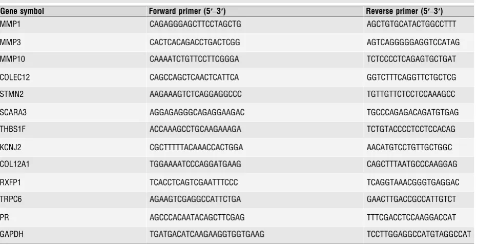
Total RNA 1lg was reverse transcribed with oligo dT random primers using MuLV reverse transcriptase (Applied Biosystems Ltd, Carlsbad, CA, USA). Paired oligonucleotide primers for amplification of genes of interest were designed using Primer 3 software (www.ncbi.nlm.nih.gov/tools/primer-blast/) against the sequences downloaded from GenBank. The primer sets used (Table 1) produced amplicons of the expected size and where possible flanked intron/exon junctions. Quantitative PCR was performed using Power SYBR Green PCR master mix (Applied Biosystems Ltd.,) and amplicon yield was monitored during cycling in a RotorGene Sequence Detector (Corbett Research Ltd.) that continually measures fluorescence caused by the bind-ing of the dye to double-stranded DNA. Pre-PCR cycle was 10 min. at 95°C followed by up to 45 cycles of 95°C for 20 sec., 58–60°C for 20 sec. and 72°Cfor 20 sec. followed by an extension at 72°C for 15 sec. The final pro-cedure involves a melt over the temperature range of 72–99°C rising by 1 degree steps with a wait for 15 sec. on the first step followed by a wait of 5 sec. for each subsequent step. The cycle at which the fluorescence reached a preset threshold (cycle threshold) was used for quantitative analy-ses. The cycle threshold in each assay was set at a level where the expo-nential increase in amplicon abundance was approximately parallel between all samples. mRNA data were expressed relative to the amount of the con-stitutively expressed housekeeping gene, GAPDH.
Affymetrix HgU133 Plus 2.0 array processing
RNA Samples were profiled by the Finnish DNA-Microarray Centre using the Affymetrix HgU133 Plus 2.0 GeneChip® (Affymetrix Inc., Santa Clara, CA, USA). Briefly, cDNA was generated from 2lg of total RNA using the GeneChip®Expression 3′-Amplification One-Cycle cDNA Syn-thesis kit, in conjunction with the GeneChip® Eukaryotic PolyA RNA Control Kit (Affymetrix Inc.). The cDNA was cleaned up using the Gene-Chip®Sample Cleanup Module and subsequently processed to generate biotin-labelled cRNA using the GeneChip® Expression 3′-Amplification IVT Labelling Kit (Affymetrix Inc.). A quantity of 25lg of labelled cRNA was fragmented using 5X fragmentation buffer and Rnase-free water at 94°C for 35 min. A quqntity of 15lg of the fragmented, biotin-labelled cRNA was made up in a hybridization cocktail and hybridized to the HgU133 Plus 2.0 array at 45°C for 16 h. Following hybridization the arrays were washed and stained using the Affymetrix Fluidics Station 450 and scanned using the Affymetrix GeneChip® Scanner 3000. All steps of the process were quality controlled by measuring yield (lg), concentration (lg/l) and 260:280 ratios via spectrophotometry using the Nanodrop ND-1000 and sample integrity using the Agilent 2100 bio-analyser (Agilent Technologies Inc.).
Results
PR regulates gene networks involved in
myometrial quiescence and extracellular matrix
integrity
[image:5.595.67.533.459.698.2]To identify genes regulated in human myometrium by liganded PR,
primary myocyte cultures were transfected with siRNA targeting PR
Table 1Sequences for PCR primers used in quantitative RT-PCR validation studies
Gene symbol Forward primer (5′–3′) Reverse primer (5′–3′)
MMP1 CAGAGGGAGCTTCCTAGCTG AGCTGTGCATACTGGCCTTT
MMP3 CACTCACAGACCTGACTCGG AGTCAGGGGGAGGTCCATAG
MMP10 CAAAATCTGTTCCTTCGGGA TCTCCCCTCAGAGTGCTGAT
COLEC12 CAGCCAGCTCAACTCATTCA GGTCTTTCAGGTTCTGCTCG
STMN2 AAGAAAGTCTCAGGAGGCCC TGTTGTTCTCCTCCAAAGCC
SCARA3 AGGAGAGGGCAGAGGAAGAC TGCCCAGAGACAGATGTGAG
THBS1F ACCAAAGCCTGCAAGAAAGA TCTGTACCCCTCCTCCACAG
KCNJ2 CGCTTTTTACAAACCACTGGA AACATGTCCTGTTGCTGGC
COL12A1 TGGAAAATCCCAGGATGAAG CAGCTTTAATGCCCAAGGAG
RXFP1 TCACCTCAGTCGAATTTCCC TCAGGTAAACGGGTGAGGAC
TRPC6 AGAAGTCGAGGCCATTCTGA GAACTTGACCGCCATTGTCT
PR AGCCCACAATACAGCTTCGAG TTTCGACCTCCAAGGACCAT
or non-targeting control siRNA, and then incubated with
medroxypro-gesterone acetate (MPA; 100 nM) for 24 hrs. Cells were lysed, mRNA
extracted and subjected to genome-wide expression profiling using
Affymetrix HgU133 Plus 2.0 GeneChip
®(Affymetrix Inc.). Targeting
PR using siRNA resulted in greater than 80% knock-down of PR-B
and PR-A expression at protein level on Western analysis (Fig. 1B).
The microarray data were filtered for significant changes in
expression (P
<
0.05 after correction for multiple testing). Overall,
354 probe sets showed a significant change in expression upon
PR-knock-down. Of these, 150 probes sets were increased and 204
decreased. When unannotated sequences and duplicate probe sets
were eliminated, 159 (57%) and 118 (43%) unique gene sequences
were increased and decreased, respectively, upon knock-down of PR.
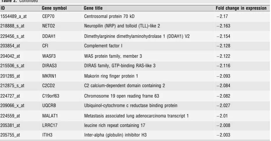
A total of 44 genes were
2-fold downregulated, of which only 11
transcripts were inhibited by
3-fold (Table 2). As PR-knock-down
decreased the expression of these genes, they represent genes
upreg-ulated in a PR-dependent manner.
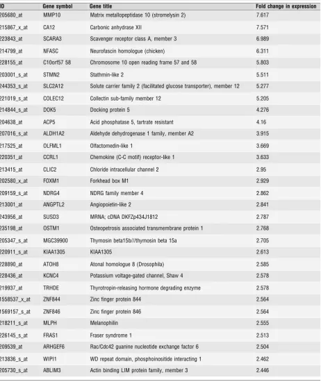
Of the 159 genes upregulated in response to PR knock-down, 79
increased by
2-fold, including 24 that were induced
3-fold
(Table 3). As PR silencing upregulated their expression, these genes
represent transcriptional targets actively repressed by PR.
Ingenuity pathways analysis (IPA) showed that PR knock-down
had the greatest effect on genes involved in cellular development,
growth and proliferation. PR-induced genes were significantly
enriched in two functional networks: (i) cellular development, growth
proliferation and death (score 17 focus molecules 12); and (ii) cellular
development, growth and proliferation, DNA replication,
recombina-tion and repair (score 15 focus molecules 11). Conversely, genes
repressed by PR were significantly involved in three networks: (i) cell
to cell signalling, inflammatory response, cellular movement (Score
21 focus molecules 15); (ii) cancer, cellular development,
haemato-logical system development (score 17, focus molecules 13); and (iii)
skeletal muscular system development and function (score 15 focus
molecules 12).
To explore the interactions between inflammatory signals and PR,
myocytes in culture were incubated for 96 hrs with PR specific siRNA
or non-targeting control siRNA, and then treated with MPA for a
fur-ther 24 hrs. In the last 6 hrs of culture, cells were incubated with
IL-1
b
(1 ng/ml) or vehicle control. We then compared the gene
expres-sion profiles of cells incubated with and without IL-1
b
in the absence
of PR knock-down, i.e. transfected with non-targeting siRNA and in
the presence of PR knock-down.
IL-1
b
activates myometrial inflammation
In cells exposed to non-targetting siRNA, IL-1
b
significantly regulated
5160 probe sets (P
<
0.05 with correction for multiple testing). Upon
elimination of unannotated sequences and duplicate probe sets, 1440
(57%) and 1892 (43%) unique gene sequences were found to be
up-and downregulated respectively. The expression of two genes,
CXCL2
and
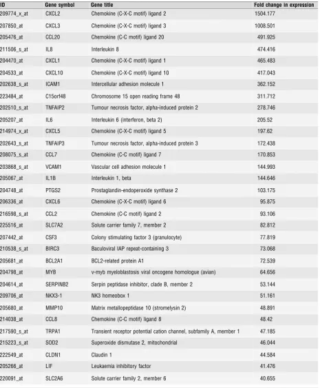
CXCL3, increased in excess of a 1000-fold. An additional 14
genes, mostly inflammatory in action, increased in expression
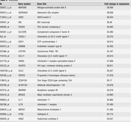
100-fold and 40 additional genes between 99- and 20-fold
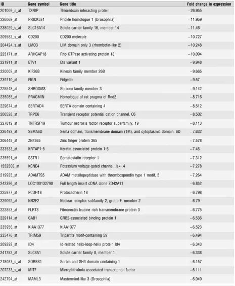
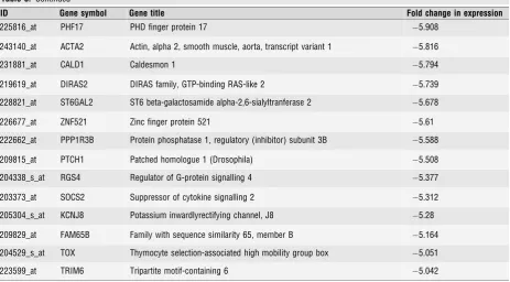
(Table 4). Of the 1892 repressed genes, 465 were inhibited
2-fold,
which included 127 and 46 transcripts regulated
3 and 5-fold
respectively (Table 5). The two probes complementary to PR mRNA
on the array showed a 1.6- and 4.8-fold reduction in total PR
tran-script levels in response to IL-1
b
stimulation (P
<
0.0002). As
antici-pated, IPA confirmed that IL-1
b
signalling has the greatest effect on
gene networks involved in inflammation, immunity, anti-microbial
response and NF-
j
B signalling.
IL-1
b
inhibits PR expression and microarray
validation
The array analysis indicated that IL-1
b
inhibits
PR
expression in
human myocytes. To validate the array, we performed Western
analy-sis to examine the regulation of PR upon MPA and IL-1
b
stimulation
in four independent primary myocyte cultures. IL-1
b
caused a
signifi-cant reduction in the expression of both PR-B and PR-A upon 6 and
24 hrs of stimulation (Fig. 1C). This was associated with increased
phosphorylation of p65, reflecting NF-
j
B activation, which peaked at
15 min., but persisted after 6 and 24 hrs (Fig. 1B). We further
vali-dated a set of selected genes regulated in a PR- and/or IL-1
b
-depen-dent manner by RTQ-PCR. These were selected arbitrarily in three
groups: genes upregulated by both PR-knock-down and IL-1
b
stimu-lation;
MMP1,
MMP3,
MMP10:
genes upregulated by
PR-knock-down;
COLEC12,
STMN2,
SCRA3,
RXFP1; and genes downregulated
by PR-knock-down
THBS1,
KCNJ2,
COL12A1
and
TRPC6
(Fig. 1D).
For these genes, the pattern of expression measured by RTQ-PCR
analysis was in keeping with the array data.
Activation of myometrial inflammation
antagonizes PR-induced gene expression
We next tested if inflammatory signal intermediates, activated by
IL-1
b
, repress PR-dependent gene expression in human myocytes. If
activation of myometrial inflammation causes ‘functional
progester-one withdrawal’, then it would be expected that IL-1
b
stimulation
would have similar effects on PR-dependent gene expression as
knock-down of the receptor. We therefore cross-referenced the
effects of IL-1
b
on the expression of the 279 unique genes regulated
upon PR-knock-down. Although we found a significant positive
corre-lation between the overall effect of PR silencing on myometrial gene
expression and IL-1
b
stimulation this correlation was weak (Fig. 2A,
Pearson’s
r
=
0.4288,
95%
confidence
interval:
0.33
–
0.52,
P
<
0.0001).
We then focused on those genes whose expression was
repressed by PR knock-down, thus induced in a PR-dependent
fash-ion. This showed a correlation with IL-1
b
-dependent gene expression
(Fig. 2B, Pearson’s
r
=
0.4420, 95% confidence interval: 0.28
–
0.58;
P
<
0.0001). The correlation became stronger if the analysis was
confined to those genes repressed
2-fold upon PR knock-down
(Pearson’s
r
=
0.52, 95% confidence interval 0.27
–
0.7,
P
<
0.003;
data not shown).
Table 2Unique genes whose expression was decreased by PR-knock-down, and whose expression is therefore upregulated by PR in the presence of progesterone. 44 genes showed changes in expression of 2-fold or more. 11 genes showed changes in expression of 3-fold or more. In each casen=4,P<0.0005, AdjustedP<0.05)
ID Gene symbol Gene title Fold change in expression
222853_at FLRT3 Fibronectin leucine-rich transmembrane protein 3 5.419
217287_s_at TRPC6 Transient receptor potential cation channel C6 5.406
202708_s_at HIST2H2BE Histone cluster 2, H2be 4.98
223877_at C1QTNF7 C1q and tumour necrosis factor related protein 7 4.545
1559114_a_at CXCR7 CXC chemokine receptor type 7 4.348
206765_at KCNJ2 Potassium inwardly rectifying channel, subfamily J, member 2 4.138
239710_at FIGN Fidgetin 4.069
201107_s_at THBS1 Thrombospondin 1 3.921
235371_at GLT8D4 Glycosyltransferase 8 domain containing 4 3.448
231186_at FLJ43390 Hypothetical LOC646113 3.196
202016_at MEST Mesoderm specific transcript homologue 3.153
233109_at COL12A1 Collagen type XII alpha-1 precursor 2.869
226610_at CENPV Centromere protein V 2.781
204837_at MTMR9 Myotubularin related protein 9 2.748
225008_at ASPH Aspartyl(asparaginyl)beta-hydroxylase 2.612
201487_at CTSC Cathepsin C 2.411
238447_at RBMS3 RNA binding motif, single stranded interacting protein 2.41
222771_s_at MYEF2 Myelin expression factor 2 2.402
211959_at IGFBP5 Insulin-like growth factor binding protein 5 2.384
220153_at ENTPD7 Ectonucleoside triphosphate diphosphohydrolase 7 2.383
224613_s_at DNAJC5 DnaJ (Hsp40) homologue, subfamily C, member 5 2.369
208903_at RPS28 Ribosomal protein S28 2.367
224767_at RPL37 Ribosomal protein L37, mRNA 2.357
205807_s_at TUFT1 Tuftelin 1 2.342
224984_at NFAT5 Nuclear factor of activated T-cells 5, tonicity-responsive 2.335
222582_at PRKAG2 Protein kinase, AMP-activated, gamma 2 non-catalytic subunit 2.314
223155_at HDHD2 Haloacid dehalogenase-like hydrolase domain containing 2 2.311
202091_at ARL2BP ADP-ribosylation factor-like 2 binding protein 2.284
213223_at RPL28 Ribosomal protein L28 2.236
213024_at TMF1 TATA element modulatory factor 1 2.217
a PR-dependent fashion), and also repressed by IL-1
b
which are
therefore co-regulated. If adjustment is made for multiple
compari-sons then 42 of 118 genes (22%) repressed by PR knock-down were
also significantly repressed by IL-1
b
(at
P
<
0.05). However, a
fur-ther 19 genes were also repressed by IL-1
b
at
P
<
0.05 where single
testing only was taken into account making a total of 61 of 188 genes
(32%).
When those genes were analysed whose expression increased
upon PR knockdown (i.e. inhibited by PR), the correlation with the
IL-1
b
-dependent transcriptional response, although still significant was
very weak (Fig. 2C, Pearson’s
r
=
0.35, 95% confidence interval: 0.2
–
0.48,
P
<
0.0001) and was entirely dependent upon the strong
induction of a single gene,
MMP10, in response to IL-1
b
and upon
PR-knock-down. Omitting MMP10 from the analysis indicated that
IL-1
b
-dependent activation of myometrial inflammation has no
signif-icant effect on the ability of PR to repress specific gene sets
(P
=
0.57).
PR does not repress pro-inflammatory gene
networks induced by IL-1
b
We next hypothesized that PR, liganded or not, may serve to repress
inflammatory genes in otherwise unstimulated myocytes. If so, PR
silencing in the absence of inflammatory stimulation should be
suffi-cient to regulate genes which are inducible by IL-1
b
. Alternatively, it
is possible that PR only confers transcriptional repression of genes
involved in myometrial inflammation upon exposure to inflammatory
cues. If this hypothesis holds true then PR knock-down should
fur-ther increase the expression of genes induced by IL-1
b
treatment.
To explore the first hypothesis, we correlated the effect of PR
knock-down in unstimulated myocytes on expression of genes which
would be activated upon IL-1
b
treatment. No correlation was found
(Fig. 2D). Of 1440 genes induced by IL-1
b
in human myocytes, PR
silencing significantly increased the expression of only 36 (0.025%)
genes, and just 15 of these by
2-fold. We then explored the second
hypothesis, but again no correlation was found (Fig. 2E).
PR-knock-down failed to significantly augment the vast majority of genes
induced by IL-1
b
stimulation. The expression of only 39 (0.027%) of
1440 IL-1
b
-induced genes was significantly enhanced upon prior PR
silencing, with transcript levels of merely 22 of these upregulated by
2-fold.
In the absence of a ‘global’ anti-inflammatory role for PR, we
speculated that this nuclear receptor may still be important for the
repression of a discrete cassette of genes encoding key inflammatory
mediators. We therefore focussed on those IL-1
b
upregulated genes
whose expression increases upon PR silencing in both stimulated and
unstimulated cells. Only seven such genes were identified from the
microarray analysis (Table 6). Furthermore, for all but one gene,
MMP10, the increase caused by IL-1
b
alone and additional increase
[image:8.595.66.532.104.346.2]in transcript levels caused by PR knock-down in the presence or
absence of IL-1
b
was modest, albeit significant. One gene, MMP1,
showed a similar pattern of expression to MMP10, but on the
micro-array analysis whilst the effect of IL-1
b
, and PR knock-down in the
presence of IL-1
b
was significant, the effect of PR knock-down alone
was significant for single testing but not when multiple testing was
taken into account. We selected this gene for independent validation
studies and found that, in those studies using qRT-PCR, the effects of
IL-1
b
, PR knock-down alone and PR knock-down in the presence of
IL-1
b
were all significant (Fig. 1D).
Table 2. Continued
ID Gene symbol Gene title Fold change in expression
1554489_a_at CEP70 Centrosomal protein 70 kD 2.17
218888_s_at NETO2 Neuropilin (NRP) and tolloid (TLL)-like 2 2.163
229456_s_at DDAH1 Dimethylarginine dimethylaminohydrolase 1 (DDAH1) V2 2.154
203854_at CFI Complement factor I 2.128
204042_at WASF3 WAS protein family, member 3 2.122
215506_s_at DIRAS3 DIRAS family, GTP-binding RAS-like 3 2.116
201285_at MKRN1 Makorin ring finger protein 1 2.093
212875_s_at C2CD2 C2 calcium-dependent domain containing 2 2.084
224727_at C19orf63 Chromosome 19 open reading frame 63 2.082
209066_x_at UQCRB Ubiquinol-cytochrome c reductase binding protein 2.027
224559_at MALAT1 Metastasis associated lung adenocarcinoma transcript 1 2.01
205381_at LRRC17 leucine rich repeat containing 17 2.008
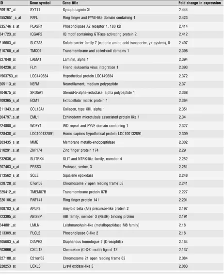
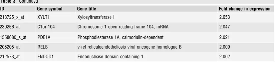
Table 3Unique genes whose expression was increased by PR-knock-down, and whose expression is therefore inhibited by liganded-PR. 24 genes showed changes in expression of 3-fold or more. 55 genes showed changes in expression of 2 to 3-fold. (In each casen=4, P<0.0005, AdjustedP<0.05)
ID Gene symbol Gene title Fold change in expression
205680_at MMP10 Matrix metallopeptidase 10 (stromelysin 2) 7.617
215867_x_at CA12 Carbonic anhydrase XII 7.571
223843_at SCARA3 Scavenger receptor class A, member 3 6.989
214799_at NFASC Neurofascin homologue (chicken) 6.311
228155_at C10orf57 58 Chromosome 10 open reading frame 57 and 58 5.803
203001_s_at STMN2 Stathmin-like 2 5.511
244353_s_at SLC2A12 Solute carrier family 2 (facilitated glucose transporter), member 12 5.277
221019_s_at COLEC12 Collectin sub-family member 12 5.205
214844_s_at DOK5 Docking protein 5 4.276
204638_at ACP5 Acid phosphatase 5, tartrate resistant 4.16
207016_s_at ALDH1A2 Aldehyde dehydrogenase 1 family, member A2 3.915
217525_at OLFML1 Olfactomedin-like 1 3.669
220351_at CCRL1 Chemokine (C-C motif) receptor-like 1 3.633
213415_at CLIC2 Chloride intracellular channel 2 2.95
202580_x_at FOXM1 Forkhead box M1 2.929
209159_s_at NDRG4 NDRG family member 4 2.862
213001_at ANGPTL2 Angiopoietin-like 2 2.841
243956_at SUSD3 MRNA; cDNA DKFZp434J1812 2.787
235198_at OSTM1 Osteopetrosis associated transmembrane protein 1 2.768
205347_s_at MGC39900 Thymosin beta15b///thymosin beta 15a 2.705
220911_s_at KIAA1305 KIAA1305 2.613
228890_at ATOH8 Atonal homologue 8 (Drosophila) 2.585
228436_at KCNC4 Potassium voltage-gated channel, Shaw 4 2.578
219937_at TRHDE Thyrotropin-releasing hormone degrading enzyme 2.578
1558537_x_at ZNF844 Zinc finger protein 844 2.564
1569157_s_at ZNF846 Zinc finger protein 846 2.564
218211_s_at MLPH Melanophilin 2.555
226145_s_at FRAS1 Fraser syndrome 1 2.513
209539_at ARHGEF6 Rac/Cdc42 guanine nucleotide exchange factor 6 2.504
213836_s_at WIPI1 WD repeat domain, phosphoinositide interacting 1 2.462
Table 3. Continued
ID Gene symbol Gene title Fold change in expression
209197_at SYT11 Synaptotagmin XI 2.444
1552651_a_at RFFL Ring finger and FYVE-like domain containing 1 2.423
235746_s_at PLA2R1 Phospholipase A2 receptor 1, 180 kD 2.414
241723_at IQGAP2 IQ motif containing GTPase activating protein 2 2.412
216603_at SLC7A8 Solute carrier family 7 (cationic amino acid transporter, y+system), 8 2.407
210768_x_at TMCO1 Transmembrane and coiled-coil domains 1 2.398
227048_at LAMA1 Laminin, alpha 1 2.394
204236_at FLI1 Friend leukaemia virus integration 1 2.393
1563753_at LOC149684 Hypothetical protein LOC149684 2.372
205113_at NEFM Neurofilament, medium polypeptide 2.37
204675_at SRD5A1 Steroid-5-alpha-reductase, alpha polypeptide 1 2.368
209365_s_at ECM1 Extracellular matrix protein 1 2.364
211343_s_at COL13A1 Collagen, type XIII, alpha 1 2.351
204797_s_at EML1 Echinoderm microtubule associated protein like 1 2.34
224800_at WDFY1 WD repeat and FYVE domain containing 1 2.327
228438_at LOC100132891 Homo sapiens hypothetical protein LOC100132891 2.309
203435_s_at MME Membrane metallo-endopeptidase 2.302
210291_s_at ZNF174 Zinc finger protein 174 2.29
232636_at SLITRK4 SLIT and NTRK-like family, member 4 2.252
207463_x_at PRSS3 Protease, serine, 3 2.251
213562_s_at SQLE Squalene epoxidase 2.248
228728_at C7orf58 Chromosome 7 open reading frame 58 2.241
225412_at TMEM87B Transmembrane protein 87B 2.227
226106_at RNF141 Ring finger protein 141 2.201
208703_s_at APLP2 Amyloid beta (A4) precursor-like protein 2 2.197
223395_at ABI3BP ABI family, member 3 (NESH) binding protein 2.191
244881_at LMLN Leishmanolysin-like (metallopeptidase M8 family) 2.18
213309_at PLCL2 Phospholipase C-like 2 2.18
205603_s_at DIAPH2 Diaphanous homologue 2 (Drosophila) 2.164
203666_at CXCL12 Chemokine (C-X-C motif) ligand 12 2.137
227188_at C21orf63 Chromosome 21 open reading frame 63 2.084
Discussion
Understanding the mechanism of the onset of human parturition is
critical for the design of strategies to predict and prevent preterm
birth. However, this requires more than simply understanding the
mechanisms of contractions. Although a range of drugs is available
which are intended to inhibit uterine contractions their clinical
effec-tiveness is disappointing. The ‘tocolytic’ reactive management of
pre-term labour is giving way to a strategy based upon prediction and
prevention, however, the only agent which currently shows promise
in preventing the onset of preterm labour is progesterone.
Progester-one appears to be effective in some woman at high risk of preterm
birth with a singleton pregnancy, but does not reduce the risk of
pre-maturity in multiple pregnancy [12]. Further development of
proges-tational agents and similar drugs for this indication requires a better
understanding of the function of progesterone and its receptor within
the uterus.
We have modelled PR-mediated functional progesterone
with-drawal by using siRNA knock-down of PR in the presence of a
syn-thetic progesterone. Comparisons were made between siRNA/PR and
control always in the presence of synthetic progesterone. We did not
compare gene expression between cells in the presence or absence
of progesterone as there is no withdrawal of progesterone in
associa-tion with human labour and therefore,
in vivo, there is always PR
ligand present. We have used MPA rather than natural progesterone
because of its greater long-term stability in culture. MPA has been
reported to have glucocorticoid receptor activity [13], but we have
found that this also applies to natural progesterone [14]. Use of
siR-NA requires the use of passaged cells in culture, as it is not possible
to obtain sufficient cells numbers for experimental purposes without
passage. Such models are widely used in investigation of myometrial
cell function, however, they do risk the possibility that cultures
become enriched for rapidly dividing fibroblast cells and there is loss
of the extracellular matrix integrity and cell-matrix interaction of the
intact tissue. We have confirmed that our cell cultures after passage
and MPA incubation are alpha-actin and oxytocin receptor positive
and that expression of PR and the PR-A:PR-B ratio is similar to that
seen in fresh myometrial tissue. A caveat to this, however, is that we
have found some patient to patient variability in PR-A:PR-B ratio in
both myometrial tissue taken at pre-labour caesarean section and
subsequent cell culture which may be dependent upon how close the
individual woman is to the onset of labour.
Progesterone is widely thought to act principally to repress
con-tractions through repressing ‘contraction-associated proteins’.
How-ever, it is now clear that progesterone plays a more complex role in
myometrial physiology during pregnancy. In the rat, progesterone is
involved in phenotypic modulation of myocytes during the synthetic
phase of myometrial differentiation in the last third of pregnancy
dur-ing which there is myometrial hypertrophy and synthesis and
deposi-tion of interstitial matrix [15]. We found that in human myocytes PR
regulates a relatively small number of genes. We did not see
signifi-cant down-regulation of classic ‘contraction-associated proteins’ by
PR. The major effect of PR upon gene expression was upon genes
concerned with cellular development, growth and proliferation. PR
also highly upregulates two ion channels; transient receptor potential
canonical-6 (TRPC-6) and the inward-rectifier potassium ion channel
KCNJ2. TRPC channels in general mediate store-operated calcium
entry. Expression of TrpC1, TrpC3, TrpC4 and TrpC6 has been
dem-onstrated in human myometrium [16]. Tonic stretch co-regulates
cal-cium entry pathways and TRPC3 and TRPC4 expression suggesting
that these receptors play a role in increasing contractility [17]. Trp6,
however, differs in that it mediates non-store operated calcium entry
and TrpC6 has been shown to play an essential role in cellular
prolif-eration in a range of cell types [18, 19].
Fibronectin leucine rich transmembrane protein-3 (FLRT-3), and
thrombospondin 1 (THBS1) were found to be highly upregulated by
PR whilst matrix metallopeptidase 10 (MMP-10) was highly
repressed. FLRT3 is a member of the fibronectin leucine rich
trans-membrane protein family which play important roles in cell matrix
adhesion. THBS1 is an adhesive glycoprotein which binds fibrinogen,
fibronectin, laminin, collagen and integrins and also mediates
cell-to-matrix interactions. MMP-10 is a member of the cell-to-matrix
metallopro-teinase family which is involved in the breakdown of extracellular
matrix. Collectively, these data point to a role for PR regulated genes
in the synthesis and maintenance of interstitial matrix in the human.
PR therefore probably acts during pregnancy more to regulate the
growth and development of the uterus than to directly inhibit
contrac-tions, which is consistent with progesterone having efficacy as a
pro-phylactic agent, but not as a tocolytic agent.
[image:11.595.67.532.103.208.2]Our principal purpose was to shed light on the inter-relationship
between PR and the myometrial inflammation. Parturition can be
thought of, at least in part, as inflammatory in nature as it is
associ-ated with upregulation of prostaglandin, cytokine and chemokine
syn-thesis with the uterus and with an influx of macrophages and
lymphocytes into the myometrium and cervix [20]. Although the
Table 3. Continued
ID Gene symbol Gene title Fold change in expression
213725_x_at XYLT1 Xylosyltransferase I 2.053
230256_at C1orf104 Chromosome 1 open reading frame 104, mRNA 2.047
1558680_s_at PDE1A Phosphodiesterase 1A, calmodulin-dependent 2.021
205205_at RELB v-rel reticuloendotheliosis viral oncogene homologue B 2.009
Table 4Unique genes whose expression was increased 20-fold or more by interleukin 1-beta. (In each casen=4,P<0.0005, Adjusted P<0.05)
ID Gene symbol Gene title Fold change in expression
209774_x_at CXCL2 Chemokine (C-X-C motif) ligand 2 1504.177
207850_at CXCL3 Chemokine (C-X-C motif) ligand 3 1008.501
205476_at CCL20 Chemokine (C-C motif) ligand 20 491.925
211506_s_at IL8 Interleukin 8 474.416
204470_at CXCL1 Chemokine (C-X-C motif) ligand 1 465.483
204533_at CXCL10 Chemokine (C-X-C motif) ligand 10 417.043
202638_s_at ICAM1 Intercellular adhesion molecule 1 362.152
223484_at C15orf48 Chromosome 15 open reading frame 48 311.712
202510_s_at TNFAIP2 Tumour necrosis factor, alpha-induced protein 2 278.746
205207_at IL6 Interleukin 6 (interferon, beta 2) 205.52
214974_x_at CXCL5 Chemokine (C-X-C motif) ligand 5 197.62
202643_s_at TNFAIP3 Tumour necrosis factor, alpha-induced protein 3 172.438
208075_s_at CCL7 Chemokine (C-C motif) ligand 7 170.853
203868_s_at VCAM1 Vascular cell adhesion molecule 1 144.993
205067_at IL1B Interleukin 1, beta 144.646
204748_at PTGS2 Prostaglandin-endoperoxide synthase 2 103.175
206336_at CXCL6 Chemokine (C-X-C motif) ligand 6 95.875
216598_s_at CCL2 Chemokine (C-C motif) ligand 2 93.106
225516_at SLC7A2 Solute carrier family 7, member 2 82.812
207442_at CSF3 Colony stimulating factor 3 (granulocyte) 77.819
210538_s_at BIRC3 Baculoviral IAP repeat-containing 3 73.068
205681_at BCL2A1 BCL2-related protein A1 72.539
204798_at MYB v-myb myeloblastosis viral oncogene homologue (avian) 64.656
204614_at SERPINB2 Serpin peptidase inhibitor, clade B, member 2 53.144
209706_at NKX3-1 NK3 homeobox 1 51.161
205680_at MMP10 Matrix metallopeptidase 10 (stromelysin 2) 48.891
214038_at CCL8 Chemokine (C-C motif) ligand 8 48.42
217590_s_at TRPA1 Transient receptor potential cation channel, subfamily A, member 1 47.185
215223_s_at SOD2 Superoxide dismutase 2, mitochondrial 46.044
222549_at CLDN1 Claudin 1 44.584
205266_at LIF Leukaemia inhibitory factor 41.476
inflammatory cell infiltrate may be a source of cytokines within the
labouring uterus it is clear that myometrium itself is a major source
of inflammatory mediators [21]. IL-1
b
concentrations rise within the
uterus in association with both term and preterm labour [22]. We
activated myometrial inflammation by incubation with IL-1
b
which
activates both NF-
j
B and AP-1 leading to upregulation of a wide
vari-ety of inflammatory genes which have been shown to be associated
with parturition [23, 24]. As would be expected we found that IL-1
b
increased expression of genes involved in immunity, inflammation,
anti-microbial response and NF-
j
B signalling.
Seminal studies of the endocrinology of parturition in sheep and
rodent models suggested that progesterone withdrawal is an early
ini-tiating factor leading to subsequent upregulation of ‘inflammatory’
type mediators, most importantly prostaglandins. This would suggest
that progesterone acts to inhibit the inflammatory biochemistry of
parturition. One hypothesis of the mechanism of human parturition
suggests that the activation of inflammation occurs late in the
pro-cess of parturition and that its principal role is the involution of the
uterus following delivery. However, there is growing evidence that
inflammation is an early initiating event in human parturition which
begins before the onset of contractions. Activation of NF-
j
B within
the uterus appears to play an important role in the onset of labour,
and in the myometrium principally regulates a group of immune/
inflammation associated genes [8, 21, 25].
[image:13.595.68.531.104.534.2]Condon
et al.
[5] have shown no activation of NF-
j
B in
my-ometrium in the third trimester of pregnancy remote from the
Table 4. Continued
ID Gene symbol Gene title Fold change in expression
205027_s_at MAP3K8 Mitogen-activated protein kkk 8 36.936
205013_s_at ADORA2A Adenosine A2a receptor 36.636
213524_s_at G0S2 G0/G1switch 2 36.454
229437_at BIC BIC transcript 36.36
209493_at PDZD2 PDZ domain containing 2 35.692
202357_s_at C2///CFB Complement component 2 factor B 35.285
823_at CX3CL1 Chemokine (C-X3-C motif) ligand 1 35.275
204224_s_at GCH1 GTP cyclohydrolase 1 34.613
204273_at EDNRB Endothelin receptor type B 34.355
207386_at CYP7B1 Cytochrome P450, 7B1 34.197
210133_at CCL11 Chemokine (C-C motif) ligand 11 32.614
231779_at IRAK2 Interleukin-1 receptor-associated kinase 2 31.006
235122_at HIVEP3 HIV type I enhancer binding protein 3 28.811
1555759_a_at CCL5 Chemokine (C-C motif) ligand 5 28.427
228186_s_at RSPO3 R-spondin 3 homologue (Xenopus laevis) 27.918
218810_at ZC3H12A Zinc finger CCCH-type containing 12A 26.17
203180_at ALDH1A3 Aldehyde dehydrogenase 1 A3 25.876
207510_at BDKRB1 Bradykinin receptor B1 23.079
225316_at MFSD2 Major facilitator superfamily domain 2 22.964
206924_at IL11 Interleukin 11 22.963
205798_at IL7R Interleukin 7 receptor 22.495
205619_s_at MEOX1 Mesenchyme homeobox 1 21.495
202902_s_at CTSS Cathepsin S 20.715
Table 5Unique genes whose expression was decreased 5-fold or more by interleukin 1-beta. (In each casen=4,P<0.0005, Adjusted P<0.05)
ID Gene symbol Gene title Fold change in expression
201009_s_at TXNIP Thioredoxin interacting protein 26.955
226069_at PRICKLE1 Prickle homologue 1 (Drosophila) 11.959
238029_s_at SLC16A14 Solute carrier family 16, member 14 11.46
209582_s_at CD200 CD200 molecule 10.727
204424_s_at LMO3 LIM domain only 3 (rhombotin-like 2) 10.248
225171_at ARHGAP18 Rho GTPase activating protein 18 10.094
221911_at ETV1 Ets variant 1 9.948
220002_at KIF26B Kinesin family member 26B 9.665
239710_at FIGN Fidgetin 9.57
225548_at SHROOM3 Shroom family member 3 9.142
235085_at PRAGMIN Homologue of rat pragma of Rnd2 8.716
229674_at SERTAD4 SERTA domain containing 4 8.512
206528_at TRPC6 Transient receptor potential cation channel, C6 8.502
227812_at TNFRSF19 Tumour necrosis factor receptor superfamily, 19 8.113
226492_at SEMA6D Sema domain, transmembrane domain (TM), and cytoplasmic domain, 6D 7.632
206448_at ZNF365 Zinc finger protein 365 7.578
233533_at KRTAP1-5 Keratin associated protein 1-5 7.45
235591_at SSTR1 Somatostatin receptor 1 7.312
1552508_at KCNE4 Potassium voltage-gated channel, Isk- 4 7.278
219935_at ADAMTS5 ADAM metallopeptidase with thrombospondin type 1 motif, 5 7.264
242396_at LOC100132798 Full length insert cDNA clone ZD42A11 6.852
225977_at PCDH18 Protocadherin 18 6.798
229092_at NR2F2 Nuclear receptor subfamily 2, group F, member 2 6.79
222853_at FLRT3 Fibronectin leucine rich transmembrane protein 3 6.775
229114_at GAB1 GRB2-associated binding protein 1 6.536
235956_at KIAA1377 KIAA1377 6.523
235476_at TRIM59 Tripartite motif-containing 59 6.494
209292_at ID4 Id-related helix-loop-helix protein Id4 6.343
241752_at SLC8A1 Solute carrier family 8, member 1 6.338
218087_s_at SORBS1 Sorbin and SH3 domain containing 1 6.157
207233_s_at MITF Microphthalmia-associated transcription factor 6.111
[image:14.595.66.532.124.689.2]onset of labour. However, in a study in which we compared tissue
samples from women taken either before or after labour at term
we found that that NF-
j
B is active in myocytes in both the upper
and lower segment of the uterus prior to the onset of labour at
term [21]. This, taken together with data which shows that
inhibi-tion of NF-
j
B in the mouse prevents normal term labour [8]
sug-gest that NF-
j
B begins to be activated in myometrium close to,
but prior to, the onset of labour at term. This concept is
supported by the results of mathematical modelling studies [26],
which explored three main hypotheses for the activation of the
human uterus at labour: functional progesterone withdrawal;
inflammatory stimulation; and oxytocin receptor activation. These
were modelled using gene expression data in pre-labour
myome-trial samples using directed graphs. It was found that
inflamma-tory activation as a primary event in parturition was highly likely,
progesterone withdrawal, as a primary event, was less likely but
plausible, and that oxytocin receptor mediated initiation was
unlikely.
We considered three non-mutually exclusive hypotheses
con-cerning the interaction between inflammation and PR in myocytes.
These were that inflammation acts to repress the function of PR;
that liganded PR acts to repress basal expression of inflammatory
mediators; or that liganded PR acts to repress stimulated
expres-sion of inflammatory mediators in myocytes. Our data shows that
activation of inflammation does act to inhibit PR function, but, in
general only in relation to genes which are upregulated, not
down-regulated by PR. Specifically, we identified a cassette of genes,
representing some 20
–
30% of genes upregulated
via
PR that are
downregulated by activation of inflammation. This is possibly a
consequence of the different ways in which PR interacts with
co-factors and the promoters of genes which are either up- or
down-regulated. There was, however, no general anti-inflammatory effect
of activated PR either upon basal or stimulated expression of
inflammatory mediators. We identified a few genes which were
oppositely regulated by inflammation or PR, most significantly the
matrix-metalloproteinases, MMP10 and MMP3, but not cytokines,
chemokines or ‘contraction-associated proteins’. The genes neuron
navigator 2, hepatocyte growth factor, squalene epoxidase, ABI
family, 3 (NESH) binding protein and xylosyltransferase I were
each found to be oppositely regulated by inflammation or PR, but
the overall effect of either IL-1
b
or PR-knock-down was very
small when compared with those effects in relation to MMP1 or
MMP10. It is doubtful that the opposite regulation of these six
genes by inflammation or PR is of significance in the context of
parturition. Interestingly, although progesterone has been shown
to inhibit IL-1
b
stimulated expression of
prostaglandin-endoperox-ide synthase type-2 and interleukin-8 in human myocytes, uterine
fibroblasts and amnion cells [11, 27] in the current study
PR-knock-down did not enhance the IL-1
b
stimulated expression of
either of these genes suggesting that the effect of progesterone in
this context is not mediated
via
PR. Progesterone itself may
nev-ertheless have a greater anti-inflammatory role within the uterus,
but mediated through receptors other than PR [14].
Our data are consistent with activation of inflammation being a
significant mediator of functional-progesterone withdrawal in the
context of the PR mediated action of progesterone. There are
sev-eral mechanisms, which may act together, by which inflammation
may repress PR function. In the present study, we found that
IL-Table 5. Continued
ID Gene symbol Gene title Fold change in expression
225816_at PHF17 PHD finger protein 17 5.908
243140_at ACTA2 Actin, alpha 2, smooth muscle, aorta, transcript variant 1 5.816
231881_at CALD1 Caldesmon 1 5.794
219619_at DIRAS2 DIRAS family, GTP-binding RAS-like 2 5.739
228821_at ST6GAL2 ST6 beta-galactosamide alpha-2,6-sialyltranferase 2 5.678
226677_at ZNF521 Zinc finger protein 521 5.61
222662_at PPP1R3B Protein phosphatase 1, regulatory (inhibitor) subunit 3B 5.588
209815_at PTCH1 Patched homologue 1 (Drosophila) 5.508
204338_s_at RGS4 Regulator of G-protein signalling 4 5.377
203373_at SOCS2 Suppressor of cytokine signalling 2 5.312
205304_s_at KCNJ8 Potassium inwardlyrectifying channel, J8 5.28
209829_at FAM65B Family with sequence similarity 65, member B 5.164
204529_s_at TOX Thymocyte selection-associated high mobility group box 5.051
[image:15.595.67.531.106.362.2]1
b
caused a significant downregulation in expression of PR itself
although not a change in the ratio of PR-B to PR-A. Mesiano
et al.
[1, 2] have suggested that it is a change in the ratio of the PR-B
to PR-A isoforms of PR which leads to functional-progesterone
withdrawal and have shown that this may be mediated by
increased prostaglandin synthesis. As would be expected, we
found that incubation with IL-1
b
leads to a large increase in
pros-taglandin-endoperoxide synthase type-2 expression. However, it is
unlikely that inhibition of PR synthesis is the sole mechanism
since at the 6-hr time point examined PR expression had reduced
by only one-third. Furthermore, the effect of IL-1
b
upon PR
regu-lated genes was only upon those upreguregu-lated, suggesting a
mech-anism other than simple receptor withdrawal. That it is likely that
NF-
j
B is activated within the uterus early in the biochemical
A B
C D
E
[image:16.595.73.521.85.492.2]events of labour suggests that the negative interaction between
NF-
j
B p65 and PR may also represent an important mechanism
of functional-progesterone withdrawal. Activation of NF-
j
B leads to
increased synthesis of its own p65 (RelA) subunit and to
increased synthesis of IL-1
b
and so to a positive feed forward
loop in which there is both synthesis and activation
(phosphoryla-tion) of p65 and therefore repression of PR. Our data supports
inhibition of PR by inflammation in general, or NF-
j
B specifically,
but suggests that the negative interaction between NF-
j
B p65 and
PR does not function in myometrium in the opposite direction as
we found no evidence of PR acting to repress NF-
j
B regulated
genes.
Much of what is currently known about the endocrinology and
biochemistry of parturition has come from animal models, however,
human parturition is unusual in that circulating progesterone levels
do not fall until after delivery [2]. Ethical considerations mostly limit
in vivo
experiments in humans to observations. Studies involving
manipulation of endocrine or inflammatory factors in humans are very
difficult to undertake
in vivo
and so need to be modelled
in vitro.
Inev-itably, no model can take into account all of the complex endocrine,
paracrine and mechanical factors which interact in the uterus
in vivo.
The model that we developed for this study was made as simple as
possible to answer our key questions: what are the genes regulated
by PR in the myocyte in the presence of progesterone and how does
PR-function and inflammation interact. Overall the data that we
pres-ent here support the concept that progesterone acts through PR in
human myocytes to mediate cellular development, growth and
prolif-eration and maintain the interstitial matrix, but that progesterone does
not play a significant anti-inflammatory role
via
PR. Conversely
inflammation does act to generally inhibit expression of genes which
are upregulated by PR. This is consistent with a hypothesis for the
mechanism of human parturition in which activation of inflammatory
mediators, likely through activation of NF-
j
B, is an early event
pre-ceding ‘functional progesterone withdrawal’.
Acknowledgements
PRB, VT, MRJ and YB are supported by the Imperial College Healthcare NHS Trust Comprehensive Biomedical Research Centre. This work was funded in part by an NIHR grant to VT. We acknowledge the help of Rachael Tribe, Kings College London in data interpretation.
Conflict of interest
The authors confirm that there are no conflicts of interest.
References
1. Zakar T, Mesiano S.How does progesterone relax the uterus in pregnancy? N Engl J Med. 2011; 364: 972–3.
2. Merlino AA, Welsh TN, Tan H,et al.Nuclear progesterone receptors in the human preg-nancy myometrium: evidence that parturition involves functional progesterone withdrawal mediated by increased expression of
proges-terone receptor-A.J Clin Endocrinol Metab. 2007; 92: 1927–33.
3. Pieber D, Allport VC, Bennett PR. Proges-terone receptor isoform A inhibits isoform B-mediated transactivation in human amnion.Eur J Pharmacol. 2001; 427: 7–11. 4. Pieber D, Allport VC, Hills F,et al.
Interac-tions between progesterone receptor
iso-forms in myometrial cells in human labour. Mol Hum Reprod. 2001; 7: 875–9. 5. Condon JC, Hardy DB, Kovaric K,
[image:17.595.64.536.137.279.2]Men-delson CR. Up-regulation of the proges-terone receptor (PR)-C isoform in labouring myometrium by activation of nuclear factor-kappaB may contribute to the onset of labour through inhibition of
Table 6Unique IL1b upregulated genes whose expression was increased in unstimulated cells by PR-knock-down (siRNA/PR FC i.e fold change), and further increased by PR-knock-down in the presence of IL1b (siRNA/PR+IL1b FC i.e fold change). (In each casen=4, P<0.0005, AdjustedP<0.05)
ID Gene Symbol Gene name Fold
increase IL1b
Fold increase siPRversuscontrol
Fold increase
siPR+IL1bv IL1balone
205680_at MMP10 Matrix metallopeptidase 10 48.9 7.6 8
204475_at MMP1 Matrix metallopeptidase 1 16 5.8* 6.4
218330_s_at NAV2 Neuron navigator 2 2.2 3.4 2.5
209960_at HGF Hepatocyte growth factor 6.2 2.6 2.8
213562_s_at SQLE Squalene epoxidase 2.7 2.2 1.8
223395_at ABI3BP ABI family, 3 (NESH) binding protein 1.6 2.2 2.3
213725_x_at XYLT1 Xylosyltransferase I 1.6 2.1 1.9
PR function. Mol Endocrinol. 2006; 20: 764–75.
6. Condon JC, Jeyasuria P, Faust JM,et al.A decline in the levels of progesterone receptor coactivators in the pregnant uterus at term may antagonize progesterone receptor func-tion and contribute to the initiafunc-tion of partu-rition.Proc Natl Acad Sci USA. 2003; 100: 9518–23.
7. Sheehan PM, Rice GE, Moses EK, Bren-necke SP.5 Beta-dihydroprogesterone and steroid 5 beta-reductase decrease in associ-ation with human parturition at term. Mol Hum Reprod. 2005; 11: 495–501. 8. Condon JC, Jeyasuria P, Faust JM,
Mendel-son CR.Surfactant protein secreted by the maturing mouse fetal lung acts as a hor-mone that signals the initiation of parturi-tion.Proc Natl Acad Sci USA. 2004; 101: 4978–83.
9. Kalkhoven E, Wissink S, van der Saag PT, van der Burg B. Negative interaction between the RelA(p65) subunit of NF-kap-paB and the progesterone receptor. J Biol Chem. 1996; 271: 6217–24.
10. Allport VC, Pieber D, Slater DM, et al.
Human labour is associated with nuclear factor-kappaB activity which mediates cyclo-oxygenase-2 expression and is involved with the ‘functional progesterone withdrawal’. Mol Hum Reprod. 2001; 7: 581–86. 11. Hardy DB, Janowski BA, Corey DR,
Mendel-son CR.Progesterone receptor plays a major antiinflammatory role in human myometrial cells by antagonism of nuclear factor-kap-paB activation of cyclooxygenase 2 expres-sion.Mol Endocrinol. 2006; 20: 2724–33. 12. Su LL, Samuel M, Chong YS.Progestational
agents for treating threatened or established
preterm labour. Cochrane Database Syst Rev. 2010; 20: CD006770.
13. Simoncini T, Mannella P, Fornari L,et al. Differential signal transduction of progester-one and medroxyprogesterprogester-one acetate in human endothelial cells. Endocrinology. 2004; 145: 5745–56.
14. Lei KY, Sooranna SR, Bennett PR, John-son MR. Glucocorticoid receptor is a key player that mediates progesterone action in human myometrium. Reproduc Sci. 2011; 18: 157A–8A.
15. Shynlova O, Tsui P, Jaffer S, Lye SJ. Inte-gration of endocrine and mechanical signals in the regulation of myometrial functions during pregnancy and labour.Eur J Obstet Gynecol Reprod Biol. 2009; 144: 2–10. 16. Dalrymple A, Slater DM, Poston L, Tribe
RM. Physiological induction of transient receptor potential canonical proteins, cal-cium entry channels, in human myometrium: influence of pregnancy, labour, and interleu-kin-1 beta.J Clin Endocrinol Metab. 2004; 89: 1291–300.
17. Dalrymple A, Mahn K, Poston L, et al.
Mechanical stretch regulates TRPC expres-sion and calcium entry in human myometrial smooth muscle cells. Mol Hum Reprod. 2007; 13: 171–9.
18. Cai R, Ding X, Zhou K, Shi Y,et al. Block-ade of TRPC6 channels induced G2/M phase arrest and suppressed growth in human gastric cancer cells.Int J Cancer. 2009; 125: 2281–7.
19. Leuner K, Kraus M, Woelfle U, et al. Reduced TRPC channel expression in psori-atic keratinocytes is associated with impaired differentiation and enhanced prolif-eration.PLoS One. 2011; 6: 14716.
20. Osman I, Young A, Ledingham MA,et al. Density and pro-inflammatory cytokine expression in human fetal membranes, decidua, cervix and myometrium before and during labour at term. Mol Hum Reprod. 2003; 9: 41–5.
21. Khanjani S, Kandola MK, Lindstrom TM,
et al. NF-kappaB regulates a cassette of immune/inflammatory genes in human preg-nant myometrium at term.J Cell Mol Med. 2011; 15: 809–24.
22. Romero R, Mazor M, Brandt F,et al. Inter-leukin-1 alpha and interInter-leukin-1 beta in pre-term and pre-term human parturition. Am J Reprod Immunol. 1992; 27: 117–23. 23. Bollapragada S, Youssef R, Jordan F,et al.
Term labour is associated with a core inflammatory response in human fetal mem-branes, myometrium, and cervix. Am J Obstet Gynecol. 2009; 200: 1–11. 24. Mittal P, Romero R, Tarca AL,et al.
Char-acterization of the myometrial transcriptome and biological pathways of spontaneous human labour at term.J Perinat Med. 2010; 38: 617–43.
25. Vora S, Abbas A, Kim CJ,et al.Nuclear fac-tor-kappa B localization and function within intrauterine tissues from term and preterm labour and cultured fetal membranes. Re-prod Biol Endocrinol. 2010; 8: 8.
26. Bisits AM, Smith R, Mesiano S, et al. Inflammatory aetiology of human myometri-al activation tested using directed graphs. PLoS Comput Biol. 2005; 1: 132–6. 27. Loudon JA, Elliott CL, Hills F, Bennett PR.