3D dissection tools in Anatomage supported
interactive human anatomy teaching
and learning
DzintraKazoka*and Mara Pilmane
R¯ıga Stradin¸š University, Department of Morphology, Kronvalda 9, Riga, Latvia Abstract. The main aim of this study is to present the usage and importance of 3D dissection tools in the teaching and learning of Anatomy and to describe and explain our experience with Anatomage Table in Human Anatomy studies at R¯ıga Stradin¸š University. In 2017–2018 two 3D dissection tools (scalpels) were used every week in work with Anatomage Table during the practical classes. As methods for collecting data were used discussions between students and teachers. Together 200 students of the Faculty of Medicine and Dentistry were involved in this study. It was possible to create incisions and cuts in order to remove and uncover different layers of organic tissues, to move deep inside step by step and find out which structures it was necessary to look for. Afterwards students showed that they were able to place the organs back and reattach the bones, muscles, blood vessels in the body and put the skin back on. Students enjoyed virtual tools in the practical classes and learned the material better. Virtual tools helped students and tutors to easily understand and memorize different anatomy structures. 3D scalpels were useful for different education activities, but the learning experience may be suitable further for the study of real materials. Key words: dissection tools, Anatomage, human anatomy, education
1 Introduction
Human Anatomy is one of the oldest subjects in medical education and there are different methods that are used for teaching and learning of it around the world [1–3]. At present, we still continue study process of human anatomy based on cadaveric dissections, but together with traditional methods, several combinations of modern methods and technologies are used. It is very important to find correct directions and reach the most effective ways in the upraising of knowledge of our students in Human Anatomy.
It is clear that teaching and learning of human body take time and may involve the use of different books, cadaver dissections, plastic models and special interactive 3D images in practical classes [4, 5]. These things help students to form a creative and productive approach, which can give them a lot of benefits for learning or self-education process [6]. New technologies and educational software are shaking up the role of educators in medical fields [7]. The educators of today should support and guide students’ activities. The advantages of the technologies and different methods can be very helpful for teachers to maximize their
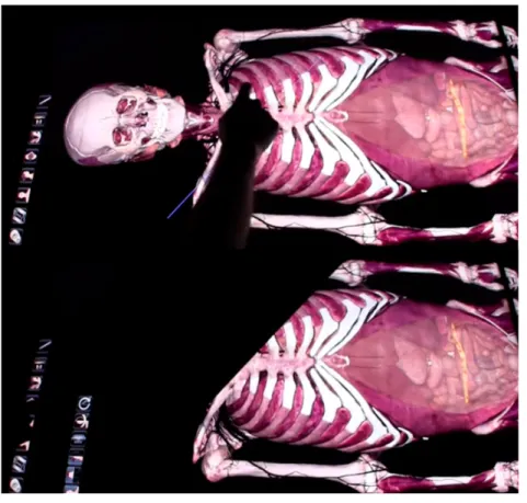
Fig. 1. The application of scalpel for straight line area – step 1 and step 2.
efforts in teaching [8–11]. It is very important that students are learning not only basics, but also how to use technologies that they have.
The Anatomage Table is life-size 3D interactive table and includes a segmented real human anatomy system. There are included virtual tools for performing an anatomical dissection and surgical approaches. It is known that the incorrect usage of scalpels is very dangerous to the patients and the surgeons [12], but in dissection the role of an incision with precision plays a very important role and it must be provided by strictly followed steps.
The main aim of this study is to present the usage and importance of 3D dissection tools in the teaching and learning of Anatomy and to describe and explain our experience with Anatomage table in Human Anatomy studies at R¯ıga Stradin¸š University.
2 Material and methods
In academic years 2017-2018 two 3D dissection tools were used every week in work with Anatomage Table (Table Application software from Anatomage, Inc. (Table EDU 4.0)) during the practical classes.
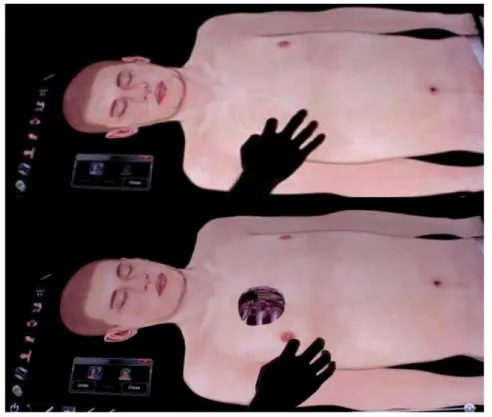
We offered and validated the usage of two virtual scalpels. The first scalpel allowed cutting according to the straight line position (Fig. 1). The second scalpel was used for freehand dissection in any area (Fig.2).
Students were divided into two groups. The first group consisted of 100 students of the Faculty of Dentistry and 100 students of the Faculty of Medicine. All students were also
Fig. 2. The application of scalpel for freehand area – step 1 and step 2.
asked if they had ever worked with the virtual scalpels as part of any prior or present course works.
At the beginning of the practical class the teacher explained students the general elements necessary to create incisions of tissue and demonstrated details of all steps involved in creating an ideal incision. Before beginning an incision, the teacher and students planned their approaches, landmarks, distances and techniques.
The students were instructed to complete their virtual dissection with discussions which contained the answers to the questions about the usage of 3D dissection tools. The questions were divided into the following categories: advantages and disadvantages of the used virtual scalpels of the Anatomage Table. All free answers were used for collecting of data.
3 Results
The students started each practical class of the dissection by repeating previous theoretical materials, skills and following the instructions. Students from Faculty of Medicine focused more on different organs systems, but students from Faculty of Dentistry focused more on the head and neck. None of the 200 students had ever worked with the virtual tools as part of prior or present courses. First two practical classes were started by students with slow incorporation of the virtual tools. The Anatomage Table offered access to life-size cross sections in different directions and allowed students to do a cross-section on the spot, using “scalpels” tools. In some cases it was a very fast process which could be done during only one anatomy class.
During the virtual cutting students trained in controlling of the directions, length and depth of all performed incisions. Virtual scalpels were utilized in a variety of ways to better
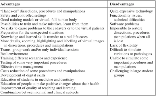
Table 1. Some advantages and disadvantages of the virtual dissection tools in Human Anatomy.
Advantages Disadvantages
“Hands-on” dissections, procedures and manipulations Safety and controlled settings
Good training models or virtual, full human body Possibilities to train and make mistakes, learn from them
No risks to cause problems to the cadavers or to the virtual patients Preparation for the unexpected situations
Knowledge and learned skills transfer to a real life cases
More details, zooming, highlighting and labelling of virtual images in dissections, procedures and manipulations
Teams, group work and/or only individual sessions Real environment
Training different scenarios and experience Testing of some very important procedures Effective time management
Costs reduction of some procedures and manipulations Development of digital skills
Education of students in medicine and dentistry
Education of people to make positive changes about their health Improvement of quality of teaching and learning
Combination between normal and clinical subjects
Quite expensive technology Functionality issues,
technical difficulties Software problems Impossible to restore
dissections, procedures manipulations when all is lost
Lack of flexibility Difficult to simulate
variations or pathologies Unable to simulate some
important procedures and manipulations
Challenging in large student groups
demonstrate layers and learn Topographical Anatomy using 3D format. For some students, watching a procedure performed by the teacher and afterwards performing the dissection by themselves with a scalpel were two very different things and experiences.
After some practical training, students became more comfortable with using virtual tools. It was possible to cut from the body surface to the inner body using a scalpel, as well as to watch images of 3D sections in the three planes. Majority of students realized that with just a click of a button they were given several options to study different parts and structures of human body. The general structures were labelled and further separated into next indentified elements. Our results of the study showed that the advantages of an adding the virtual scalpels to Human Anatomy course outweighed the disadvantages (Table1).
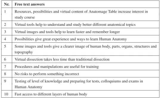
During discussions students’ answers as well as their feedback were used to explore details of an effectiveness of virtual tools in study process (Table2). Students of both groups noted these activities to be very beneficial in classes.
Students performed several digital procedures on life-size male or female virtual cadavers that were viewed from different angles and tissue levels. Different types of cuts were used by students to look into the body. It was possible to create incisions and to remove and uncover different layers of tissues, to move deep inside step by step and to understand which structures it was necessary to look for. Afterwards students showed that they were able to place the organs back and reattach the bones, muscles, blood vessels in the body and put the skin back on. They increased or decreased areas of interest from superficial until deep levels of structures. Students enjoyed the virtual tools in the practical classes and learned theoretical material better. Majority of students performed work with the first type or straight scalpel because it was more useful for dissection or slicing.
All students highlighted or removed from the view direct or specific body parts. According to these procedures, the variations and/or abnormalities were compared with normal examples. Possibilities of virtual dissection allowed students to limit or to reduce any mistakes immediately. At the end of the practical class students performed reset of virtual body to its initial position and original state.
Table 2. The most common answers of students obtained after discussions about virtual tools at the end of practical classes.
Nr. Free text answers
1 Resources, possibilities and virtual content of Anatomage Table increase interest in study course
2 Virtual tools help to understand and study better different anatomical topics 3 Virtual images and tools help to learn faster and remember longer
4 Possibilities give great experience and ways to learn Human Anatomy
5 Some images and tools give a clearer image of human body, parts, organs, structures and topography
6 Virtual dissection takes less time than traditional dissection 7 Procedures and manipulations are useful for training 8 No risks to perform something incorrect
9 Testing of level of knowledge and preparing for tests, colloquiums and exams in Human Anatomy
10 Fast access to different layers of human body
Virtual dissection was excellent for slicing with virtual scalpels, identification of special anatomical structures and the understanding of spatial relationships among organs. It was a new direction to see and interact with the same anatomy, as well as the access to a collection of normal and abnormal cases. Scalpels and their usage helped and allowed students to see structures from different positions and levels.
4 Discussion
Many of the world‘s medical schools and Universities use new visualization systems or technologies for medical education [13, 14]. Virtual 3D Anatomage Table is one of technology that can teach students different dissection procedures and can determine their levels of knowledge and competencies before they make real manipulations [15]. It allows students to repeat and return to the same tasks or steps of procedures or topics several times. Virtual 3D screen and possibilities can duplicate dissection or operative fields, support training and reduce some needs for expensive models in study process [16–18].
There is very limited information in the literature in regard to how the virtual scalpels of the Anatomage Table impact on students’ teaching and learning. Several authors presented students’ learning in different ways [19] and relationships between learning-teaching of anatomy [20]. Our students liked the ability to rotate, dissect and manipulate the virtual cadaver to better understand topography and cross-sections in Human Anatomy. The use of the “reset” button and possibility to return to previous steps allowed the students to repeat, train and make dissections of different regions and parts of the body several times. The tutors served as guides for students throughout the teaching and learning process. Students worked together in pairs or teams, thus forming a reality that was more similar to surgical environment [21].
The most significant challenge for students in learning Human Anatomy is direct understanding of body in dimensions and how different structures fit together [22]. There are many ways to improve the field of education with Anatomage Table using 3D dissection tools.
These new virtual things give possibility to make Anatomage supported training experiences available for students and tutors.
Can all students learn through the use of virtual tools in dissection of the human body? One way to determine this is to introduce the classes taught completely via virtual dissection and compare the results with those gained when the same classes are taught in a traditional way.
Several authors have shown that many technologies and virtual simulations can be used in medical education [23,24]. But, definitely, they should not replace direct communication or face-to-face learning.
Our experience shows that virtual instruments can develop real learning and teaching environment for students and tutors. It is clear that technologies and virtual simulators will play a great and an important role in future, and quantitative measurements of competencies would have to be part of the education system [25–27]. Some dissection procedures or manipulations consist of a series of tasks and steps and different virtual instruments can be used.
In some cases virtual scalpels can be used for faster procedures, manipulations and dissection of the human body. Digital images can show some details of structures in more precise ways [28].
Students can remove away different layers, structures and understand precisely connections and relationships between structures. Virtual tools and demonstrations of visual images of performed manipulations can be included into study course creating a self-training environment. Students can perform not only individual sessions, but they can learn together and these possibilities are some of the most effective ways to learn Human Anatomy [29,30]. Virtual scalpels also offer students the possibilities to practice responding to unexpected situations or scenarios [31].
We believe that the teaching and learning of Human Anatomy through the use of 3D virtual tools can be rather positive and productive accomplishment. One future area of this study will be to look at quantifiable data, comparing tests or exam scores from the students of previous years who had the virtual tools and dissection in their courses. Further studies are suggested to determine if a virtual experience can be a substitute for real dissections, or may serve as an enhancement to them.
5 Conclusion
Virtual dissections offered students the opportunity to prepare for and follow up on their human anatomy course. Virtual tools help students and teachers to understand and memorize better different anatomy structures. 3D dissection scalpels are useful for different teaching and learning activities, but the learning experience may be further enhanced by providing opportunity for the study of real materials.
References
[1] P. Benly, J. Pharm. Sci. Res. 6, 242–43 (2014)
[2] A.W. Keedy, J.C. Durack, P. Sandhu, E.M. Chen, P.S.O’ Sullivan, R.S. Breiman, Anat. Sci. Educ. 4, 84–91 (2011)
[3] D.S. Barry, F. Marzouk, K. Chulak-Oglu, D. Bennett, P. Tierney, Anat. Sci. Educ. 9(1), 90–96 (2016)
[5] J.R. Fredieu, J. Kerbo, M. Herron, R. Klatte, M. Cooke, Medical Sci. Educator 25, 183–194 (2015)
[6] P.K. Ganguly, Open Med. Educ. J. 3, 5–10 (2010)
[7] K. Sugand, P. Abrahams, A. Khurana, Anat. Sci. Educ. 3, 83–93 (2010) [8] J. Collins, BMJ 337, 665 (2008)
[9] L. Donnelly, D. Patten, P. White, G. Finn, Med. Teach. 31, 553 (2009) [10] K. Yammine, C. Violato, Anat. Sci. Educ. 8, 525–538 (2015)
[11] T. Lewis, B. Burnett, R. Tunstall, P. Abrahams, Clin. Anat. 27(3), 313–20 (2014) [12] P. Ruisoto Palomera, J.A. Juanes Méndez, A. Prats Galino, Comput. Hum. Behav. 31,
446 (2014)
[13] M. Hackett, M. Proctor, J. Sci. Educ. Technol. 25(4), 641–54 (2016) [14] H. Hoffman, D. Vu, Acad. Med. 72(12), 1076–1081 (1997)
[15] C.F. Smith, H.S. Clin. Anat. 24, 113–19 (2011)
[16] M. Zilverschoon, K.L. Vincken, R.L. A.W. Bleys, J. Biomed. Inf. 65, 58–75 (2017) [17] J. Sweitzer, Sci. 36(2), 41–44 (1996)
[18] N. Hoyek, Ch. Collet, F. Rienzo, M. Almeida, A. Guillot, Anat. Sci. Educ. 7(6), 430–437 (2014)
[19] D.I. Newble, N.J. Entwistle, Med. Educ. 20(3), 162–75 (1986) [20] K.M. Patel, B.J. Moxham, Clin. Anat. 21, 182–89 (2008) [21] M. Estai, S. Bunt, Ann. Anat. 208, 151–57 (2016)
[22] R. Jaiswal, S. Sathe, V. Gajbhiye, R. Sathe, Int. J. Anat. Res. 3(2), 1103–08 (2015) [23] P. Bradley, Med. Educ. 40(3), 254–62 (2006)
[24] J.P. Akpan, T. Andre, J. Comput. Math. Sci. Teaching 19(3), 297–313 (2000) [25] K. Wilkinson, P. Barter, J. Pedagogic Development 6(1), 14–23 (2016)
[26] J. Qualter, F. Sculli, A. Oliker, Z. Napier, S. Lee, J. Garcia, S. Frenkel, V. Harbik, M. Triola, Stud. Health Technol. Inf. 173, 359–61 (2012)
[27] T. Greenhalgh, BMJ 322(7277), 40–44 (2001)
[28] R. Trelease, A. Rosset, Anat. Sci. Educ. 1(2), 50–5 (2008) [29] B. Preim, P. Saalfeld, Computers and Graphics 71, 132–53 (2018)
[30] Z. Merchant, E.T. Goetz, L. Cifuentes, W. Keeney-Kennicutt, T.J. Davis, Comput. Education 70, 29–40 (2014)