2-(4-Methylphenyl)acetohydrazide
A.S. Praveen,aJerry P. Jasinski,b* Shannon T. Krauss,b H. S. Yathirajanaand B. Narayanac
a
Department of Studies in Chemistry, University of Mysore, Manasagangotri, Mysore 570 006, India,bDepartment of Chemistry, Keene State College, 229 Main Street, Keene, NH 03435-2001, USA, andcDepartment of Studies in Chemistry, Mangalore
University, Mangalagangotri 574 199, India Correspondence e-mail: [email protected]
Received 16 November 2012; accepted 21 November 2012
Key indicators: single-crystal X-ray study;T= 173 K; mean(C–C) = 0.003 A˚; Rfactor = 0.050;wRfactor = 0.151; data-to-parameter ratio = 14.1.
In the title compound, C9H12N2O, the dihedral angle between
the benzene ring and the mean plane of the acetohydrazide group is 88.2 (7). In the crystal, N—H O hydrogen bonds and weak C—H O interactions link the molecules into infinite ribbons along [001].
Related literature
For hydrazides as precursors in the synthesis of heterocyclic systems, see: Narayanaet al.(2005). For related structures, see: Hanifet al.(2007); Liu & Gao (2012); Fun et al.(2011). For standard bond lengths, see: Allenet al.(1987).
Experimental
Crystal data
C9H12N2O Mr= 164.21 Monoclinic,P21=c a= 15.4261 (16) A˚
b= 6.2618 (7) A˚ c= 9.2073 (10) A˚
= 106.651 (12)
V= 852.09 (16) A˚3
CuKradiation
= 0.69 mm1
0.320.220.08 mm
Data collection
Agilent Xcalibur (Eos, Gemini) diffractometer
Absorption correction: multi-scan (CrysAlis RED; Agilent, 2012) Tmin= 0.746,Tmax= 1.000
4845 measured reflections 1675 independent reflections 1359 reflections withI> 2(I) Rint= 0.028
Refinement
R[F2> 2(F2)] = 0.050 wR(F2) = 0.151 S= 1.07 1675 reflections 119 parameters 3 restraints
H atoms treated by a mixture of independent and constrained refinement
max= 0.26 e A˚
3
min=0.21 e A˚
[image:1.610.315.564.292.340.2]3
Table 1
Hydrogen-bond geometry (A˚ ,).
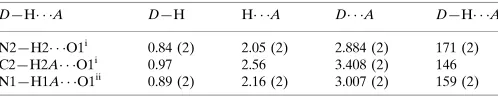
D—H A D—H H A D A D—H A
N2—H2 O1i
0.84 (2) 2.05 (2) 2.884 (2) 171 (2)
C2—H2A O1i
0.97 2.56 3.408 (2) 146
N1—H1A O1ii 0.89 (2) 2.16 (2) 3.007 (2) 159 (2)
Symmetry codes: (i)x;yþ1
2;z12; (ii)x;yþ12;zþ12.
Data collection: CrysAlis PRO(Agilent, 2012); cell refinement:
CrysAlis PRO; data reduction: CrysAlis RED (Agilent, 2012); program(s) used to solve structure: SHELXS97 (Sheldrick, 2008); program(s) used to refine structure:SHELXL97(Sheldrick, 2008); molecular graphics:SHELXTL(Sheldrick, 2008); software used to prepare material for publication:SHELXTL.
ASP thanks the UOM for research facilities. JPJ acknowl-edges the NSF–MRI program (grant No. CHE1039027) for funds to purchase the X-ray diffractometer.
Supplementary data and figures for this paper are available from the IUCr electronic archives (Reference: NG5307).
References
Agilent (2012). CrysAlis PRO and CrysAlis RED. Agilent Technologies, Yarnton, England.
Allen, F. H., Kennard, O., Watson, D. G., Brammer, L., Orpen, A. G. & Taylor, R. (1987).J. Chem. Soc. Perkin Trans. 2, pp. S1–19.
Fun, H.-K., Quah, C. K., Malladi, S. M. V. A. & Isloor, A. M. (2011).Acta Cryst.E67, o165.
Hanif, M., Qadeer, G., Rama, N. H., Farman, M. & Ruzˇicˇka, A. (2007).Acta Cryst.E63, o4828.
Liu, G. & Gao, J. (2012).Acta Cryst.E68, o1969.
Narayana, B., Ashalatha, B. V., Vijayaraj, K. K., Fernandes, J. & Sarojini, B. K. (2005).Bioorg. Med. Chem.13, 4638–4644.
Sheldrick, G. M. (2008).Acta Cryst.A64, 112–122. Structure Reports
Online
supporting information
Acta Cryst. (2012). E68, o3467 [doi:10.1107/S160053681204799X]
2-(4-Methylphenyl)acetohydrazide
A.S. Praveen, Jerry P. Jasinski, Shannon T. Krauss, H. S. Yathirajan and B. Narayana
S1. Comment
Hydrazides are useful precursors in the synthesis of several related heterocyclic systems (Narayana et al., 2005). The
crystal structures of some similar hydrazides, viz., 2-(4-methoxyphenoxy)acetohydrazide (Liu & Gao, 2012),
2-(3-meth-oxyphenyl)acetohydrazide (Hanif et al., 2007) and 2-(4-methylphenoxy)acetohydrazide (Fun et al., 2011) have been
reported. In view of the importance of hydrazides, the crystal structure of title compound (I) is reported.
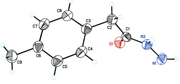
In the title compound, C9H12N2O, the dihedral angle between the mean planes of the benzene ring (C3–C8) and
acetohydrazide group (O1/C1/N2/N1) is 88.2 (7)° (Fig. 1). Bond lengths are in normal ranges (Allen et al., 1987). In the
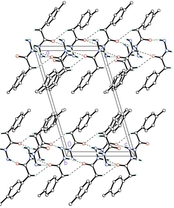
crystal N—H···O hydrogen bonds and weak C—H···O intermolecular interactions link the molecules into infinite ribbons
along [001] (Fig. 2, Table 1).
S2. Experimental
To a solution of methyl (4-methylphenyl)acetate (2 g, 12.18 mmol) in methanol (20 mL), hydrazine hydrate (2 mL) was
added and the reaction mixture was stirred at room temperature for 6 hours (Fig. 3). After the completion of the reaction
methanol was removed under vacuum, added water, precipitated solid was filtered and dried. The single crystal was
grown from mixture methanol: water (2:1) by slow evaporation method and yield of the compound was 91%. (m.p.:
426-428 K).
S3. Refinement
H1A, H1B and H2 were restrained with DFIX = 0.86 (2)Å. All the H atoms were placed in their calculated positions and
then refined using the riding model with Atom—H lengths of 0.93Å (CH), 0.97Å (CH2), 0.96Å (CH3) or 0.86Å (NH).
Isotropic displacement parameters for these atoms were set to 1.19-1.21 (CH, CH2), 1.49 (CH3) or 1.20 (NH) times Ueq of
Figure 1
Molecular structure of the title compound showing the atom labeling scheme and 50% probability displacement
Figure 2
Packing diagram of the title compound viewed along the b axis. Dashed lines indicate N—H···O hydrogen bonds and
weak C—H···O intermolecular interactions linking molecules into infinite 1-D chains along [001]. The remaining H
[image:4.610.132.483.70.491.2]atoms have been removed for clarity.
Figure 3
Crystal data
C9H12N2O Mr = 164.21 Monoclinic, P21/c Hall symbol: -P 2ybc a = 15.4261 (16) Å b = 6.2618 (7) Å c = 9.2073 (10) Å β = 106.651 (12)° V = 852.09 (16) Å3 Z = 4
F(000) = 352 Dx = 1.280 Mg m−3
Cu Kα radiation, λ = 1.54184 Å Cell parameters from 1566 reflections θ = 3.0–72.3°
µ = 0.69 mm−1 T = 173 K Chunk, colorless 0.32 × 0.22 × 0.08 mm
Data collection
Agilent Xcalibur (Eos, Gemini) diffractometer
Radiation source: Enhance (Cu) X-ray Source Graphite monochromator
Detector resolution: 16.0416 pixels mm-1 ω scans
Absorption correction: multi-scan (CrysAlis RED; Agilent, 2012) Tmin = 0.746, Tmax = 1.000
4845 measured reflections 1675 independent reflections 1359 reflections with I > 2σ(I) Rint = 0.028
θmax = 72.5°, θmin = 3.0° h = −18→19
k = −7→4 l = −10→11
Refinement
Refinement on F2 Least-squares matrix: full R[F2 > 2σ(F2)] = 0.050 wR(F2) = 0.151 S = 1.07 1675 reflections 119 parameters 3 restraints
Primary atom site location: structure-invariant direct methods
Secondary atom site location: difference Fourier map
Hydrogen site location: inferred from neighbouring sites
H atoms treated by a mixture of independent and constrained refinement
w = 1/[σ2(F
o2) + (0.0882P)2 + 0.1271P] where P = (Fo2 + 2Fc2)/3
(Δ/σ)max < 0.001 Δρmax = 0.26 e Å−3 Δρmin = −0.21 e Å−3
Special details
Geometry. All esds (except the esd in the dihedral angle between two l.s. planes) are estimated using the full covariance matrix. The cell esds are taken into account individually in the estimation of esds in distances, angles and torsion angles; correlations between esds in cell parameters are only used when they are defined by crystal symmetry. An approximate (isotropic) treatment of cell esds is used for estimating esds involving l.s. planes.
Refinement. Refinement of F2 against ALL reflections. The weighted R-factor wR and goodness of fit S are based on F2, conventional R-factors R are based on F, with F set to zero for negative F2. The threshold expression of F2 > σ(F2) is used only for calculating R-factors(gt) etc. and is not relevant to the choice of reflections for refinement. R-factors based on F2 are statistically about twice as large as those based on F, and R- factors based on ALL data will be even larger.
Fractional atomic coordinates and isotropic or equivalent isotropic displacement parameters (Å2)
x y z Uiso*/Ueq
H1B −0.0656 (15) 0.105 (3) 0.093 (2) 0.048* N2 0.03002 (10) 0.2537 (2) 0.05112 (18) 0.0336 (4) H2 0.0399 (15) 0.293 (3) −0.030 (2) 0.040* C1 0.09783 (12) 0.1991 (3) 0.17011 (19) 0.0319 (4) C2 0.19169 (12) 0.2268 (3) 0.1528 (2) 0.0370 (4) H2A 0.1873 0.2917 0.0553 0.044* H2B 0.2199 0.0878 0.1551 0.044* C3 0.25012 (11) 0.3654 (3) 0.27779 (19) 0.0340 (4) C4 0.22379 (12) 0.5730 (3) 0.2991 (2) 0.0381 (4) H4 0.1707 0.6279 0.2343 0.046* C5 0.27565 (12) 0.6990 (3) 0.4157 (2) 0.0394 (4) H5 0.2562 0.8363 0.4292 0.047* C6 0.35650 (12) 0.6232 (3) 0.5132 (2) 0.0381 (4) C7 0.38281 (12) 0.4170 (3) 0.4904 (2) 0.0402 (5) H7 0.4366 0.3630 0.5538 0.048* C8 0.33043 (12) 0.2894 (3) 0.3747 (2) 0.0373 (4) H8 0.3495 0.1515 0.3621 0.045* C9 0.41413 (15) 0.7621 (4) 0.6381 (2) 0.0518 (6) H9A 0.3783 0.8125 0.7009 0.078* H9B 0.4365 0.8817 0.5944 0.078* H9C 0.4642 0.6802 0.6985 0.078*
Atomic displacement parameters (Å2)
U11 U22 U33 U12 U13 U23
O1 0.0437 (8) 0.0512 (8) 0.0323 (7) −0.0037 (6) 0.0144 (6) 0.0019 (5) N1 0.0299 (8) 0.0442 (9) 0.0452 (10) −0.0016 (6) 0.0115 (7) −0.0051 (7) N2 0.0311 (8) 0.0391 (8) 0.0321 (8) −0.0006 (6) 0.0113 (6) 0.0010 (6) C1 0.0347 (9) 0.0320 (8) 0.0298 (9) −0.0025 (6) 0.0105 (7) −0.0047 (6) C2 0.0328 (9) 0.0492 (10) 0.0307 (9) −0.0012 (7) 0.0119 (7) −0.0038 (7) C3 0.0293 (8) 0.0435 (10) 0.0310 (9) −0.0022 (7) 0.0113 (7) 0.0004 (7) C4 0.0288 (9) 0.0460 (10) 0.0392 (10) 0.0037 (7) 0.0095 (7) 0.0048 (7) C5 0.0327 (9) 0.0409 (10) 0.0468 (11) −0.0015 (7) 0.0151 (8) −0.0014 (8) C6 0.0303 (9) 0.0484 (10) 0.0374 (10) −0.0060 (7) 0.0126 (7) −0.0032 (8) C7 0.0301 (9) 0.0496 (11) 0.0388 (10) 0.0012 (7) 0.0065 (7) 0.0040 (8) C8 0.0340 (9) 0.0386 (9) 0.0398 (10) 0.0027 (7) 0.0115 (8) 0.0021 (7) C9 0.0403 (11) 0.0650 (14) 0.0498 (12) −0.0083 (9) 0.0121 (10) −0.0157 (10)
Geometric parameters (Å, º)
C2—H2B 0.9700 C9—H9A 0.9600 C3—C8 1.387 (2) C9—H9B 0.9600 C3—C4 1.393 (3) C9—H9C 0.9600
N2—N1—H1A 106.6 (15) C3—C4—H4 119.5 N2—N1—H1B 106.3 (15) C4—C5—C6 121.05 (17) H1A—N1—H1B 102 (2) C4—C5—H5 119.5 C1—N2—N1 123.05 (15) C6—C5—H5 119.5 C1—N2—H2 120.6 (15) C7—C6—C5 117.77 (17) N1—N2—H2 116.0 (15) C7—C6—C9 121.12 (17) O1—C1—N2 122.32 (16) C5—C6—C9 121.11 (18) O1—C1—C2 121.82 (16) C6—C7—C8 121.30 (16) N2—C1—C2 115.86 (15) C6—C7—H7 119.3 C1—C2—C3 111.39 (14) C8—C7—H7 119.3 C1—C2—H2A 109.3 C3—C8—C7 120.79 (17) C3—C2—H2A 109.3 C3—C8—H8 119.6 C1—C2—H2B 109.3 C7—C8—H8 119.6 C3—C2—H2B 109.3 C6—C9—H9A 109.5 H2A—C2—H2B 108.0 C6—C9—H9B 109.5 C8—C3—C4 118.18 (16) H9A—C9—H9B 109.5 C8—C3—C2 121.29 (16) C6—C9—H9C 109.5 C4—C3—C2 120.52 (15) H9A—C9—H9C 109.5 C5—C4—C3 120.90 (16) H9B—C9—H9C 109.5 C5—C4—H4 119.5
N1—N2—C1—O1 −2.6 (3) C3—C4—C5—C6 1.2 (3) N1—N2—C1—C2 177.00 (15) C4—C5—C6—C7 −0.6 (3) O1—C1—C2—C3 −55.6 (2) C4—C5—C6—C9 178.63 (17) N2—C1—C2—C3 124.79 (16) C5—C6—C7—C8 −0.1 (3) C1—C2—C3—C8 120.96 (18) C9—C6—C7—C8 −179.37 (18) C1—C2—C3—C4 −58.4 (2) C4—C3—C8—C7 0.4 (3) C8—C3—C4—C5 −1.1 (3) C2—C3—C8—C7 −179.03 (16) C2—C3—C4—C5 178.30 (16) C6—C7—C8—C3 0.3 (3)
Hydrogen-bond geometry (Å, º)
D—H···A D—H H···A D···A D—H···A
N2—H2···O1i 0.84 (2) 2.05 (2) 2.884 (2) 171 (2) C2—H2A···O1i 0.97 2.56 3.408 (2) 146 N1—H1A···O1ii 0.89 (2) 2.16 (2) 3.007 (2) 159 (2)