Effects of Particle Size on Cell Function and Morphology in Titanium and Nickel
6
0
0
Full text
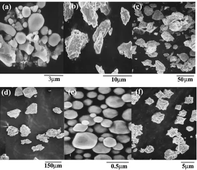
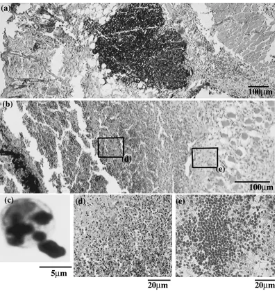
(2) Effects of Particle Size on Cell Function and Morphology in Titanium and Nickel. 3053. Fig. 1 SEM images of Ti 2 µm (a), 10 µm (b), 45 µm (c) and 150 µm (d) particles and Ni 0.4 µm (e) and 5 µm (f) particles.. 2.2.3 Superoxide anion 6 Superoxide anion (O− 2 ) production per 10 neutrophils was assayed by measuring the superoxide dismutase-inhibiting reduction of equine ferricytochrome C (550 nm). Specimens were incubated at 37◦ C for 5 min and the reaction was inhibited by adding PMA (139 mM).16–18) 2.2.4 Cytokines (TNF-α and IL-1β) Tumor necrosis factor-alpha (TNF-α) and interleukin-1 beta (IL-1β) production per 106 neutrophils in the supernatant was measured using ELISA kits (Endogen, Inc. USA). For the measurement of 2.2.1 ∼ 4, the values are expressed as means ± standard deviation (n = 6). Data were analyzed by Student’s t-test19) with the level of significance set at 5%. 2.3 SEM observation Neutrophils mixed with HBSS containing metallic particles were fixed in Karnovskys’ solution (pH 7.4), postfixed in 0.1-mol/L cacodylate buffer and 0.1 M-osmium acid for 1 h, dehydrated and coated with Pt–Pd. Morphological changes were observed by scanning electron microscopy (SEM: HITACHI S-4300, Tokyo, Japan). 2.4 Animal experiments Wistar rats aged between 11 and 12 weeks (weight 350–380 g) were anesthetized with diethyl ether (Wako Pure Chemical Industries, Osaka, Japan), then pentobarbital sodium (30 mg/kg; NEMBUTAL INJECTION, Dainabot, Osaka, Japan) was injected into the abdominal cavity. Ti or. Ni particles were inserted into the subcutaneous connective tissue in the abdominal region. The wounds were then sutured. After one week of implantation the rats were sacrificed and blocks of connective tissue were extracted. After fixation with 10% neutral buffered formalin, the tissue blocks were conventionally embedded in paraffin, then sectioned and stained with hematoxylin-eosin. The specimens were histopathologically observed using an optical microscope (ZEISS, Axioskop, Germany). 3. Results Figure 1 shows the SEM images of various sizes of Ti and Ni particles used for the experiment. The nominal size was 2 µm (a), 10 µm (b), 45 µm (c), 150 µm (d) for Ti particles and 0.4 µm (e), 5 µm (f) for Ni particles, respectively. Figure 2 shows a histological image of rat soft tissue inserted with pure titanium particles of 2 µm (a) and nickel particles of 0.4 µm (b) for one week. The agglomeration of small black dots in the center area of Fig. 2(a) are Ti particles. Numerous inflammatory cells were observed in the surroundings. The macrophages or neutrophils show the degenerative changes in morphology. The enlarged view in Fig. 2(c) shows that Ti particles were phagocytized into the cytoplasm by an inflammatory cell. Figure 2(b) shows that Ni particles in the lower left area caused strong inflammation and necrosis. Figure 2(d) shows necrosis surrounding the region inserted Ni particles and (e) shows the strong inflammatory response.
(3) 3054. K. Tamura, N. Takashi, R. Kumazawa, F. Watari and Y. Totsuka. Fig. 2 Tissue response to particles inserted in subcutaneous tissue of rats for one week (HE stain). (a) 2 µm Ti, (b) 0.4 µm Ni, (c) and (d), (e) enlargement of (a) and (b). The agglomeration of 2 µm Ti (a) particles is seen in the center and 0.4 µm Ni particles (b) in the lower left. A neutrophil might phagocytize the 2 µm Ti (c). Ni particles caused necrosis (d) and strong inflammation (e).. by infiltration of lymphocytes and histiocytes, approximately 0.6–0.7 mm apart from Ni particles. Figure 3 shows the SEM image of human neutrophils exposed to Ti and Ni particles in HBSS solution. Figure 3(a) shows the control neutrophil in HBSS. When a neutrophil was stimulated by 2 µm Ti particles, the surface changed to smoother (b) by the transformation of the cell membrane. Figure 3(c) show that a neutrophil may extends its pseudopod to phagocytize a Ti particle. The morphology of neutrophils exposed to Ni particles were often transformed (d) or destructed (e). Figure 3(f) is the typical SEM image for the case of 10 µm Ti particles or larger. 45 µm Ti particles are observed in the top of Fig. 3(f) and the form of neutrophils in the lower left changed very little. Figure 4 shows the survival rate of neutrophils in the Ti and Ni particle solution. HBSS solution was the control. Although significant difference from control was not observed in all Ti particle solutions, the smallest value was 2 µm and followed by 10 µm. The other Ti particles had no difference from control. Ni particles showed the lower survival rate signifi-. cantly differed from the HBSS. The survival rate was clearly lower in the 0.4 µm Ni particles than 5 µm. Figure 5 shows the value of LDH in each particle solution containing neutrophils. The difference between each Ti particle and the control was significant except 150 µm. Levels of LDH were significantly higher in the 2 µm than the other sizes. LDH showed tendency to increase as the Ti particle size became smaller. The Ni group showed higher values than Ti. They also had the tendency to increase as the particle size became smaller. Figure 6 shows the quantity of superoxide anion produced from neutrophils in the solutions of Ti and Ni particles. Ti group showed the significant increase from HBSS. The 2 µm particle showed the largest value. The other size is slightly higher than control. On the other hand, Ni had significantly much smaller values from HBSS and the smallest in the 0.4 µm size. Figure 7 shows the amount of TNF-α released from neutrophils in HBSS containing metallic particles. The TNF-α levels increased only in HBSS containing 2 µm Ti particles..
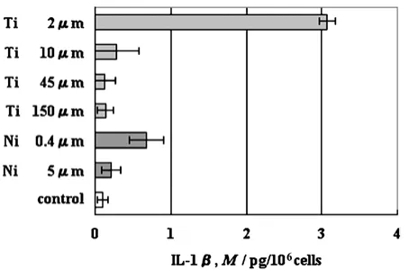
(4) Effects of Particle Size on Cell Function and Morphology in Titanium and Nickel. 3055. Fig. 3 SEM image of human neutrophils exposed to particles: a normal neutrophil in HBSS (a), neutrophil stimulated by Ti (b), a neutrophil phagocytizing 2 µm Ti particle (c), a neutrophil transformed (d) or broken down (e) with Ni particles, and neutrophils associated with 45 µm Ti particles (f).. Fig. 4 Survival rate of neutrophils in the solutions of Ti and Ni particles.. Fig. 5 LDH released from human neutrophils on the exposure to Ti and Ni particles.. No significant changes were associated with any other Ti and Ni particles. Figure 8 shows the amount of IL-1β released from neutrophils in HBSS containing metal particles. The IL-1β levels. Fig. 6 Superoxide anion production of human neutrophils exposed to Ti and Ni particles.. Fig. 7 Amount of TNF-α released from human neutrophils exposed to Ti and Ni particles.. significantly increased only in HBSS containing the 2 µm Ti particles. The other Ti and Ni particles released slightly but they had not much difference from the control..
(5) 3056. K. Tamura, N. Takashi, R. Kumazawa, F. Watari and Y. Totsuka. Fig. 8 Amount of IL-1β released from human neutrophils exposed to Ti and Ni particles.. 4. Discussion 4.1 Probe cells The most representative leukocytes with the function of phagocytosis are neutrophils and macrophages. In response to foreign objects, neutrophils react first, followed then by macrophages. Neutrophils account the largest proportion (about 54–65%) of human leucocytes. Their function is initially a non-specific reaction to foreign objects, whereas macrophages have a more complex function. Macrophages have a specific relationship with individual cytokines and respond to certain cytokines emitted from neutrophils after they have reacted with foreign objects. Our results showed that Ti fine particles, especially when they are smaller than cell size, cause inflammatory reaction in vivo and in vitro. To investigate the initial and simple reaction of cells to fine particles we used human neutrophils20) rather than macrophages for monitoring the effect of Ti and Ni particles in this study. 4.2 Cell disruption Cytotoxicity of Ti and Ni particles was examined by biochemical functional analysis as well as by microscopic morphology observation. LDH is an intracellular enzyme involved in the glycolytic pathway. The LDH value increases when the cell membrane is destroyed. Therefore LDH is the representative indication of cell disruption. From this point of view, the results of the cell survival rates (Fig. 4) and LDH production (Fig. 5) showed the very good accordance. LDH values were increased as the survival rate was lowered. Both results had qualitatively the very good correspondence each other in orders to particle size and particle group of Ti and Ni. 4.3 Cytofunctional reaction on exposition to particles Neutrophils produce several kinds of active oxygen such as hydroxyradical, hydrogen peroxide, and superoxide anion. The superoxide dismutase-inhibiting reduction of equine ferricytochrome C method, which was used in the present study, detect superoxide anion (O− 2 ). Superoxide anions are released from intracellular organs and from the cell membrane of neutrophils when the latter is stimulated. The amount of superoxide produced by neutrophils (Fig. 6) significantly increased, especially in 2 µm Ti and slightly in the other sizes of Ti particles. These results suggest that Ti particles stimulate neu-. trophils and its effect depends on the size of the particles. On the other hand, Ni produced much less superoxide (Fig. 6), which seems to contradict its cytotoxical effects. This could be explained partly due to the low survival rate of neutrophils (Fig. 4) caused by disruption in Ni particle solution, which might surpass the stimulatory effect of Ni. The biochemical analysis indicated that the TNF-α and IL1β release was significantly increased only in the solution of 2 µm Ti particles throughout the solutions in the different size of particles of Ti and Ni. One of the causes of TNF-α release from neutrophils occurs when the foreign matter is taken up by the cells. Our SEM observation (Fig. 3(c)) showed that only 2 µm particles in Ti group were phagocytized by neutrophils of about 5–10 µm in diameter. IL-1β is released when neutrophils are stimulated by foreign matter and it causes the inflammation reaction cascade. We may assume that the 2 µm Ti particles stimulate neutrophils and make them release superoxide and IL-1β. The 2 µm Ti particles are also phagocytized by neutrophils, which then produces superoxide anions again and TNF-α. The phagocytosis is difficult for the 10 µm, 45 µm and 150 µm Ti particles and the above effects were absent in the present results. The 0.4 µm and 5 µm Ni particles showed the different results from Ti, but they also had some tendency that small particles had the stronger influence. 4.4 Size dependence of cell toxicity We examined particles that were smaller (2 µm) and larger (10 µm, 45 µm, and 150 µm) than the neutrophils, to determine the relationship between cell and particle size on cytotoxicity. 2 µm Ti particles stimulated the neutrophil activity and increased the production of superoxide anions (Fig. 6), while Ti particles larger than 10 µm did very little. Only 2 µm Ti particles clearly released TNF-α (Fig. 7) and IL-1β (Fig. 8). All the results of cell survival rate (Fig. 4), LDH production (Fig. 5), superoxide production (Fig. 6) and TNF-α (Fig. 7), IL-1β (Fig. 8) release had the good accordance that they have the size dependence and the size effect becomes more remarkable as the size becomes smaller.11) This effect is especially pronounced when particles are smaller than the cell size, although there are some differences in the effects between Ti and Ni. Neutrophils may phagocytize Ti particles when the particles are smaller than the cell size of 5–10 µm. The increased quantity of TNF-α (Fig. 7) by phagocytosis caused neutrophil to further activation, resulting in inflammation. An increased superoxide content in vivo may affect the cell circumference and damage the DNA. These fine Ti particles may cause cytotoxicity, although the macroscopic size of Ti implant was quite biocompatible. 4.5 Cytotoxicity of Ti and Ni Titanium is an insoluble, chemically stable metal and the most frequently used as implant and plate in vivo. On the other hand, nickel is dissolved into the surrounding tissues where it works highly toxic, although Ni–Ti alloy is often used as implant and orthodontics wire. The SEM observations showed that the morphology of.
(6) Effects of Particle Size on Cell Function and Morphology in Titanium and Nickel. some neutrophils was partially deformed in the 2 µm Ti particle solution and it had very little change in the particles of larger sizes (Fig. 3). We had confirmed by ICP-AES (Inductively Coupled Plasma-Atomic Emission Spectrometry) that the dissolution from Ti particles was under the limit of detection (ICPS-8100, Shimazu, Tokyo, Japan). Therefore the biofunctional change detected in the present study was not caused by Ti ions but mostly by Ti particles. Figure 4 showed that cell survival rate of neutrophils mixed with Ni particles was significantly lower. Figure 5 showed that LDH values in Ni were significantly higher. These biochemical analyses indicated that a part of neutrophils were destroyed by exposure to Ni particle solution. Cell morphology shown in Fig. 3 confirmed that neutrophils were disrupted with Ni particles. To summarize the results described above, the smaller change in release of superoxide anion (Fig. 6), cell survival rate (Fig. 4), LDH value (Fig. 5) and the deformed morphology (Fig. 3) suggest that Ti particles do not destroy the cell membrane, but they rather stimulate human neutrophils. However the effect is pronounced if Ti particles size is smaller than neutrophils. Since the dissolution into Ti ions is neglected in the usual condition, the stimulation mainly occurs because of the size effect of Ti particles. On the other hand, the decreased cell survival rate (Fig. 4), the increased LDH activity (Fig. 5) and the disrupted morphology in the SEM observation (Fig. 3) suggest that Ni is cytotoxic to neutrophils. This toxicity is originated principally from Ni ions derived by dissolution and is affected secondarily by its size effect. 4.6 Relationship to in vivo findings We have observed the tissue reactions to various sizes of Ti particles inserted into the soft tissue of rats. When Ti particles smaller than cells were inserted (Fig. 2), numerous inflammatory cells appeared around the agglomerated particles. In vitro cytotoxicity findings discussed above were closely related to the in vivo results, where a long-term inflammatory reaction was stimulated by 2 µm Ti particles. Since the dissolution from particles was undetectable in vitro, the main effect about Ti in rats appears to be caused by particles rather than by ions. These results in vivo can be explained by the increased amount of superoxide, TNF-α and IL-1β release and subsequent cytotoxic stimulation in the soft tissue around the inserted titanium particles that partly resulted from phagocytosis. Ni showed a severe cytotoxicity. We examined after one day after Ni insertion (Fig. 2). This caused the strong inflammation and necrosis partially occurred. Our previous results of macro size Ni inserted in soft tissue clearly showed that necrosis and inflammation depends on the distance from Ni surface which is also the function of concentration of dissolved Ni ions as it was analyzed by X-ray scanning analytical microscope (XSAM).14) The strong toxicity of Ni is mainly originated from it’s dissolved ions. 5. Conclusion The present study clearly showed the cytotoxicity due to fine particles and its size dependence using human neutrophils. 3057. in vitro and soft tissue in vivo. (1) All the results of cell survival rate, LDH production, superoxide anion production, TNF-α and IL-1β release, and SEM observation are in accordance each other in that they have particle size dependence and its effect becomes remarkable as the size becomes smaller. (2) Ti has a cell stimulatory effect, while Ni high dissolution rate has much more toxicity to lead to cell disruption and tissue necrosis. (3) Different release of TNF-α observed only in 2 µm Ti particles is closely related to the phagocytosis by neutrophils. Acknowledgements The authors thank Dr. Takafumi Domon, Dr. Tadashi Iizuka, Dr. Yoshinobu Nodasaka and Dr. Takuo Tsutiya, who belong to Hokkaido University, Graduate School of Dental Medicine, for the valuable advices and preparation of SEM observation. REFERENCES 1) F. Watari, A. Yokoyama, F. Saso, M. Uo and T. Kawasaki: Composites 28B (1997) 5–11. 2) H. Matsuno, A. Yokoyama, F. Watari, M. Uo and T. Kawasaki: Biomaterials 22(11) (2001) 1253–1262. 3) J. Ryhane, M. Kallioinen, J. Tuukkanen, E. Niemela, P. Sandvik and W. Serlo: J. Biomed. Mater. Res. 41 (1998) 481–8M. 4) H. G. Penman and P. A. Ring: J. Bone Jt Ssurg. 66-B (1984) 632–4. 5) K. Takamura, K. Hayashi, N. Ishinishi, T. Yamada and Y. Sugioka: J. Biomed. Mater. Res. 28 (1994) 583–9. 6) F. Takeshita, H. Takata, Y. Ayukawa and T. Suetsugu: Biomaterials 18 (1997) 21–5. 7) M. Uo, F. Watari, A. Yokoyama, H. Matsumoto and T. Kawasaki: Biomaterials 20 (1999) 47–55. 8) Y.-K. Kim, H.-H. Yeo and S.-C. Lim: J. Oral Maxillofac. Surg. 55 (1997) 322–326. 9) A. Rosenberg, K. W. Grätz and H. F. Sailer: J. Oral Maxillofac. Surg. 22 (1993) 185–188. 10) Y. Tanimura, R. Kumazawa, F. Watari, N. Inoue and Y. Totsuka: J. of Oral and Maxillofacial Surgery 46(11) (2000) 750. 11) R. Kumazawa, F. Watari and Y. Totsuka: J. of the Japanese Society for Dental Materials and Devices 6 (2001) 20. 12) M. Uo, F. Watari, A. Yokoyama, H. Matuno and T. Kawasaki: Biomaterials 22 (2001) 677–685. 13) M. Uo, F. Watari, A. Yokoyama, H. Matuno and T. Kawasaki: Biomaterials 20 (1999) 747–755. 14) H. Matuno, M. Uo, F. Watari, A. Yokoyama and T. Kawasaki: Biomaterials 22 (2001) 1784–1794. 15) K. Fujie: Japanese J. Conservative Dentistry 37(4) (1994) 1279–1293. 16) W. Sakamoto, K. Fujie, H. Honda, T. Ogiwara and M. Mino: International J. Vit Nurtr. 60-3 (1991) 338–342. 17) B. M. Bahior, R. S. Kipnes and J. T. Curnulte: J. Clin Invest. 52 (1973) 741–744. 18) K. Yagi and M. Nakano: Active Oxygen species, (Ishiyaku Publishers, Japan, 1994) pp. 139–145 19) R. K. Rieglman and R. P. Hirsch: Studying a study and Testing a testHow to read the Health Science Literature-Third edition, (Medical Science International, USA, 1996) pp. 243–248 20) H. Baumann and J. Glauldie: Immunol Today 15(1) (1994) 74–80..
(7)
Figure

Related documents