research communications
Acta Cryst.(2016). E72, 675–682 http://dx.doi.org/10.1107/S2056989016005958
675
Received 31 March 2016 Accepted 10 April 2016
Edited by W. T. A. Harrison, University of Aberdeen, Scotland
Keywords:crystal structure; benzamide; hydrogen bonding.
CCDC references:1473261; 1473260; 1473259
Supporting information:this article has supporting information at journals.iucr.org/e
Crystal structures of three
3,4,5-trimethoxy-benzamide-based derivatives
Ligia R. Gomes,a,bJohn Nicolson Low,c* Catarina Oliveira,dFernando Cagidedand
Fernanda Borgesd
aREQUIMTE, Departamento de Quı´mica e Bioquı´mica, Faculdade de Cieˆncias da Universidade do Porto, Rua do Campo
Alegre, 687, P-4169-007, Porto, Portugal,bFP-ENAS-Faculdade de Cieˆncias de Sau´de, Escola Superior de Sau´de da UFP, Universidade Fernando Pessoa, Rua Carlos da Maia, 296, P-4200-150 Porto, Portugal,cDepartment of Chemistry, University of Aberdeen, Meston Walk, Old Aberdeen AB24 3UE, Scotland, anddCIQ/Departamento de Qumica e Bioqumica, Faculdade de Cieˆncias, Universidade do Porto, 4169-007 Porto, Portugal. *Correspondence e-mail: [email protected]
The crystal structures of three benzamide derivatives,viz. N -(6-hydroxyhexyl)-3,4,5-trimethoxybenzamide, C16H25NO5, (1), N
-(6-anilinohexyl)-3,4,5-trimeth-oxybenzamide, C22H30N2O4, (2), and N
-(6,6-diethoxyhexyl)-3,4,5-trimethoxy-benzamide, C20H33NO6, (3), are described. These compounds differ only in the
substituent at the end of the hexyl chain and the nature of these substituents determines the differences in hydrogen bonding between the molecules. In each molecule, the m-methoxy substituents are virtually coplanar with the benzyl ring, while thep-methoxy substituent is almost perpendicular. The carbonyl O atom of the amide rotamer is transrelated with the amidic H atom. In each structure, the benzamide N—H donor group and O acceptor atoms link the molecules intoC(4) chains. In1, a terminal –OH group links the molecules into a
C(3) chain and the combined effect of theC(4) andC(3) chains is a ribbon made up of screw relatedR22(17) rings in which the O—H chain lies in the centre
of the ribbon and the trimethoxybenzyl groups forms the edges. In 2, the combination of the benzamideC(4) chain and the hydrogen bond formed by the terminal N—H group to an O atom of the 4-methoxy group link the molecules into a chain ofR2
2
(17) rings. In3, the molecules are linked only byC(4) chains.
1. Chemical context
Phenolic acids are widely distributed in the plant kingdom and exist in significant quantities in the human diet (e.g. in fruits and vegetables). Like other phenolic compounds they are recognized for their health benefits, which are linked to their biological properties, particularly anti-oxidant, anti-inflam-matory and anticancer properties (Benfeito et al., 2013, Roleiraet al., 2015, Garridoet al., 2013, Teixeiraet al., 2013). Within this framework, our project has been focused on the synthesis of new molecules based on the benzoic acid scaffold. Accordingly, herein we describe the syntheses and structures of three new benzamide derivatives,viz. N -(6-hydroxyhexyl)-3,4,5-trimethoxybenzamide (1) N -(6-anilinohexyl)-3,4,5-tri-methoxybenzamide (2) and N -(6,6-diethoxyhexyl)-3,4,5-tri-methoxybenzamide (3).
2. Structural commentary
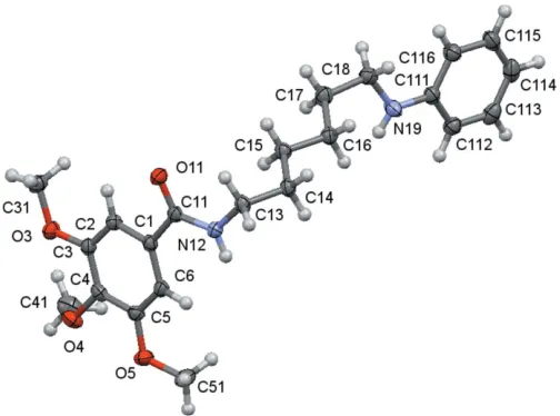
The molecular structures of compounds1,2and3are shown in Figs. 1–3. The molecules consist of a trimethoxybenzamide ‘head’ that is linked to a six-carbon-atom alkyl chain ‘tail’ that ends with different functional groups: a hydroxyl group for1, a
phenylamino group for2and a diethoxy group for3. In spite of having the same ‘head’ and ‘tail’, the differences observed for their molecular conformations are not only due to the different ‘end tail’ functional groups. Thus, the analysis of the molecular conformations will be performed on a comparative basis encompassing the following: (i) the relative positions of the methoxy substituents on the aromatic ring; (ii) the conformation of the amide unit and (iii) the conformation of the alkyl chain. The specifics of the substituents at the end of the alkyl chain determine the differences in the supra-molecular structures, as discussed in the next section.
Them-methoxy substituents are virtually co-planar with the benzene ring and aretransrelated with respect to thep-carbon atom of the ring [the maximum deviation of the methoxy carbon atom to the best plane of the phenyl ring is 0.238 (1) A˚ in2], while thep-methoxy group is nearly perpendicular [the minimum deviation of the methoxy carbon atom to the best plane of the benzene ring being 0.923 (2) A˚ , also in2]. These relative positions agree with previous predictions of theoret-ical calculations for the stabilization energies for methoxy-group conformations attached to aromatic rings (Tsuzuki et al., 2002), which suggested that, while co-planarity is the most stable conformation when there is only one methoxy substit-uent on the aromatic ring, the perpendicular conformation
may appear as an alternative one when twovicinalmethoxy groups are present. According to these authors, this spatial arrangement is stabilized by a short C—H O contact between the neighbouring groups. As can be seen in Tables 4, 5 and 6, the shortest distances between a methyl H atom and a
vicinalmethoxy O atom are 2.44, 2.33 and 2.37 A˚ for1,2and
3, respectively, which do suggest the possibility of a very weak interaction.
In the amide rotamer, the carbonyl oxygen atom is in atrans
position with respect to the hydrogen atom of the amidic nitrogen atom for all compounds, and so, the trimethoxy phenyl group is also trans related to the alkyl chain. The rotation of the trimethoxy phenyl substituent with respect to the amide rotamer around the C11—C1 bond may be eval-uated by the N12—C11—C1—C6 torsion angle, whose values are given in Tables 1–3. The mean planes between the C1 benzene ring and the mean plane of the three atoms O11, C11 and N12 are 35.1 (3), 12.00 (16) and 20.19 (14), respectively,
for1,2and3, showing that the substituent in2is significantly
676
Gomeset al. C16H25NO5, C22H30N2O4and C20H33NO6 Acta Cryst.(2016). E72, 675–682
[image:2.610.314.566.70.257.2]research communications
Figure 1
A view of the asymmetric unit of (1) with the atom-numbering scheme. Displacement ellipsoids are drawn at the 70% probability level.
Figure 2
[image:2.610.47.296.219.335.2]A view of the asymmetric unit of (2) with the atom-numbering scheme. Displacement ellipsoids are drawn at the 70% probability level.
Figure 3
[image:2.610.311.563.553.722.2]less distorted than in the others. In 1and in2, the sense of rotation is anticlockwise.
The freedom of rotation around the N—C(alkyl) bond together with the regular tetrahedral geometry of the
sp3-hybridized carbon atoms allows the molecules to acquire
very different conformational profiles for the alkyl chain as is observed in the C11—N12—C13—C14 torsion angles [129.1 (3) for1,112.80 (13) for2and 114.65 (12)for
3], as well as the direction of the alkyl chain with respect to the N12—C13 bond, which primarily affects the relative position of the alkyl ‘tail’ with respect to the benzamide moiety. Considering the disposition of the amide rotamer: in1and in3
the alkyl chain is directed backwards from the amide plane and in 2 forward from that plane. This affects the general shape of the molecules, as can be better visualized in Figs. 7–9. So, in spite of the consistent zigzag shape of the remaining alkyl chain those molecules have entirely different spatial arrangements.
3. Supramolecular features
3.1. Hydrogen Bonding and short contacts
Tables 4, 5 and 6 show the hydrogen-bonding details for1,2
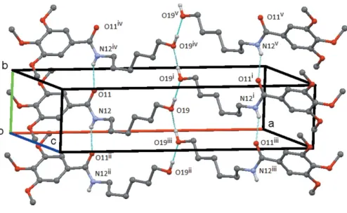
and3, respectively. In each compound, the amide group forms the common C(4) chain motif by an N—H O hydrogen bond. In1, the N12—-H12 O11 chain runs parallel to theb
axis and adjacent molecules are at unit translation along this axis. The O19—-H19 O19 hydrogen bond links the
mol-ecules into aC(3) chain formed by the action of the twofold screw axis at (1
2,y, 3
4). These two chains link the molecules to
form a ribbon made up of screw-relatedR2
2(17) rings, which
runs parallel to thebaxis with the O—H chain running up the centre of the ribbon and the trimethoxybenzyl groups forming the edges (Fig. 4). In 2, both the N12—H12 O11
research communications
Acta Cryst.(2016). E72, 675–682 Gomeset al. C
[image:3.610.42.295.73.394.2]16H25NO5, C22H30N2O4and C20H33NO6
677
Table 1
Selected torsion angles () for1.
C31—O3—C3—C4 176.7 (2) C6—C1—C11—N12 35.6 (3) C31—O3—C3—C2 3.5 (4) C11—N12—C13—C14 129.1 (3) C41—O4—C4—C5 108.9 (3) N12—C13—C14—C15 177.5 (2) C41—O4—C4—C3 74.4 (3) C13—C14—C15—C16 65.7 (3) C51—O5—C5—C4 175.7 (2) C14—C15—C16—C17 173.9 (2) C51—O5—C5—C6 3.6 (4) C15—C16—C17—C18 174.4 (2) C13—N12—C11—C1 171.3 (2) C16—C17—C18—O19 177.9 (2) C2—C1—C11—N12 149.3 (2)
Table 2
Selected torsion angles () for2.
C31—O3—C3—C2 0.16 (17) C2—C1—C11—N12 167.30 (11) C31—O3—C3—C4 178.57 (11) C11—N12—C13—C14 112.80 (13) C41—O4—C4—C3 67.59 (16) N12—C13—C14—C15 66.85 (14) C41—O4—C4—C5 118.62 (13) C13—C14—C15—C16 179.75 (11) C51—O5—C5—C6 11.14 (18) C14—C15—C16—C17 175.06 (11) C51—O5—C5—C4 170.38 (11) C15—C16—C17—C18 175.02 (11) C13—N12—C11—C1 179.22 (10) C111—N19—C18—C17 172.76 (11) C6—C1—C11—N12 13.05 (17) C16—C17—C18—N19 67.90 (15)
Table 3
Selected torsion angles () for
3.
C31—O3—C3—C2 9.59 (16) C2—C1—C11—N12 158.58 (10) C31—O3—C3—C4 171.49 (10) C6—C1—C11—N12 19.07 (15) C41—O4—C4—C5 61.51 (15) C11—N12—C13—C14 114.65 (12) C41—O4—C4—C3 124.05 (12) N12—C13—C14—C15 175.72 (9) C51—O5—C5—C6 9.66 (17) C13—C14—C15—C16 67.27 (13) C51—O5—C5—C4 171.35 (11) C14—C15—C16—C17 175.71 (10) C13—N12—C11—C1 170.25 (10) C15—C16—C17—C18 177.76 (10)
Table 4
Hydrogen-bond geometry (A˚ ,) for1.
D—H A D—H H A D A D—H A
O19—H19 O19i 0.92 (4) 1.86 (4) 2.7799 (14) 176 (4)
N12—H12 O11ii 0.77 (3) 2.15 (3) 2.859 (3) 153 (3) C18—H18B O11iii 0.99 2.64 3.614 (3) 168
C41—H41B O3 0.98 2.44 3.010 (3) 117
Symmetry codes: (i)xþ1;yþ1 2;zþ
3
2; (ii)x;y1;z; (iii)x;yþ 3 2;zþ
1 2.
Table 5
Hydrogen-bond geometry (A˚ ,) for2.
Cgis the centroid of the C111–C116 ring.
D—H A D—H H A D A D—H A
N12—H12 O11i 0.867 (17) 2.052 (17) 2.9051 (14) 167.9 (15)
N19—H19 O4i 0.855 (17) 2.106 (17) 2.9436 (15) 166.3 (15) C6—H6 O11i 0.95 2.33 3.2356 (15) 159
C41—H41C O3 0.98 2.33 2.9287 (18) 119 C112—H112 O4i 0.95 2.65 3.3845 (16) 134
C13—H13A Cgii 0.99 2.64 3.5272 (15) 148
C31—H31C Cgiii 0.98 2.62 3.5205 (16) 152
Symmetry codes: (i) x;yþ1 2;z
1
2; (ii) xþ1;yþ1;zþ1; (iii)
x;y1 2;zþ
3 2.
Table 6
Hydrogen-bond geometry (A˚ ,) for3.
D—H A D—H H A D A D—H A
N12—H12 O11i 0.856 (16) 2.169 (16) 2.9890 (13) 160.2 (14) C6—H6 O11i 0.95 2.34 3.2549 (14) 162
C15—H15B O18ii 0.99 2.49 3.4239 (14) 157
Symmetry codes: (i)x;yþ3 2;z
1
2; (ii)x;yþ 1 2;z
[image:3.610.317.564.537.689.2]1 2.
Figure 4
Compound 1: view of the ribbon structure formed by the N12— H12 O11 and O19—H19 O19 hydrogen bonds. Hydrogen atoms not involved in the hydrogen bonding are omitted. Symmetry codes: (i)x+ 1,
y+1 2,z+
3
2; (ii)x,y1,z+ 1; (iii)x+ 1,y 1 2,z+
3 2; (iv) x+ 1,y+ 1,z+ 1; (v)x+ 1,y+3
and N19—H19 O4 hydrogen bonds link the molecules into a chain of R2
2(17) rings, which are bridged by the C11—N12
bond. This chain runs parallel to thecaxis and is formed by the action of the c-glide plane at 1/4 along thebaxis (Fig. 5). In
3, the N12—H12 O11 hydrogen bond links the molecules into aC(4) chain, which runs parallel to thecaxis and which is formed by the action of thec-glide plane at 3/4 along theb
axis, Fig. 6. Possible weak C—H O interactions are detailed in the relevant Tables 4–6.
3.2. Hirshfeld Surfaces
Hirshfeld surfaces were generated using Crystal Explorer 3.1 (Wolff et al., 2012) mapped over dnorm for the title
compounds. The contact distancesdianddefrom the Hirshfeld
surface to the nearest atom inside and outside, respectively, were used to analyse the intermolecular interactions through the mapping ofdnormand the plot ofdiversusdeprovides
two-dimensional fingerprint plots (Rohlet al., 2008) that are used to summarize those contacts. Figs. 7–9 are views of the Hirshfeld surfaces mapped overdnorm for1, 2and3
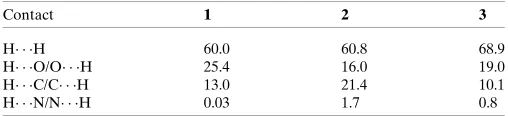
respec-tively. Since the molecules have a six-atom alkyl chain, most of the contacts are H H contacts. Leaving these aside, the remaining surface highlights the red areas that indicate contact points for the atoms participating in the (O/N/C)— H O intermolecular interactions. There are also significant contributions of C—H C contacts, as can be visualized in the figures for each compound. The percentages of (O/N/C)— H O and C—H C contacts are listed in Table 7.
In all three compounds, red spots near the amide indicate the N(amide)—H O hydrogen bonds that connect the amide groups in the classic fashion, making a C(4) chain for all
678
Gomeset al. C16H25NO5, C22H30N2O4and C20H33NO6 Acta Cryst.(2016). E72, 675–682
research communications
Figure 5
Compound2: the chain of rings formed by the interaction of the N12— H12 O11 and N19—H19 O4 hydrogen bonds. This chain extends along thecaxis and is generated by thec-glideplane aty=1
4. Hydrogen atoms not involved in the hydrogen bonding are omitted. Symmetry codes: (i)x,y1
2,z 1
2; (ii)x,y+ 1 2,z+
[image:4.610.47.295.71.271.2] [image:4.610.313.567.93.151.2]1 2.
Figure 6
Compound 2: the simple C(4) chain formed by the N12—H12 O11 hydrogen bond. This chain extends along thecaxis and is generated by thecglideplane aty=3
4. Hydrogen atoms not involved in the hydrogen bonding are omitted. Symmetry codes: (i)x,y3
2,z 1
2; (ii)x,y 1 2, z+1
2.
Table 7
The percentages of (O/N/C)–H O and C—H C contacts.
Contact 1 2 3
H H 60.0 60.8 68.9
[image:4.610.300.560.402.730.2] [image:4.610.46.546.409.722.2]H O/O H 25.4 16.0 19.0 H C/C H 13.0 21.4 10.1 H N/N H 0.03 1.7 0.8
Figure 7
[image:4.610.46.296.526.686.2]compounds. In2and3, there are two pairs of red spots at the amide environment indicating that, in these structures, the carbonyl oxygen atom acts as the receptor for another H contact (the C6—H6 O11 contact).
The classical O(hydroxy)–H O hydrogen bond is located at the chain ‘tail’ in1and is identified by two red spots indi-cating that the oxygen atom O19 acts as donor and acceptor making the C(3) chain. The red spots in structure 2 show another two hydrogen bonds: one of these involves the amine nitrogen atom of the end ‘tail’ phenylamine residue and the other also indicates the involvement of thep-methoxy group located at the trimethoxybenzamide ‘head’. This behaviour contrasts with that observed for1and3, in which the methoxy groups are not involved in classical hydrogen bonding.
The full fingerprint (FP) plots showing various crystal packing interactions are given in Figs. 10–12; the contributions from various contacts, listed in Table 7, were selected by the partial analysis of these plots. The FP plots show, for all compounds, a pair of long sharp spikes characteristic of a
research communications
Acta Cryst.(2016). E72, 675–682 Gomeset al. C
[image:5.610.312.566.69.316.2]16H25NO5, C22H30N2O4and C20H33NO6
679
Figure 8
View of the Hirshfeld surface mapped overdnormfor2.
Figure 9
[image:5.610.47.296.75.345.2]View of the Hirshfeld surface mapped overdnormfor3.
Figure 10
The full fingerprint (FP) plot showing various crystal packing interactions for1. Dark blue corresponds to the low frequency of occurrence of adi/de pair, while light blue indicates a higher frequency for the occurrence.
Figure 11
[image:5.610.315.566.458.707.2] [image:5.610.47.300.558.728.2]strong hydrogen bond, in an area near 1.7–1.8 A˚ . The symmetry of the upper left/down right spikes is an indication for the balance between the donor and acceptor character of the atoms involved in the hydrogen bonding, as seen before. They correspond to the N—H O and O—H O contacts. Thede/dipoints corresponding to H H interactions appear
around the hydrogen atom van der Waals radius of 1.20 A˚ . The wings in the graphical representation of 2 indicate that C—H interactions are more relevant in this crystal structure, highlighting the contribution of the C—H
interaction (Table 5) involving the phenylamide residue of the ‘tail’. Structure 2 also displays the biggest percentage of H C/C H contacts: besides the C—H contacts with the aromatic ring that define the supramolecular structure for all compounds, in2the benzene ring of the phenylamine forms an extra interaction of this kind
4. Database survey
A search made in the February 2016 version of the Cambridge Structural Database, (Groom et al., 2016), revealed the exis-tence of 37 structures (containing 48 unique molecules) featuring the 3,4,5-trisubstituted benzamide scaffold.
ortho-C atom C2 was selected such that the amino N atom N12 was trans to it and in the following survey it is
trans-related torsion angles which are discussed. The analysis of the torsion angles for theo-C atoms of the benzyl ring and the N atom of the benzamide group showed two distinct populations about 180in the angular ranges180 to145with a median
value of152.5and 136–171with a median value of 149.2.
The value of 179.3 for HESLEX,N,N -(heptane-2,6-diyl)-N0-(3,4,5-methoxybenzoyl)thiourea (Dillen et al., 2006) is
unusual: if this is excluded, then the lower limit for the negative range is172. The methyl groups attached to atoms
C3 and C5 are inclined to the benzyl ring in the range20 to 24with a median values close to 0. This excludes a molecule
with a C5 methoxy torsion angle of 85.9: PIDTEC, 4- hydroxy-3,5-diethoxybenzaldehyde-3,4,5-trimethoxybenzoyl-hydrazone monohydrate (Sunet al., 2007). The methyl groups attached to atoms C4 are inclined to the benzyl ring in the ranges63 to122with a median values close to90. Of
these 48 molecules, 16 participate in N—H OC(4) chains similar to those in the present compounds. In these structures, the torsion angles for thetrans o-C atoms of the benzyl ring and the N atom of the benzamide group showed that, as above, the torsion angles lie in two populations: one in the range153 to145and the other in the very similar positive
range 142 to 165with median values of147.6and 148.1,
respectively. The value for this torsion angle for1,149.3 (3)
lies within the negative range, those for2,167.27 (12)and 3, 158.58 (10) lie outside this range.The results of the database searches are included in the supporting information.
5. Synthesis and crystallization
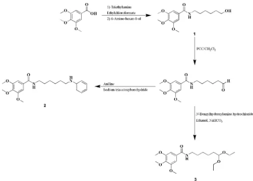
The title benzoic derivatives were obtained in moderate-to-high yieldsviathe synthetic strategy described in the Scheme below. Compound 1 was obtained from 3,4,5-trimethoxy-benzoic acid by an amidation reaction using ethylchloro-formate as coupling agent. After oxidation of compound 1
alcohol function to an aldehyde, compounds (2) and (3) could be obtained. Compound 2 was synthesized by a reductive amination reaction using sodium triacetoxyborohydride as reducing agent. Compound 3 was synthesized using an ethanolic solution ofN-benzylhydroxylamine hydrochloride.
1: N-(6-hydroxyhexyl)-3,4,5-trimethoxybenzamide (1). Overall yield 82%; m.p. 393–399 K; crystallization solvent: ethyl acetate, to yield colourless needles.
680
Gomeset al. C16H25NO5, C22H30N2O4and C20H33NO6 Acta Cryst.(2016). E72, 675–682
[image:6.610.46.296.69.308.2] [image:6.610.313.564.520.700.2]research communications
Figure 12
2: N-(6-anilinohexyl)-3,4,5-trimethoxybenzamide (2). Overall yield 51%; m.p. 376–388 K; crystallization solvent: ethyl acetate to yield colourless laths
3: N-(6,6-diethoxyhexyl)-3,4,5-trimethoxybenzamide (3). Overall yield 50%; m.p. 364–374 K; crystallization solvents: chloroform/n-hexane to yield colourless needles.
6. Refinement
Crystal data, data collection and structure refinement details are summarized in Table 8. The N—H and O—H hydrogen atoms were located in difference Fourier maps and freely refined. The C-bound H atoms were included in calculated positions and treated as riding: C—H(aromatic) = 0.95 A˚ and C—H2(methylene) = 0.99 A˚ with Uiso = 1.2Ueq(C), C—
H(methyl) = 0.98 A˚ withUiso= 1.5Ueq(C).
Acknowledgements
The authors thank the staff at the National Crystallographic Service, University of Southampton, for the data collection, help and advice (Coles & Gale, 2012), and the Foundation for Science and Technology (FCT) of Portugal (QUI/UI0081/ 2015) for financial support. Grants to CO (SFRH/BD/88773/ 2012) and FC (SFRH/BPD/72923/2010) are supported by FCT, POPH and QREN.
References
Agilent (2014). CrysAlis PRO. Agilent Technologies UK Ltd, Yarnton, England.
Benfeito, S., Oliveira, C., Soares, P., Fernandes, C., Silva, T., Teixeira, J. & Borges, F. (2013).Mitochondrion,13, 427–435.
Coles, S. J. & Gale, P. A. (2012).Chem. Sci.3, 683–689.
Dillen, J., Woldu, M. G. & Koch, K. R. (2006).Acta Cryst.E62, o5225– o5227.
Dolomanov, O. V., Bourhis, L. J., Gildea, R. J., Howard, J. A. K. & Puschmann, H. (2009).J. Appl. Cryst.42, 339–341.
Garrido, J. & Borges, F. (2013).Food. Res. Int.54, 1844–1858. Groom, C. R., Bruno, I. J., Lightfoot, M. P. & Ward, S. C. (2016).Acta
Cryst. B72, 171–179.
Hu¨bschle, C. B., Sheldrick, G. M. & Dittrich, B. (2011).J. Appl. Cryst.
44, 1281–1284.
Macrae, C. F., Edgington, P. R., McCabe, P., Pidcock, E., Shields, G. P., Taylor, R., Towler, M. & van de Streek, J. (2006).J. Appl. Cryst.39, 453–457.
McArdle, P., Gilligan, K., Cunningham, D., Dark, R. & Mahon, M. (2004).CrystEngComm,6, 303–309.
Oszla´nyi, G. & Su¨to, A. (2004).Acta Cryst.A60, 134–141.
Rigaku (2012).CrystalClear-SM Expert. Rigaku Corporation, Tokyo, Japan.
Rohl, A. L., Moret, M., Kaminsky, W., Claborn, K., McKinnon, J. J. & Kahr, B. (2008).Cryst. Growth Des.8, 4517–4525.
Roleira, F. M. F., Tavares-da-Silva, E. J., Varela, C. L., Costa, S. C., Silva, T., Garrido, J. & Borges, F. (2015).Food Chem.183, 235–258. Sheldrick, G. M. (2015a).Acta Cryst.A71, 3–8.
Sheldrick, G. M. (2015b).Acta Cryst.C71, 3–8.
research communications
Acta Cryst.(2016). E72, 675–682 Gomeset al. C
[image:7.610.48.564.95.402.2]16H25NO5, C22H30N2O4and C20H33NO6
681
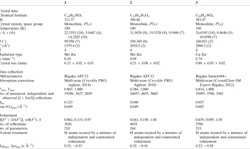
Table 8
Experimental details.
1 2 3
Crystal data
Chemical formula C16H25NO5 C22H30N2O4 C20H33NO6
Mr 311.37 386.48 383.47
Crystal system, space group Monoclinic,P21/c Monoclinic,P21/c Monoclinic,P21/c
Temperature (K) 100 100 100
a,b,c(A˚ ) 22.3351 (18), 5.0467 (4), 14.2265 (10)
11.5626 (8), 19.5328 (9), 9.5488 (7) 24.6345 (18), 8.4646 (5), 10.0598 (7) () 99.956 (7) 109.369 (8) 100.851 (2)
V(A˚3) 1579.4 (2) 2034.5 (2) 2060.2 (2)
Z 4 4 4
Radiation type MoK MoK CuK
(mm1) 0.10 0.09 0.74
Crystal size (mm) 0.150.020.01 0.250.080.02 0.800.050.02
Data collection
Diffractometer Rigaku AFC12 Rigaku AFC12 Rigaku Saturn944+ Absorption correction Multi-scan (CrysAlis PRO;
Agilent, 2014)
Multi-scan (CrysAlis PRO; Agilent, 2014)
Multi-scan (CrystalClear-SM Expert; Rigaku, 2012)
Tmin,Tmax 0.803, 1.000 0.384, 1.000 0.814, 1.000
No. of measured, independent and observed [I> 2(I)] reflections
19396, 3627, 2039 26057, 4655, 3869 18993, 3706, 3362
Rint 0.123 0.040 0.037
(sin/ )max(A˚
1) 0.649 0.649 0.602
Refinement
R[F2> 2(F2)],wR(F2),S 0.062, 0.133, 0.97 0.041, 0.105, 1.04 0.035, 0.095, 1.05
No. of reflections 3626 4652 3706
No. of parameters 210 264 253
H-atom treatment H atoms treated by a mixture of independent and constrained refinement
H atoms treated by a mixture of independent and constrained refinement
H atoms treated by a mixture of independent and constrained refinement
max,min(e A˚
3) 0.25,0.33 0.32,0.18 0.23,0.28
Spek, A. L. (2009).Acta Cryst.D65, 148–155.
Sun, Y.-F., Sun, X.-Z., Li, J.-K. & Zheng, Z.-B. (2007).Acta Cryst.E63, o2180–o2181.
Teixeira, J., Silva, T., Andrade, P. B. & Borges, F. (2013).Curr. Med.
Chem.20, 2939–2952.
Tsuzuki, S., Houjou, H., Nagawa, & Hiratani, K. (2002).J. Chem. Soc.
Perkin Trans. 2, pp. 1271–1273.
Wolff, S. K., Grimwood, D. J., McKinnon, J. J., Turner, M. J., Jayatilaka, D. & Spackman, M. A. (2012).Crystal Explorer. The University of Western Australia.
682
Gomeset al. C16H25NO5, C22H30N2O4and C20H33NO6 Acta Cryst.(2016). E72, 675–682
supporting information
sup-1 Acta Cryst. (2016). E72, 675-682
supporting information
Acta Cryst. (2016). E72, 675-682 [doi:10.1107/S2056989016005958]
Crystal structures of three 3,4,5-trimethoxybenzamide-based derivatives
Ligia R. Gomes, John Nicolson Low, Catarina Oliveira, Fernando Cagide and Fernanda Borges
Computing details
For all compounds, data collection: CrystalClear-SM Expert (Rigaku, 2012). Cell refinement: CrysAlis PRO (Agilent,
2014) for (1), (2); CrystalClear-SM Expert (Rigaku, 2012) for (3). Data reduction: CrysAlis PRO (Agilent, 2014) for (1),
(2); CrystalClear-SM Expert (Rigaku, 2012) for (3). Program(s) used to solve structure: SHELXT (Sheldrick, 2015a),
PLATON (Spek, 2009), Flipper 25 (Oszlányi & Sütő, 2004) and OLEX2 (Dolomanov et al., 2009). for (1); SHELXT
(Sheldrick, 2015a), PLATON (Spek, 2009), Flipper 25 (Oszlányi & Sütő, 2004) and OLEX2 (Dolomanov et al., 2009) for
(2); OSCAIL (McArdle et al., 2004) and SHELXT (Sheldrick, 2015a) for (3). For all compounds, program(s) used to
refine structure: OSCAIL (McArdle et al., 2004), ShelXle (Hübschle et al., 2011) and SHELXL2014 (Sheldrick, 2015b);
molecular graphics: Mercury (Macrae et al., 2006); software used to prepare material for publication: OSCAIL (McArdle
et al., 2004), SHELXL2014 (Sheldrick, 2015b) and PLATON (Spek, 2009).
(1) N-(6-Hydroxyhexyl)-3,4,5-trimethoxybenzamide
Crystal data
C16H25NO5
Mr = 311.37
Monoclinic, P21/c
a = 22.3351 (18) Å b = 5.0467 (4) Å c = 14.2265 (10) Å β = 99.956 (7)° V = 1579.4 (2) Å3
Z = 4
F(000) = 672 Dx = 1.309 Mg m−3
Mo Kα radiation, λ = 0.71073 Å Cell parameters from 4347 reflections θ = 2.8–27.5°
µ = 0.10 mm−1
T = 100 K Needle, colourless 0.15 × 0.02 × 0.01 mm
Data collection
Rigaku AFC12 diffractometer
Radiation source: Rotating Anode Detector resolution: 28.5714 pixels mm-1
profile data from ω–scans
Absorption correction: multi-scan
(CrysAlis PRO; Agilent, 2014)
Tmin = 0.803, Tmax = 1.000
19396 measured reflections 3627 independent reflections 2039 reflections with I > 2σ(I) Rint = 0.123
θmax = 27.5°, θmin = 1.9°
h = −29→28 k = −6→6 l = −18→18
Refinement
Refinement on F2
Least-squares matrix: full R[F2 > 2σ(F2)] = 0.062
wR(F2) = 0.133
S = 0.97
3626 reflections 210 parameters 0 restraints
supporting information
sup-2 Acta Cryst. (2016). E72, 675-682
H atoms treated by a mixture of independent and constrained refinement
w = 1/[σ2(F
o2) + (0.0434P)2 + 1.2336P]
where P = (Fo2 + 2Fc2)/3
(Δ/σ)max = 0.003
Δρmax = 0.25 e Å−3
Δρmin = −0.33 e Å−3
Special details
Geometry. All esds (except the esd in the dihedral angle between two l.s. planes) are estimated using the full covariance matrix. The cell esds are taken into account individually in the estimation of esds in distances, angles and torsion angles; correlations between esds in cell parameters are only used when they are defined by crystal symmetry. An approximate (isotropic) treatment of cell esds is used for estimating esds involving l.s. planes.
Fractional atomic coordinates and isotropic or equivalent isotropic displacement parameters (Å2)
x y z Uiso*/Ueq
supporting information
sup-3 Acta Cryst. (2016). E72, 675-682
C31 0.19093 (12) 0.9255 (6) −0.19180 (19) 0.0230 (7) H31A 0.1762 1.0234 −0.2508 0.034* H31B 0.2294 0.8380 −0.1968 0.034* H31C 0.1972 1.0487 −0.1377 0.034* C41 0.01986 (12) 0.6026 (6) −0.14884 (19) 0.0222 (6) H41A −0.0146 0.5805 −0.2011 0.033* H41B 0.0410 0.7684 −0.1577 0.033* H41C 0.0051 0.6081 −0.0879 0.033* C51 0.07898 (12) −0.0741 (6) 0.09227 (18) 0.0216 (6) H51A 0.0404 −0.1652 0.0935 0.032* H51B 0.0887 0.0406 0.1484 0.032* H51C 0.1114 −0.2053 0.0929 0.032*
Atomic displacement parameters (Å2)
U11 U22 U33 U12 U13 U23
O3 0.0264 (10) 0.0215 (12) 0.0182 (9) −0.0036 (9) −0.0009 (8) 0.0066 (8) O4 0.0197 (10) 0.0133 (11) 0.0209 (9) 0.0013 (8) −0.0071 (8) −0.0020 (8) O5 0.0166 (9) 0.0164 (11) 0.0200 (9) −0.0060 (8) −0.0013 (7) 0.0035 (8) O11 0.0216 (10) 0.0107 (10) 0.0249 (10) −0.0038 (8) −0.0031 (8) 0.0000 (8) O19 0.0322 (11) 0.0154 (12) 0.0186 (10) 0.0008 (9) −0.0110 (8) −0.0003 (9) N12 0.0202 (12) 0.0120 (14) 0.0181 (12) −0.0017 (11) −0.0055 (9) −0.0032 (10) C1 0.0166 (13) 0.0096 (15) 0.0150 (12) 0.0015 (11) 0.0018 (10) −0.0025 (10) C2 0.0198 (14) 0.0115 (14) 0.0161 (12) −0.0012 (12) 0.0052 (11) 0.0001 (11) C3 0.0215 (14) 0.0137 (15) 0.0122 (12) 0.0035 (12) 0.0017 (10) 0.0014 (11) C4 0.0175 (14) 0.0141 (14) 0.0136 (12) 0.0019 (12) −0.0020 (10) −0.0027 (11) C5 0.0161 (13) 0.0105 (14) 0.0166 (13) −0.0005 (11) 0.0028 (10) −0.0014 (11) C6 0.0192 (13) 0.0100 (14) 0.0164 (13) 0.0010 (11) 0.0031 (11) 0.0003 (11) C11 0.0154 (13) 0.0102 (14) 0.0168 (13) −0.0002 (11) 0.0015 (10) −0.0022 (11) C13 0.0187 (14) 0.0178 (16) 0.0166 (13) −0.0016 (12) −0.0045 (11) 0.0020 (11) C14 0.0209 (14) 0.0163 (15) 0.0178 (13) −0.0032 (12) −0.0001 (11) −0.0010 (12) C15 0.0261 (15) 0.0130 (15) 0.0155 (13) −0.0023 (12) −0.0004 (11) −0.0004 (11) C16 0.0217 (14) 0.0140 (15) 0.0183 (13) 0.0001 (12) 0.0005 (11) 0.0004 (11) C17 0.0205 (14) 0.0146 (16) 0.0175 (13) −0.0004 (12) 0.0006 (11) −0.0011 (11) C18 0.0217 (14) 0.0203 (16) 0.0161 (13) 0.0000 (13) −0.0018 (11) −0.0006 (12) C31 0.0238 (15) 0.0230 (17) 0.0229 (14) 0.0000 (13) 0.0064 (12) 0.0075 (13) C41 0.0232 (15) 0.0157 (16) 0.0251 (14) 0.0015 (13) −0.0031 (12) 0.0028 (12) C51 0.0226 (14) 0.0184 (16) 0.0229 (14) −0.0044 (13) 0.0017 (11) 0.0055 (12)
Geometric parameters (Å, º)
supporting information
sup-4 Acta Cryst. (2016). E72, 675-682
O19—C18 1.431 (3) C16—H16A 0.9900 O19—H19 0.92 (4) C16—H16B 0.9900 N12—C11 1.335 (3) C17—C18 1.510 (4) N12—C13 1.459 (3) C17—H17A 0.9900 N12—H12 0.77 (3) C17—H17B 0.9900 C1—C2 1.393 (3) C18—H18A 0.9900 C1—C6 1.394 (3) C18—H18B 0.9900 C1—C11 1.498 (3) C31—H31A 0.9800 C2—C3 1.395 (3) C31—H31B 0.9800 C2—H2 0.9500 C31—H31C 0.9800 C3—C4 1.396 (4) C41—H41A 0.9800 C4—C5 1.393 (3) C41—H41B 0.9800 C5—C6 1.399 (3) C41—H41C 0.9800 C6—H6 0.9500 C51—H51A 0.9800 C13—C14 1.509 (4) C51—H51B 0.9800 C13—H13A 0.9900 C51—H51C 0.9800 C13—H13B 0.9900
supporting information
sup-5 Acta Cryst. (2016). E72, 675-682
C14—C13—H13A 109.4 O4—C41—H41B 109.5 N12—C13—H13B 109.4 H41A—C41—H41B 109.5 C14—C13—H13B 109.4 O4—C41—H41C 109.5 H13A—C13—H13B 108.0 H41A—C41—H41C 109.5 C13—C14—C15 113.5 (2) H41B—C41—H41C 109.5 C13—C14—H14A 108.9 O5—C51—H51A 109.5 C15—C14—H14A 108.9 O5—C51—H51B 109.5 C13—C14—H14B 108.9 H51A—C51—H51B 109.5 C15—C14—H14B 108.9 O5—C51—H51C 109.5 H14A—C14—H14B 107.7 H51A—C51—H51C 109.5 C16—C15—C14 114.3 (2) H51B—C51—H51C 109.5
C6—C1—C2—C3 −0.9 (4) C3—C4—C5—C6 −1.3 (4) C11—C1—C2—C3 −175.9 (2) C2—C1—C6—C5 −2.3 (4) C31—O3—C3—C4 176.7 (2) C11—C1—C6—C5 172.6 (2) C31—O3—C3—C2 −3.5 (4) O5—C5—C6—C1 −175.8 (2) C1—C2—C3—O3 −176.8 (2) C4—C5—C6—C1 3.4 (4) C1—C2—C3—C4 3.0 (4) C13—N12—C11—O11 7.0 (4) C41—O4—C4—C5 108.9 (3) C13—N12—C11—C1 −171.3 (2) C41—O4—C4—C3 −74.4 (3) C2—C1—C11—O11 32.3 (4) O3—C3—C4—O4 1.2 (4) C6—C1—C11—O11 −142.8 (3) C2—C3—C4—O4 −178.6 (2) C2—C1—C11—N12 −149.3 (2) O3—C3—C4—C5 177.9 (2) C6—C1—C11—N12 35.6 (3) C2—C3—C4—C5 −1.9 (4) C11—N12—C13—C14 129.1 (3) C51—O5—C5—C4 −175.7 (2) N12—C13—C14—C15 177.5 (2) C51—O5—C5—C6 3.6 (4) C13—C14—C15—C16 65.7 (3) O4—C4—C5—O5 −5.3 (4) C14—C15—C16—C17 173.9 (2) C3—C4—C5—O5 178.0 (2) C15—C16—C17—C18 −174.4 (2) O4—C4—C5—C6 175.5 (2) C16—C17—C18—O19 177.9 (2)
Hydrogen-bond geometry (Å, º)
D—H···A D—H H···A D···A D—H···A
O19—H19···O19i 0.92 (4) 1.86 (4) 2.7799 (14) 176 (4)
N12—H12···O11ii 0.77 (3) 2.15 (3) 2.859 (3) 153 (3)
C18—H18B···O11iii 0.99 2.64 3.614 (3) 168
C41—H41B···O3 0.98 2.44 3.010 (3) 117
Symmetry codes: (i) −x+1, y+1/2, −z+3/2; (ii) x, y−1, z; (iii) x, −y+3/2, z+1/2.
(2) N-(6-Anilinohexyl)-3,4,5-trimethoxybenzamide
Crystal data
C22H30N2O4
Mr = 386.48
Monoclinic, P21/c
a = 11.5626 (8) Å b = 19.5328 (9) Å c = 9.5488 (7) Å β = 109.369 (8)°
V = 2034.5 (2) Å3
Z = 4
F(000) = 832 Dx = 1.262 Mg m−3
supporting information
sup-6 Acta Cryst. (2016). E72, 675-682
µ = 0.09 mm−1
T = 100 K
Lath, colourless 0.25 × 0.08 × 0.02 mm
Data collection
Rigaku AFC12 diffractometer
Radiation source: Rotating Anode Confocal mirrors, VHF Varimax
monochromator
Detector resolution: 28.5714 pixels mm-1
profile data from ω–scans
Absorption correction: multi-scan
(CrysAlis PRO; Agilent, 2014)
Tmin = 0.384, Tmax = 1.000
26057 measured reflections 4655 independent reflections 3869 reflections with I > 2σ(I) Rint = 0.040
θmax = 27.5°, θmin = 1.9°
h = −15→15 k = −25→25 l = −12→11
Refinement
Refinement on F2
Least-squares matrix: full R[F2 > 2σ(F2)] = 0.041
wR(F2) = 0.105
S = 1.04 4652 reflections 264 parameters 0 restraints
Hydrogen site location: mixed
H atoms treated by a mixture of independent and constrained refinement
w = 1/[σ2(F
o2) + (0.0498P)2 + 0.7227P]
where P = (Fo2 + 2Fc2)/3
(Δ/σ)max < 0.001
Δρmax = 0.32 e Å−3
Δρmin = −0.17 e Å−3
Special details
Geometry. All esds (except the esd in the dihedral angle between two l.s. planes) are estimated using the full covariance matrix. The cell esds are taken into account individually in the estimation of esds in distances, angles and torsion angles; correlations between esds in cell parameters are only used when they are defined by crystal symmetry. An approximate (isotropic) treatment of cell esds is used for estimating esds involving l.s. planes.
Fractional atomic coordinates and isotropic or equivalent isotropic displacement parameters (Å2)
x y z Uiso*/Ueq
supporting information
sup-7 Acta Cryst. (2016). E72, 675-682
H13A 0.6882 0.3072 0.8527 0.021* H13B 0.6367 0.3450 0.9685 0.021* C14 0.57161 (11) 0.38743 (6) 0.75829 (14) 0.0180 (3) H14A 0.6426 0.4178 0.7688 0.022* H14B 0.5435 0.3694 0.6556 0.022* C15 0.46909 (11) 0.42972 (6) 0.78092 (14) 0.0182 (3) H15A 0.4970 0.4483 0.8832 0.022* H15B 0.3978 0.3996 0.7704 0.022* C16 0.42906 (11) 0.48867 (6) 0.67131 (14) 0.0186 (3) H16A 0.5016 0.5166 0.6756 0.022* H16B 0.3942 0.4700 0.5695 0.022* C17 0.33424 (12) 0.53406 (7) 0.70390 (15) 0.0220 (3) H17A 0.2597 0.5065 0.6917 0.026* H17B 0.3669 0.5487 0.8090 0.026* C18 0.29791 (12) 0.59736 (6) 0.60736 (14) 0.0206 (3) H18A 0.2467 0.6272 0.6468 0.025* H18B 0.3726 0.6232 0.6116 0.025* C31 0.15479 (12) 0.18615 (7) 1.18125 (15) 0.0213 (3) H31A 0.1426 0.2335 1.1457 0.032* H31B 0.2383 0.1808 1.2510 0.032* H31C 0.0957 0.1751 1.2315 0.032* C41 −0.01587 (12) 0.06541 (8) 0.79790 (18) 0.0299 (3) H41A −0.0621 0.0225 0.7856 0.045* H41B −0.0427 0.0911 0.7045 0.045* H41C −0.0305 0.0928 0.8764 0.045* C51 0.29185 (13) 0.07190 (7) 0.54292 (15) 0.0270 (3) H51A 0.2716 0.1175 0.4990 0.040* H51B 0.2615 0.0371 0.4655 0.040* H51C 0.3810 0.0676 0.5881 0.040* C111 0.17742 (11) 0.62986 (6) 0.35170 (14) 0.0182 (3) C112 0.10342 (11) 0.61135 (7) 0.20818 (14) 0.0207 (3) H112 0.0895 0.5643 0.1826 0.025* C113 0.05055 (12) 0.66108 (7) 0.10363 (15) 0.0237 (3) H113 0.0006 0.6478 0.0067 0.028* C114 0.06936 (12) 0.73016 (7) 0.13832 (16) 0.0248 (3) H114 0.0320 0.7642 0.0664 0.030* C115 0.14307 (12) 0.74837 (7) 0.27873 (16) 0.0241 (3) H115 0.1571 0.7955 0.3033 0.029* C116 0.19717 (12) 0.69942 (6) 0.38473 (15) 0.0206 (3) H116 0.2482 0.7132 0.4808 0.025*
Atomic displacement parameters (Å2)
U11 U22 U33 U12 U13 U23
supporting information
sup-8 Acta Cryst. (2016). E72, 675-682
N12 0.0199 (5) 0.0155 (5) 0.0164 (6) −0.0004 (4) 0.0087 (4) 0.0000 (4) N19 0.0273 (6) 0.0138 (5) 0.0216 (6) −0.0013 (4) 0.0039 (5) 0.0010 (4) C1 0.0176 (5) 0.0150 (6) 0.0162 (6) 0.0027 (4) 0.0062 (5) 0.0036 (5) C2 0.0188 (6) 0.0154 (6) 0.0159 (6) 0.0014 (5) 0.0066 (5) 0.0002 (5) C3 0.0176 (6) 0.0168 (6) 0.0175 (6) 0.0030 (5) 0.0083 (5) 0.0036 (5) C4 0.0171 (6) 0.0125 (6) 0.0213 (7) 0.0004 (4) 0.0059 (5) 0.0028 (5) C5 0.0210 (6) 0.0146 (6) 0.0165 (6) 0.0035 (5) 0.0060 (5) −0.0001 (5) C6 0.0198 (6) 0.0169 (6) 0.0165 (6) 0.0025 (5) 0.0091 (5) 0.0022 (5) C11 0.0175 (5) 0.0148 (6) 0.0161 (6) 0.0033 (4) 0.0068 (5) 0.0025 (5) C13 0.0170 (6) 0.0175 (6) 0.0212 (7) 0.0002 (5) 0.0090 (5) 0.0029 (5) C14 0.0194 (6) 0.0165 (6) 0.0203 (7) −0.0004 (5) 0.0095 (5) 0.0029 (5) C15 0.0186 (6) 0.0174 (6) 0.0202 (7) 0.0003 (5) 0.0085 (5) 0.0019 (5) C16 0.0202 (6) 0.0180 (6) 0.0184 (7) 0.0012 (5) 0.0076 (5) 0.0017 (5) C17 0.0237 (6) 0.0231 (6) 0.0212 (7) 0.0053 (5) 0.0098 (5) 0.0044 (5) C18 0.0229 (6) 0.0183 (6) 0.0207 (7) 0.0029 (5) 0.0075 (5) 0.0000 (5) C31 0.0209 (6) 0.0254 (7) 0.0213 (7) −0.0025 (5) 0.0120 (5) −0.0036 (5) C41 0.0184 (6) 0.0284 (7) 0.0415 (9) −0.0027 (5) 0.0082 (6) −0.0028 (6) C51 0.0351 (7) 0.0280 (7) 0.0206 (7) −0.0021 (6) 0.0130 (6) −0.0066 (6) C111 0.0177 (6) 0.0175 (6) 0.0217 (7) 0.0013 (5) 0.0097 (5) 0.0019 (5) C112 0.0218 (6) 0.0207 (6) 0.0217 (7) 0.0000 (5) 0.0100 (5) −0.0014 (5) C113 0.0205 (6) 0.0330 (7) 0.0195 (7) 0.0026 (5) 0.0090 (5) 0.0014 (6) C114 0.0241 (6) 0.0274 (7) 0.0270 (8) 0.0075 (5) 0.0139 (6) 0.0109 (6) C115 0.0277 (7) 0.0175 (6) 0.0316 (8) 0.0030 (5) 0.0157 (6) 0.0049 (5) C116 0.0224 (6) 0.0179 (6) 0.0229 (7) 0.0004 (5) 0.0093 (5) −0.0001 (5)
Geometric parameters (Å, º)
supporting information
sup-9 Acta Cryst. (2016). E72, 675-682
C6—H6 0.9500 C112—H112 0.9500 C13—C14 1.5224 (17) C113—C114 1.389 (2) C13—H13A 0.9900 C113—H113 0.9500 C13—H13B 0.9900 C114—C115 1.376 (2) C14—C15 1.5179 (16) C114—H114 0.9500 C14—H14A 0.9900 C115—C116 1.3821 (19) C14—H14B 0.9900 C115—H115 0.9500 C15—C16 1.5212 (17) C116—H116 0.9500 C15—H15A 0.9900
supporting information
sup-10 Acta Cryst. (2016). E72, 675-682
C13—C14—H14A 108.6 C111—C112—H112 119.8 C15—C14—H14B 108.6 C112—C113—C114 120.91 (13) C13—C14—H14B 108.6 C112—C113—H113 119.5 H14A—C14—H14B 107.6 C114—C113—H113 119.5 C14—C15—C16 112.81 (10) C115—C114—C113 118.74 (12) C14—C15—H15A 109.0 C115—C114—H114 120.6 C16—C15—H15A 109.0 C113—C114—H114 120.6 C14—C15—H15B 109.0 C114—C115—C116 121.23 (13) C16—C15—H15B 109.0 C114—C115—H115 119.4 H15A—C15—H15B 107.8 C116—C115—H115 119.4 C17—C16—C15 112.09 (10) C115—C116—C111 120.49 (13) C17—C16—H16A 109.2 C115—C116—H116 119.8 C15—C16—H16A 109.2 C111—C116—H116 119.8
C6—C1—C2—C3 1.73 (18) C13—N12—C11—C1 179.22 (10) C11—C1—C2—C3 −177.93 (11) C6—C1—C11—O11 −167.75 (11) C31—O3—C3—C2 −0.16 (17) C2—C1—C11—O11 11.89 (17) C31—O3—C3—C4 178.57 (11) C6—C1—C11—N12 13.05 (17) C1—C2—C3—O3 176.80 (11) C2—C1—C11—N12 −167.30 (11) C1—C2—C3—C4 −1.87 (18) C11—N12—C13—C14 −112.80 (13) C41—O4—C4—C3 67.59 (16) N12—C13—C14—C15 66.85 (14) C41—O4—C4—C5 −118.62 (13) C13—C14—C15—C16 −179.75 (11) O3—C3—C4—O4 −4.78 (17) C14—C15—C16—C17 −175.06 (11) C2—C3—C4—O4 174.01 (11) C15—C16—C17—C18 175.02 (11) O3—C3—C4—C5 −178.49 (11) C111—N19—C18—C17 172.76 (11) C2—C3—C4—C5 0.30 (18) C16—C17—C18—N19 67.90 (15) C51—O5—C5—C6 −11.14 (18) C18—N19—C111—C116 7.44 (18) C51—O5—C5—C4 170.38 (11) C18—N19—C111—C112 −174.08 (11) O4—C4—C5—O5 6.04 (16) N19—C111—C112—C113 −179.39 (12) C3—C4—C5—O5 179.96 (11) C116—C111—C112—C113 −0.86 (18) O4—C4—C5—C6 −172.51 (11) C111—C112—C113—C114 −0.06 (19) C3—C4—C5—C6 1.41 (18) C112—C113—C114—C115 0.76 (19) C2—C1—C6—C5 −0.02 (18) C113—C114—C115—C116 −0.52 (19) C11—C1—C6—C5 179.62 (11) C114—C115—C116—C111 −0.41 (19) O5—C5—C6—C1 −179.95 (11) N19—C111—C116—C115 179.61 (12) C4—C5—C6—C1 −1.55 (18) C112—C111—C116—C115 1.09 (18) C13—N12—C11—O11 0.05 (18)
Hydrogen-bond geometry (Å, º)
Cg is the centroid of the C111–C116 ring.
D—H···A D—H H···A D···A D—H···A
N12—H12···O11i 0.867 (17) 2.052 (17) 2.9051 (14) 167.9 (15)
N19—H19···O4i 0.855 (17) 2.106 (17) 2.9436 (15) 166.3 (15)
C6—H6···O11i 0.95 2.33 3.2356 (15) 159
supporting information
sup-11 Acta Cryst. (2016). E72, 675-682
C13—H13A···Cgii 0.99 2.64 3.5272 (15) 148
C31—H31C···Cgiii 0.98 2.62 3.5205 (16) 152
Symmetry codes: (i) x, −y+1/2, z−1/2; (ii) −x+1, −y+1, −z+1; (iii) −x, y−1/2, −z+3/2.
(3) N-(6,6-Diethoxyhexyl)-3,4,5-trimethoxybenzamide
Crystal data
C20H33NO6
Mr = 383.47
Monoclinic, P21/c
a = 24.6345 (18) Å b = 8.4646 (5) Å c = 10.0598 (7) Å β = 100.851 (2)° V = 2060.2 (2) Å3
Z = 4
F(000) = 832 Dx = 1.236 Mg m−3
Cu Kα radiation, λ = 1.5418 Å
Cell parameters from 18993 reflections θ = 3.7–68.3°
µ = 0.74 mm−1
T = 100 K Needle, colourless 0.80 × 0.05 × 0.02 mm
Data collection
Rigaku Saturn944+ diffractometer
Radiation source: Sealed Tube Confocal monochromator
Detector resolution: 22.2222 pixels mm-1
profile data from ω–scans
Absorption correction: multi-scan
(CrystalClear-SM Expert; Rigaku, 2012)
Tmin = 0.814, Tmax = 1.000
18993 measured reflections 3706 independent reflections 3362 reflections with I > 2σ(I) Rint = 0.037
θmax = 68.2°, θmin = 3.7°
h = −29→28 k = −10→10 l = −8→11
Refinement
Refinement on F2
Least-squares matrix: full R[F2 > 2σ(F2)] = 0.035
wR(F2) = 0.095
S = 1.05 3706 reflections 253 parameters 0 restraints
Hydrogen site location: mixed
H atoms treated by a mixture of independent and constrained refinement
w = 1/[σ2(F
o2) + (0.0504P)2 + 0.704P]
where P = (Fo2 + 2Fc2)/3
(Δ/σ)max = 0.004
Δρmax = 0.23 e Å−3
Δρmin = −0.28 e Å−3
Special details
Geometry. All esds (except the esd in the dihedral angle between two l.s. planes) are estimated using the full covariance matrix. The cell esds are taken into account individually in the estimation of esds in distances, angles and torsion angles; correlations between esds in cell parameters are only used when they are defined by crystal symmetry. An approximate (isotropic) treatment of cell esds is used for estimating esds involving l.s. planes.
Fractional atomic coordinates and isotropic or equivalent isotropic displacement parameters (Å2)
x y z Uiso*/Ueq
supporting information
sup-12 Acta Cryst. (2016). E72, 675-682
supporting information
sup-13 Acta Cryst. (2016). E72, 675-682
H18A 0.4279 0.1870 0.4843 0.033* H18B 0.4536 0.0120 0.4988 0.033* C182 0.46903 (6) 0.13715 (18) 0.67851 (15) 0.0368 (3) H18C 0.5043 0.1814 0.6645 0.055* H18D 0.4760 0.0429 0.7357 0.055* H18E 0.4493 0.2159 0.7229 0.055*
Atomic displacement parameters (Å2)
U11 U22 U33 U12 U13 U23
O3 0.0169 (4) 0.0288 (5) 0.0197 (4) 0.0044 (3) 0.0051 (3) −0.0042 (3) O4 0.0213 (4) 0.0219 (4) 0.0258 (5) 0.0092 (3) −0.0019 (4) −0.0044 (3) O5 0.0329 (5) 0.0246 (4) 0.0216 (5) 0.0120 (4) 0.0065 (4) 0.0075 (3) O11 0.0182 (4) 0.0221 (4) 0.0131 (4) 0.0027 (3) 0.0032 (3) 0.0022 (3) O18 0.0215 (4) 0.0241 (4) 0.0199 (4) 0.0029 (3) 0.0046 (3) −0.0016 (3) O19 0.0260 (5) 0.0182 (4) 0.0241 (5) 0.0061 (3) 0.0057 (4) 0.0015 (3) N12 0.0197 (5) 0.0192 (5) 0.0127 (5) 0.0055 (4) 0.0035 (4) 0.0021 (4) C1 0.0139 (5) 0.0148 (5) 0.0158 (6) −0.0014 (4) 0.0005 (4) −0.0028 (4) C2 0.0157 (5) 0.0174 (5) 0.0146 (6) −0.0016 (4) 0.0014 (4) −0.0022 (4) C3 0.0146 (5) 0.0181 (5) 0.0185 (6) −0.0017 (4) 0.0029 (4) −0.0067 (4) C4 0.0173 (6) 0.0153 (5) 0.0228 (6) 0.0027 (4) 0.0002 (5) −0.0039 (4) C5 0.0229 (6) 0.0160 (5) 0.0174 (6) 0.0012 (4) 0.0003 (5) 0.0003 (4) C6 0.0184 (6) 0.0170 (5) 0.0159 (6) 0.0004 (4) 0.0035 (4) −0.0012 (4) C11 0.0141 (5) 0.0148 (5) 0.0144 (6) −0.0029 (4) 0.0031 (4) −0.0001 (4) C13 0.0185 (6) 0.0195 (6) 0.0159 (6) 0.0056 (4) 0.0035 (4) 0.0014 (4) C14 0.0193 (6) 0.0206 (6) 0.0170 (6) 0.0028 (5) 0.0027 (4) 0.0020 (5) C15 0.0220 (6) 0.0181 (6) 0.0176 (6) 0.0016 (5) 0.0047 (5) −0.0002 (4) C16 0.0230 (6) 0.0192 (6) 0.0184 (6) 0.0023 (5) 0.0052 (5) 0.0012 (5) C17 0.0220 (6) 0.0195 (6) 0.0197 (6) 0.0021 (5) 0.0057 (5) −0.0003 (5) C18 0.0192 (6) 0.0183 (6) 0.0215 (6) 0.0031 (4) 0.0047 (5) 0.0000 (5) C31 0.0202 (6) 0.0405 (7) 0.0187 (6) 0.0043 (5) 0.0056 (5) −0.0041 (5) C41 0.0209 (6) 0.0321 (7) 0.0272 (7) 0.0078 (5) −0.0029 (5) −0.0017 (5) C51 0.0333 (7) 0.0288 (6) 0.0192 (6) 0.0083 (5) 0.0057 (5) 0.0070 (5) C110 0.0304 (7) 0.0231 (6) 0.0253 (7) 0.0039 (5) 0.0021 (5) 0.0056 (5) C111 0.0338 (8) 0.0244 (7) 0.0391 (8) 0.0077 (6) 0.0014 (6) 0.0037 (6) C181 0.0264 (7) 0.0269 (6) 0.0292 (7) −0.0011 (5) 0.0078 (5) −0.0020 (5) C182 0.0327 (8) 0.0382 (8) 0.0360 (8) −0.0058 (6) −0.0028 (6) 0.0007 (6)
Geometric parameters (Å, º)
supporting information
sup-14 Acta Cryst. (2016). E72, 675-682
O18—C181 1.4302 (15) C18—H18 1.0000 O19—C18 1.4085 (14) C31—H31A 0.9800 O19—C110 1.4319 (15) C31—H31B 0.9800 N12—C11 1.3390 (15) C31—H31C 0.9800 N12—C13 1.4602 (14) C41—H41A 0.9800 N12—H12 0.856 (16) C41—H41B 0.9800 C1—C2 1.3932 (16) C41—H41C 0.9800 C1—C6 1.3938 (16) C51—H51A 0.9800 C1—C11 1.5024 (15) C51—H51B 0.9800 C2—C3 1.3901 (16) C51—H51C 0.9800 C2—H2 0.9500 C110—C111 1.5023 (18) C3—C4 1.3986 (17) C110—H11A 0.9900 C4—C5 1.3985 (17) C110—H11B 0.9900 C5—C6 1.3909 (16) C111—H11C 0.9800 C6—H6 0.9500 C111—H11D 0.9800 C13—C14 1.5268 (16) C111—H11E 0.9800 C13—H13A 0.9900 C181—C182 1.507 (2) C13—H13B 0.9900 C181—H18A 0.9900 C14—C15 1.5284 (15) C181—H18B 0.9900 C14—H14A 0.9900 C182—H18C 0.9800 C14—H14B 0.9900 C182—H18D 0.9800 C15—C16 1.5214 (16) C182—H18E 0.9800
supporting information
sup-15 Acta Cryst. (2016). E72, 675-682
C1—C6—H6 120.3 H51A—C51—H51B 109.5 O11—C11—N12 122.70 (10) O5—C51—H51C 109.5 O11—C11—C1 120.36 (10) H51A—C51—H51C 109.5 N12—C11—C1 116.88 (10) H51B—C51—H51C 109.5 N12—C13—C14 110.54 (9) O19—C110—C111 107.35 (11) N12—C13—H13A 109.5 O19—C110—H11A 110.2 C14—C13—H13A 109.5 C111—C110—H11A 110.2 N12—C13—H13B 109.5 O19—C110—H11B 110.2 C14—C13—H13B 109.5 C111—C110—H11B 110.2 H13A—C13—H13B 108.1 H11A—C110—H11B 108.5 C13—C14—C15 113.47 (9) C110—C111—H11C 109.5 C13—C14—H14A 108.9 C110—C111—H11D 109.5 C15—C14—H14A 108.9 H11C—C111—H11D 109.5 C13—C14—H14B 108.9 C110—C111—H11E 109.5 C15—C14—H14B 108.9 H11C—C111—H11E 109.5 H14A—C14—H14B 107.7 H11D—C111—H11E 109.5 C16—C15—C14 114.66 (9) O18—C181—C182 108.10 (11) C16—C15—H15A 108.6 O18—C181—H18A 110.1 C14—C15—H15A 108.6 C182—C181—H18A 110.1 C16—C15—H15B 108.6 O18—C181—H18B 110.1 C14—C15—H15B 108.6 C182—C181—H18B 110.1 H15A—C15—H15B 107.6 H18A—C181—H18B 108.4 C15—C16—C17 111.13 (9) C181—C182—H18C 109.5 C15—C16—H16A 109.4 C181—C182—H18D 109.5 C17—C16—H16A 109.4 H18C—C182—H18D 109.5 C15—C16—H16B 109.4 C181—C182—H18E 109.5 C17—C16—H16B 109.4 H18C—C182—H18E 109.5 H16A—C16—H16B 108.0 H18D—C182—H18E 109.5 C18—C17—C16 113.51 (10)
supporting information
sup-16 Acta Cryst. (2016). E72, 675-682
O5—C5—C6—C1 177.74 (10) C18—O19—C110—C111 −179.38 (10) C4—C5—C6—C1 −1.20 (17) C18—O18—C181—C182 −160.85 (10) C2—C1—C6—C5 −0.72 (16)
Hydrogen-bond geometry (Å, º)
D—H···A D—H H···A D···A D—H···A
N12—H12···O11i 0.856 (16) 2.169 (16) 2.9890 (13) 160.2 (14)
C6—H6···O11i 0.95 2.34 3.2549 (14) 162
C15—H15B···O18ii 0.99 2.49 3.4239 (14) 157