Crystal structure of
3-methyl-1-phenyl-6-propylamino-1
H
-pyrazolo[3,4-
b
]-pyridine-5-carbonitrile
Jerry P. Jasinski,aMehmet Akkurt,bShaaban K.
Mohamed,c,d* Hajjaj H. M. Abdu-Allaheand Mustafa R. Albayatif
a
Department of Chemistry, Keene State College, 229 Main Street, Keene, NH 03435-2001, USA,bDepartment of Physics, Faculty of Sciences, Erciyes University, 38039 Kayseri, Turkey,cChemistry and Environmental Division, Manchester Metropolitan University, Manchester M1 5GD, England,dChemistry Department, Faculty of Science, Minia University, 61519 El-Minia, Egypt, eDepartment of Pharmaceutical Organic Chemistry, Faculty of Pharmacy, Assiut University, 71526 Assiut, Egypt, andfKirkuk University, College of Science, Department of Chemistry, Kirkuk, Iraq. *Correspondence e-mail:
Received 10 September 2015; accepted 11 September 2015
Edited by W. T. A. Harrison, University of Aberdeen, Scotland
In the title compound, C17H17N5, the dihedral angle between the 1H-pyrazolo[3,4-b]pyridine ring system (r.m.s. deviation = 0.001 A˚ ) and the attached phenyl group is 2.56 (6). The propylamino side chain has a contorted conformation [Car— N—C—C =77.97 (16) and N—C—C—C =57.37 (17)].
An intramolecular C—H N interaction closes anS(6) ring. In the crystal, inversion dimers linked by pairs of N—H N hydrogen bonds generateR2
2
(12) loops. Aromatic–stacking interactions [centroid–centroid distance = 3.5726 (8) A˚ ] are also observed.
Keywords:crystal structure; pyrazolo[3,4-b]pyridine; amination; nucleo-philic substitution.
CCDC reference:1423566
1. Related literature
For the chemistry of pyrazolo[3,4-b]pyridines, see: Ha¨ufel & Breitmaier (1974); El-emary (2007); Dodiyaet al.(2013). For a similar structure, see: Wang & Zhu (2006).
2. Experimental
2.1. Crystal data
C17H17N5
Mr= 291.36 Monoclinic,P21=c
a= 5.1450 (2) A˚ b= 15.1359 (7) A˚ c= 19.5828 (9) A˚ = 96.547 (4)
V= 1515.05 (12) A˚3
Z= 4
MoKradiation = 0.08 mm1
T= 296 K
0.440.220.18 mm
2.2. Data collection
Agilent Xcalibur Eos Gemini diffractometer
Absorption correction: multi-scan (CrysAlis PRO; Agilent, 2014) Tmin= 0.795,Tmax= 1.000
10807 measured reflections 5031 independent reflections 3834 reflections withI> 2(I) Rint= 0.025
2.3. Refinement
R[F2> 2(F2)] = 0.055
wR(F2) = 0.143 S= 1.03 5031 reflections 205 parameters
H atoms treated by a mixture of independent and constrained refinement
max= 0.39 e A˚3 min=0.19 e A˚3
Table 1
Hydrogen-bond geometry (A˚ ,).
D—H A D—H H A D A D—H A C17—H17 N3 0.93 2.33 2.9823 (18) 127 N4—H4N N5i 0.87 (2) 2.20 (2) 3.0326 (17) 160.1 (18) Symmetry code: (i)x1;yþ1;zþ1.
Data collection: CrysAlis PRO (Agilent, 2014); cell refinement: CrysAlis PRO; data reduction: CrysAlis PRO; program(s) used to solve structure:SHELXS2014(Sheldrick, 2008); program(s) used to refine structure:SHELXL2014(Sheldrick, 2015); molecular graphics: ORTEP-3 for Windows(Farrugia, 2012); software used to prepare material for publication:PLATON(Spek, 2009).
Acknowledgements
JPJ acknowledges the NSF–MRI program (grant No. CHE-1039027) for funds to purchase the X-ray diffractometer.
data reports
o766
Jasinskiet al. doi:10.1107/S2056989015017004 Acta Cryst.(2015).E71, o766–o767University, for his advice on the synthetic method.
Supporting information for this paper is available from the IUCr electronic archives (Reference: HB7504).
References
Agilent (2014).CrysAlis PRO. Agilent Technologies Ltd, Yarnton, England.
Chem.16, 400–417.
El-emary, T. I. (2007).J. Chin. Chem. Soc.54, 507–518. Farrugia, L. J. (2012).J. Appl. Cryst.45, 849–854.
Ha¨ufel, J. & Breitmaier, E. (1974).Angew. Chem. Int. Ed. Engl.13, 604–606. Sheldrick, G. M. (2008).Acta Cryst.A64, 112–122.
supporting information
sup-1
Acta Cryst. (2015). E71, o766–o767supporting information
Acta Cryst. (2015). E71, o766–o767 [doi:10.1107/S2056989015017004]
Crystal structure of 3-methyl-1-phenyl-6-propylamino-1
H
-pyrazolo[3,4-b
]pyridine-5-carbonitrile
Jerry P. Jasinski, Mehmet Akkurt, Shaaban K. Mohamed, Hajjaj H. M. Abdu-Allah and Mustafa
R. Albayati
S1. Comment
Pyrazolo[3,4-b]pyridines (Häufel & Breitmaier, 1974; El-Emary et al., 2007; Dodiya et al., 2013) are attractive targets in organic synthesis and are being extensively investigated because of their wide range of biological activities. We are
interested in the synthesis of 6-amino derivatives of 3-methyl-1-phenyl-6-chloro-1H-pyrazolo[3,4-b ]pyridine-5-carbo-nitrile for potential biological interest.
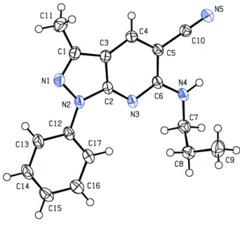
The molecular structure of the title compound is shown in Fig. 1. The 1H-pyrazolo[3,4-b]pyridine ring system (N1— N3/C1—C6) is essentially planar [r.m.s. deviation = 0.001 Å]. It forms a dihedral angle of 2.56 (6) ° with the attached
phenyl ring (C12—C17). All bond lengths and bond angles in the title compound are comparable with those of a similar
structure previously published (Wang & Zhu, 2006).
In the crystal, pairs of N—H···O hydrogen bonds connect the molecules to each other, forming centrosymmetric dimers
with R2
2(12) motifs (Table 1, Fig. 2). π-π stacking interactions between the dimers [Cg1···Cg3(-1 + x, y, z) = 3.5726 (8) Å]
between the centroids of the phenyl and pyrazole rings of the molecules] are also observed.
S2. Experimental
A mixture of 3-methyl-1-phenyl-6-chloro-1H-pyrazolo[3,4-b]pyridine-5-carbonitrile (0.268 g, 1 mmol) and propyl amine (1.2 ml, 12 mmol) was heated to 323 K overnight with constant stirring. The reaction mixture was cooled to room
temperature and taken up in dichloromethane, washed with 5% aq. NaHCO3, water and then with brine. The organic layer
was dried over anhydrous MgSO4 and the solvent was removed under reduced pressure. The crude product was
recrystallized from aqueous ethanol to give the title compound as colourless prisms (0.2 g, 69% yield); Rf = 0.25
(hexane:ethyl acetate, 4:1).
S3. Refinement
The hydrogen atoms attached to carbon atoms were positioned geometrically and constrained to ride on their parent
atoms, with carbon hydrogen bond distances of 0.93 - 0.97 Å. Uiso(H) values were set to a multiple of Ueq(C) with 1.5 for
CH3 and 1.2 for CH and CH2, respectively. Reflection (-1 1 1) was affected by the beam stop and was omitted from the
Figure 1
supporting information
[image:5.610.134.470.66.506.2]sup-3
Acta Cryst. (2015). E71, o766–o767Figure 2
A part of N—H···O dimers viewed down b axis. H atoms not involved in H bonding are omitted for clarity.
3-Methyl-1-phenyl-6-propylamino-1H-pyrazolo[3,4-b]pyridine-5-carbonitrile
Crystal data
C17H17N5
Mr = 291.36
Monoclinic, P21/c
Hall symbol: -P 2ybc
a = 5.1450 (2) Å
b = 15.1359 (7) Å
c = 19.5828 (9) Å
β = 96.547 (4)°
V = 1515.05 (12) Å3
Z = 4
F(000) = 616
Dx = 1.277 Mg m−3
Mo Kα radiation, λ = 0.71073 Å Cell parameters from 2809 reflections
θ = 4.1–32.4°
µ = 0.08 mm−1
Agilent Xcalibur Eos Gemini diffractometer
Radiation source: Enhance (Mo) X-ray Source Graphite monochromator
Detector resolution: 16.0416 pixels mm-1
ω scans
Absorption correction: multi-scan (CrysAlis PRO; Agilent, 2014)
Tmin = 0.795, Tmax = 1.000
10807 measured reflections 5031 independent reflections 3834 reflections with I > 2σ(I)
Rint = 0.025
θmax = 32.7°, θmin = 3.4°
h = −7→7
k = −21→19
l = −29→17
Refinement
Refinement on F2
Least-squares matrix: full
R[F2 > 2σ(F2)] = 0.055
wR(F2) = 0.143
S = 1.03 5031 reflections 205 parameters 0 restraints
Hydrogen site location: mixed
H atoms treated by a mixture of independent and constrained refinement
w = 1/[σ2(F
o2) + (0.0575P)2 + 0.5443P]
where P = (Fo2 + 2Fc2)/3
(Δ/σ)max < 0.001
Δρmax = 0.39 e Å−3
Δρmin = −0.19 e Å−3
Special details
Geometry. Bond distances, angles etc. have been calculated using the rounded fractional coordinates. All su's are estimated from the variances of the (full) variance-covariance matrix. The cell e.s.d.'s are taken into account in the estimation of distances, angles and torsion angles
Refinement. Refinement on F2 for ALL reflections except those flagged by the user for potential systematic errors.
Weighted R-factors wR and all goodnesses of fit S are based on F2, conventional R-factors R are based on F, with F set to
zero for negative F2. The observed criterion of F2 > σ(F2) is used only for calculating -R-factor-obs etc. and is not relevant
to the choice of reflections for refinement. R-factors based on F2 are statistically about twice as large as those based on F,
and R-factors based on ALL data will be even larger.
Fractional atomic coordinates and isotropic or equivalent isotropic displacement parameters (Å2)
x y z Uiso*/Ueq
supporting information
sup-5
Acta Cryst. (2015). E71, o766–o767C13 0.5115 (3) 0.27319 (9) 0.18042 (8) 0.0340 (4) C14 0.7117 (3) 0.21152 (10) 0.18464 (9) 0.0386 (4) C15 0.7886 (3) 0.16777 (9) 0.24520 (9) 0.0356 (4) C16 0.6611 (3) 0.18531 (10) 0.30210 (8) 0.0334 (4) C17 0.4609 (3) 0.24712 (9) 0.29935 (7) 0.0301 (4)
H4 −0.43410 0.53170 0.26680 0.0330*
H4N −0.206 (4) 0.4127 (12) 0.4777 (10) 0.041 (5)*
H7A 0.10150 0.33880 0.53730 0.0370*
H7B 0.26340 0.34310 0.47430 0.0370*
H8A 0.05460 0.21790 0.42670 0.0400*
H8B 0.18220 0.19630 0.50130 0.0400*
H9A −0.22310 0.20520 0.54190 0.0700*
H9B −0.23880 0.13680 0.48150 0.0700*
H9C −0.35420 0.23200 0.46870 0.0700*
H11A −0.20430 0.56570 0.13330 0.0670*
H11B −0.38750 0.48550 0.11120 0.0670*
H11C −0.11980 0.49580 0.08100 0.0670*
H13 0.46060 0.30210 0.13910 0.0410*
H14 0.79530 0.19950 0.14600 0.0460*
H15 0.92450 0.12700 0.24780 0.0430*
H16 0.71030 0.15520 0.34290 0.0400*
H17 0.37710 0.25850 0.33810 0.0360*
Atomic displacement parameters (Å2)
U11 U22 U33 U12 U13 U23
N1—N2 1.3930 (15) C13—C14 1.386 (2)
N1—C1 1.3143 (18) C14—C15 1.376 (2)
N2—C2 1.3732 (15) C15—C16 1.382 (2)
N2—C12 1.4148 (15) C16—C17 1.388 (2)
N3—C2 1.3366 (15) C4—H4 0.9300
N3—C6 1.3406 (15) C7—H7A 0.9700
N4—C6 1.3463 (17) C7—H7B 0.9700
N4—C7 1.4556 (18) C8—H8A 0.9700
N5—C10 1.144 (2) C8—H8B 0.9700
C1—C3 1.421 (2) C9—H9A 0.9600
C1—C11 1.490 (2) C9—H9B 0.9600
C2—C3 1.4055 (18) C9—H9C 0.9600
C3—C4 1.395 (2) C11—H11A 0.9600
C4—C5 1.3802 (19) C11—H11B 0.9600
N4—H4N 0.87 (2) C11—H11C 0.9600
C5—C10 1.4330 (19) C13—H13 0.9300
C5—C6 1.4437 (16) C14—H14 0.9300
C7—C8 1.523 (2) C15—H15 0.9300
C8—C9 1.510 (3) C16—H16 0.9300
C12—C17 1.3869 (19) C17—H17 0.9300
C12—C13 1.394 (2)
N2—N1—C1 106.80 (11) C3—C4—H4 121.00
N1—N2—C2 110.43 (10) C5—C4—H4 121.00
N1—N2—C12 118.66 (10) N4—C7—H7A 109.00
C2—N2—C12 130.87 (10) N4—C7—H7B 109.00
C2—N3—C6 115.13 (10) C8—C7—H7A 109.00
C6—N4—C7 123.36 (11) C8—C7—H7B 109.00
N1—C1—C3 110.86 (12) H7A—C7—H7B 108.00
N1—C1—C11 121.48 (13) C7—C8—H8A 109.00
C3—C1—C11 127.65 (14) C7—C8—H8B 109.00
N2—C2—N3 126.71 (11) C9—C8—H8A 109.00
N2—C2—C3 106.27 (11) C9—C8—H8B 109.00
N3—C2—C3 127.03 (11) H8A—C8—H8B 108.00
C1—C3—C2 105.65 (12) C8—C9—H9A 109.00
C1—C3—C4 136.75 (13) C8—C9—H9B 109.00
C2—C3—C4 117.60 (12) C8—C9—H9C 110.00
C3—C4—C5 117.40 (12) H9A—C9—H9B 109.00
C7—N4—H4N 116.6 (13) H9A—C9—H9C 109.00
C6—N4—H4N 119.6 (13) H9B—C9—H9C 109.00
C4—C5—C10 119.53 (11) C1—C11—H11A 109.00
C4—C5—C6 120.45 (11) C1—C11—H11B 109.00
C6—C5—C10 120.02 (12) C1—C11—H11C 109.00
supporting information
sup-7
Acta Cryst. (2015). E71, o766–o767N4—C7—C8 114.17 (12) C12—C13—H13 120.00
C7—C8—C9 113.92 (13) C14—C13—H13 120.00
N5—C10—C5 179.66 (15) C13—C14—H14 120.00
N2—C12—C13 118.95 (12) C15—C14—H14 120.00 C13—C12—C17 119.75 (12) C14—C15—H15 120.00 N2—C12—C17 121.30 (12) C16—C15—H15 120.00 C12—C13—C14 119.66 (14) C15—C16—H16 119.00 C13—C14—C15 120.95 (15) C17—C16—H16 119.00 C14—C15—C16 119.09 (14) C12—C17—H17 120.00 C15—C16—C17 121.11 (14) C16—C17—H17 120.00 C12—C17—C16 119.43 (13)
C1—N1—N2—C2 −0.22 (14) N1—C1—C3—C2 0.00 (16) C1—N1—N2—C12 177.73 (11) N3—C2—C3—C1 179.11 (12) N2—N1—C1—C3 0.13 (15) N2—C2—C3—C1 −0.15 (14) N2—N1—C1—C11 −178.72 (13) N3—C2—C3—C4 −1.3 (2) C2—N2—C12—C17 0.2 (2) N2—C2—C3—C4 179.47 (12) N1—N2—C12—C17 −177.28 (12) C1—C3—C4—C5 179.97 (16) C12—N2—C2—N3 3.4 (2) C2—C3—C4—C5 0.51 (19) N1—N2—C2—C3 0.23 (13) C3—C4—C5—C6 0.41 (18) N1—N2—C2—N3 −179.03 (11) C3—C4—C5—C10 −179.87 (12) C2—N2—C12—C13 −179.14 (12) C10—C5—C6—N3 179.49 (11) C12—N2—C2—C3 −177.40 (12) C4—C5—C6—N3 −0.79 (18) N1—N2—C12—C13 3.40 (17) C4—C5—C6—N4 179.43 (12) C2—N3—C6—C5 0.14 (17) C10—C5—C6—N4 −0.29 (17) C6—N3—C2—C3 0.91 (18) N4—C7—C8—C9 −57.37 (17) C2—N3—C6—N4 179.92 (11) N2—C12—C13—C14 178.16 (12) C6—N3—C2—N2 −180.00 (12) C17—C12—C13—C14 −1.2 (2) C7—N4—C6—C5 −175.32 (11) N2—C12—C17—C16 −178.48 (12) C7—N4—C6—N3 4.89 (18) C13—C12—C17—C16 0.8 (2) C6—N4—C7—C8 −77.97 (16) C12—C13—C14—C15 0.4 (2) C11—C1—C3—C2 178.77 (14) C13—C14—C15—C16 0.8 (2) C11—C1—C3—C4 −0.7 (3) C14—C15—C16—C17 −1.1 (2) N1—C1—C3—C4 −179.50 (16) C15—C16—C17—C12 0.3 (2)
Hydrogen-bond geometry (Å, º)
D—H···A D—H H···A D···A D—H···A
C17—H17···N3 0.93 2.33 2.9823 (18) 127
N4—H4N···N5i 0.87 (2) 2.20 (2) 3.0326 (17) 160.1 (18)