AMNIOTIC FLUID EMBOLISM (AFE) DURING LABOR RESULTING IN BRAIN DAMAGE, KING
ABDUL AZIZ HOSPITAL, JEDDAH, SAUDI ARABIA
1,*
Sana Al-Ahmari,
2Asseil Bossei,
3
Hanan Al
1Medical intern, Ibn Sina National College ISNC, Jeddah, Saudi Arabia
26th year Bachelor of Medicine, Bachelor of Surgery Student at ISNC, Jeddah, Saudi Arabia
3Obstetrics and Gynecology Resident
4Obstetrics and Gynecology consultant, King Abdulazez
ARTICLE INFO ABSTRACT
A 27 year old primigravida underwent caesarean section of labour. Intra
Subsequently, she developed respiratory failure requiring mechanical ventilation and dilated left ventricle
(AFE), a rare complication of pregnancy with a variable presentation, ranging from cardiac arrest and death through to mild degrees of organ system dysfunction
differential diagnosis includes pre
and pulmonary embolism. The course of these events was so rapid and catastrophic, which was consistent with AFE. Thus, we r
treatment of AFE by referring to up
of fetal elements in the maternal circulation is non
cardiovascular collapse by mechanical obstruction of the pulmonary circulation. It is now thought that a combination of left ventricular dysfunction and acute lung injury occur, with activation of several of the clotting factors. An immun
therapy and treatment is supportive. The mortality of the condition remains high.
Copyright © 2018, Sana Al-Ahmari et al. This is an open use, distribution, and reproduction in any medium, provided
INTRODUCTION
A 27-year-old female primigravida admitted through emergency room in labor. She was unbooked in our hospital with gestational age 39 weeks. She had history of chronic hypertension on medication (Lisinopril 5 mg BID
during pregnancy, she then shifted to (Coversyl 5 mg OD). She had history of Osteoporosis. She is known case of bronchial asthma and she is smoker. Laboratory findings: ABO group A+, WBC 14.03 k/ul, RBC 4.14 m/ul, HGB
28.9%, MCV 69.8 fL, MCH 22.9 pg/cell,
RDW 15.3%, PLT 380 k/uL, PT 9.8 sec, APTT 25.4 sec, INR 0.9%. On Examination, Blood pressure was 128/79
Pulse rate was 87 per min. Abdominal examination
Fundal height is 39 weeks, cephalic, estimated weight 3.5 kg and fetal movement was positive.
*Corresponding author: Sana Al-Ahmari,
Medical intern, Ibn Sina National College ISNC, Jeddah, Saudi Arabia
DOI: https://doi.org/10.24941/ijcr.32086.08.2018
ISSN: 0975-833X
Article History: Received 10th May, 2018 Received in revised form 22nd June, 2018
Accepted 20th July, 2018
Published online 31st August, 2018
Citation: Sana Al-Ahmari, Asseil Bossei, Areej Badawood, Mohammed Shata, Hanan Al Labor Resulting In Brain Damage, King Abdul Aziz Hospital, Jeddah, Saudi Arabia Key Words:
Amniotic Fluid Embolism, Brain Anoxia, Pregnancy, Cardiorespiratory Arrest.
CASE STUDY
AMNIOTIC FLUID EMBOLISM (AFE) DURING LABOR RESULTING IN BRAIN DAMAGE, KING
ABDUL AZIZ HOSPITAL, JEDDAH, SAUDI ARABIA
Asseil Bossei,
1Areej Badawood,
2Mohammed Shata,
Hanan Al-Ghamdi and
4Azza Khafaji
Medical intern, Ibn Sina National College ISNC, Jeddah, Saudi Arabia
6th year Bachelor of Medicine, Bachelor of Surgery Student at ISNC, Jeddah, Saudi Arabia Obstetrics and Gynecology Resident - R3, King Abdulazez Hospital, Jeddah, Saudi Arabia
Obstetrics and Gynecology consultant, King Abdulazez Hospital, Jeddah, Saudi Arabia
ABSTRACT
A 27 year old primigravida underwent caesarean section because of fetal distress following induction of labour. Intra-operative patient become dyspneic , lost conscious and unrecordable blood pressure. Subsequently, she developed respiratory failure requiring mechanical ventilation and dilated left ventricle . cardiac arrest followed after these events. The likely diagnosis was amniotic fluid embolism (AFE), a rare complication of pregnancy with a variable presentation, ranging from cardiac arrest and death through to mild degrees of organ system dysfunction with or without coagulopathy. The differential diagnosis includes pre-eclamptic toxaemia/pregnancy
and pulmonary embolism. The course of these events was so rapid and catastrophic, which was consistent with AFE. Thus, we report this case precisely and review pathophysiology, diagnosis, treatment of AFE by referring to up-to-date literatures. There is no diagnostic test for AFE; the finding of fetal elements in the maternal circulation is non-specific. Historically, AFE was
cardiovascular collapse by mechanical obstruction of the pulmonary circulation. It is now thought that a combination of left ventricular dysfunction and acute lung injury occur, with activation of several of the clotting factors. An immunological basis for these effects is postulated. There is no specific therapy and treatment is supportive. The mortality of the condition remains high.
open access article distributed under the Creative Commons Attribution provided the original work is properly cited.
old female primigravida admitted through She was unbooked in our hospital with gestational age 39 weeks. She had history of chronic hypertension on medication (Lisinopril 5 mg BID TAB) and during pregnancy, she then shifted to (Coversyl 5 mg OD). She had history of Osteoporosis. She is known case of bronchial Laboratory findings: ABO group A+, WBC 14.03 k/ul, RBC 4.14 m/ul, HGB 9.5 g/dl, HCT pg/cell, MCHC 32.9, 380 k/uL, PT 9.8 sec, APTT 25.4 sec, INR 0.9%. On Examination, Blood pressure was 128/79 mmhg. examination revealed, is 39 weeks, cephalic, estimated weight 3.5 kg
Medical intern, Ibn Sina National College ISNC, Jeddah, Saudi
.08.2018
Vaginal examination revealed cervix was soft, Os dilated 3 cm, Membrane intact and Vertex
admitted to delivery room started IV fluid Dextrose and Connected to continuous fetal monitor (CTG). CTG showed reactive fetal heart. On patient
mild abdominal contraction. Advised sedation Pethidine 100 mg IM and Plasil 10 mg IM.
effaced, Membrane bulging in the cervix, vertex
Artificial rupture of membrane (ARM) was done, thick meconium after ARM immediately patient become dyspneic and couldn’t lay down in bed suddenly lost conscious and became unrecordable blood pressure, CPR started and code blue announced. CPR continued for 10 min. the patient picked up and checked fetal heart beats were Bradycardic 90
per min. Emergency caesarean section done. Initial diagnosis, Amniotic Fluid Embolism (AFE). Post
and Neurologist consultation done. Echo cardiology showed: E.F 20% and dysfunction dilated left ventr
ventilator and shifted to Intensive care unit (ICU) and covered by antibiotics. ICU doctors tried to wean of the tube but not responded.
International Journal of Current Research
Vol. 10, Issue, 08, pp.73040-73044, August, 2018
Ahmari, Asseil Bossei, Areej Badawood, Mohammed Shata, Hanan Al-Ghamdi, Azza Khafaji. Amniotic Fluid Embolism (AFE) During Labor Resulting In Brain Damage, King Abdul Aziz Hospital, Jeddah, Saudi Arabia”, International Journal of Current Research
AMNIOTIC FLUID EMBOLISM (AFE) DURING LABOR RESULTING IN BRAIN DAMAGE, KING
Mohammed Shata,
Medical intern, Ibn Sina National College ISNC, Jeddah, Saudi Arabia
6th year Bachelor of Medicine, Bachelor of Surgery Student at ISNC, Jeddah, Saudi Arabia Hospital, Jeddah, Saudi Arabia Hospital, Jeddah, Saudi Arabia
because of fetal distress following induction operative patient become dyspneic , lost conscious and unrecordable blood pressure. Subsequently, she developed respiratory failure requiring mechanical ventilation and dilated left
. cardiac arrest followed after these events. The likely diagnosis was amniotic fluid embolism (AFE), a rare complication of pregnancy with a variable presentation, ranging from cardiac arrest and with or without coagulopathy. The eclamptic toxaemia/pregnancy-induced hypertension, anaphylaxis and pulmonary embolism. The course of these events was so rapid and catastrophic, which was eport this case precisely and review pathophysiology, diagnosis, date literatures. There is no diagnostic test for AFE; the finding specific. Historically, AFE was thought to induce cardiovascular collapse by mechanical obstruction of the pulmonary circulation. It is now thought that a combination of left ventricular dysfunction and acute lung injury occur, with activation of several of ological basis for these effects is postulated. There is no specific therapy and treatment is supportive. The mortality of the condition remains high.
ribution License, which permits unrestricted
examination revealed cervix was soft, Os dilated 3-4 Vertex at pelvic brim. The patient admitted to delivery room started IV fluid Dextrose and Connected to continuous fetal monitor (CTG). CTG showed reactive fetal heart. On patient review at 6:00 am there was mild abdominal contraction. Advised sedation Pethidine 100 mg IM and Plasil 10 mg IM. PV: os dilated 7-8 cm 80% effaced, Membrane bulging in the cervix, vertex -3 station. Artificial rupture of membrane (ARM) was done, thick conium after ARM immediately patient become dyspneic and couldn’t lay down in bed suddenly lost conscious and became unrecordable blood pressure, CPR started and code blue announced. CPR continued for 10 min. the patient picked eats were Bradycardic 90-80 beat min. Emergency caesarean section done. Initial diagnosis, Amniotic Fluid Embolism (AFE). Post- operative Cardiologist and Neurologist consultation done. Echo cardiology showed: E.F 20% and dysfunction dilated left ventricle. Patient was on ventilator and shifted to Intensive care unit (ICU) and covered by antibiotics. ICU doctors tried to wean of the tube but not
INTERNATIONAL JOURNAL OF CURRENT RESEARCH
Amniotic Fluid Embolism (AFE) During
After 12 days, post-operative tracheostomy done and the patient shifted to Gyne ward in Unconscious state, not oriented and blindness spastic limbs because of brain damage. Pelvi-abdominal ultrasonography revealed Hepatomegaly, Gall bladder stone, Minimal right pleural effusion and Bilateral grade 1 renal nephropathy. Non-contrast CT scan of the brain revealed; Symmetrically dilated supratentorial ventricular system with prominent cortical sulci, sylvian fissures and basal cisterns.
DISCUSSION
According to recent large population-based studies,( Ray, 2004; Society for Maternal-Fetal Medicine, 2016; Kramer et al., 2006; Abenhaim et al., 2008; Pallasmaa et al., 2008; Knight et al., 2008) the incidence of AFE, which includes both fatal and nonfatal cases, ranges between 1 in 12,953 deliveries in the United States,(10) to 1 in 56,500 in the United Kingdom (Knight et al., 2008). Based on those studies, the pooled estimated incidence of AFE would be 1 in 15,200 deliveries (95% confidence interval (CI), 1 in 13,900 to 1 in 16,700 deliveries) in North America (Gilbert et al., 1999),(8),(10) and 1 in 53,800 deliveries (95% CI, 1 in 48,800 to 1 in 59,900 deliveries) in Europe (Samuelsson et al., 2007; Pallasmaa et al., 2008; Knight, 2008). There were no available data on incidence of AFE in other regions of the world. The true incidence of AFE, however, is difficult to determine because the diagnosis of this syndrome remains one of exclusion, with possible underreporting of nonfatal cases. On the other hand, it is possible that AFE is over-diagnosed for medico-legal reasons, since this complication is widely considered to be an unavoidable cause of maternal death. AFE most often occurs related to labor and delivery or after uterine manipulation, and is characterized by development of severe dyspnea and hypoxemia, followed by seizures and cardiovascular collapse or arrest. Those who survive the initial event may develop disseminated intravascular coagulation and ARDS (Vanden Hoek et al., 2010). The mechanism of amniotic fluid embolism involves traumatic opening of uterine vessels, as suggested by data from a U.S. registry of cases, where 78% of the patients had ruptured membranes and several had just undergone intrauterine procedures (Tamura et al., 2014). Fetal squamous cells are found in the maternal pulmonary circulation at autopsy, but this finding is not specific for the syndrome as fetal cells may be recovered from pulmonary artery catheters in symptom-free patients with other diagnoses (Clark et al., 1986). Hemodynamically, the mechanism involves the acute development of pulmonary hypertension followed by left ventricular dysfunction (Vanden Hoek, 2010).These effects may be caused by constituents of amniotic fluid, including leukotrienes and arachidonic acid metabolites. In view of some similarities to anaphylaxis it has been suggested that the disorder be renamed anaphylactoid syndrome of pregnancy (Tamura et al., 2014). Recent evidence suggests a possible pathogenic and prognostic role for low C1 esterase inhibitor levels (Tamura et al., 2014). Radiographically, the patients usually develop bilateral pulmonary infiltrates. If the fetus survives the initial event, it should be promptly delivered. In cases of imminent maternal demise, emergency postmortem or periresuscitative cesarean section should be performed, as in other instances of cardiopulmonary resuscitation in pregnancy. (Lapinsky, 2015). The maternal mortality rate has been reported as high as 86% but in more recent reports, as low as 22%. 47 Amniotic fluid embolism may account for 14% of all maternal deaths (Perry et al., 1998).
After thorough literature search we found that few authors have proposed that two clinical forms of AFE exist typical and atypical. Uszynski also documented that symptoms vary in both forms of AFE (Davies et al., 1992). Typical, (classic) with three phases: Phase 1-respiratory and circulatory disorders, Phase 2-coagulation disturbances of maternal hemostasis, Phase 3- acute renal failure and acute respiratory distress syndrome (ARDS), and leading to cardiopulmonary collapse. Atypical: In contrast to typical embolism, cardiopulmonary collapse does not occur in atypical embolism but the first symptom is life threatening hemorrhage due to DIC. Atypical embolism was observed during caesarean section or immediately after it, in cases of profound rupture of uterine cervix, as well as in the course of placenta abruption and in association with induced midtrimester abortion (Davies
et al., 1992; Martin, 1996). The most significant pathologic
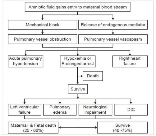
findings at autopsy are limited to the lungs. Grossly, the lungs show evidence of pulmonary edema (in 70% of the cases). Alveolar hemorrhage and pulmonary embolism of amniotic fluid materials are present; the presence of embolic particles is essential for diagnosis, but on histology they may be missed because of their small size (Gilbert et al., 1999). In 1995, Clarke (Clarke et al., 1995) suggested that the syndrome arose from an immune rather than embolic process. Amniotic fluid embolism is caused by fetal antigens in the amniotic fluid stimulating a cascade of endogenous immune mediators, producing a reaction similar to anaphylaxis. Biochemical mediators found in the amniotic fluid are thought to trigger the main features of anaphylactoid reaction with multisystem involvement. The fetal products found in suspension in the amniotic fluid are responsible only for the minor effects caused by the actual mechanical obstruction. Clarke(1995) suggested that the syndrome of acute peripartum hypoxia, hemodynamic collapse, and coagulopathy should be described as anaphylactoid syndrome of pregnancy and not AFE. There are also striking similarities between clinical and hemodynamic findings in AFE and septic shock, which suggests a common pathological mechanism. Gei and Hankins (2000) proposed a pathophysiological co urse. Figure 1.
The initial respiratory reaction possibly begins with transient pulmonary vasospasm (Abenhaim et al., 2008) Although this possible transient vasospasm has not been documented (probably because the signs and symptoms appear so abruptly in a seemingly healthy person who is not monitored via invasive methods), vasospasm may be caused by amniotic microemboli that trigger the release of arachidonic acid metabolites, (Clark, 1990) and leads to pulmonary hypertension, intrapulmonary shunting, bronchoconstriction, and severe hypoxia (Reeves et al., 1983). Exactly which components of amniotic fluid actually cause this effect is unknown, but Clarke (1990) suggested that abnormal components such as meconium may play a role. Many experts (Reeves et al., 1983; Hammerschidt et al., 1984; Azegami et al., 1986; E1 Maradny et al., 1985; Lunetta, 1996; Fineschi et al., 1998) speculate that maternal mediators also may have an influence. The second manifestation includes negative inotropism and left ventricular failure resulting in increasing pulmonary edema and hypotension quickly leading to shock. The third manifestation is a neurological response to the respiratory and hemodynamic injury, which may include seizures, confusion, or coma (Azegami et al., 1986). This coagulopathy is thought to be precipitated by several procoagulant components of amniotic fluid, most notably thromboplastin, which initiates the extrinsic pathway of the
clotting cascade and results in excessive fibrinolytic activity (Azegami et al., 1986). Depending upon the magnitude of the events and the maternal physiological reserve, the patient may not recover from the injury. During this convalescent period, patient may die as a consequence of the severe lung or brain injury, multi-organ failure, or from an infection
intensive care unit (E1 Maradny et al., 1995)
Figure 1. Postulated mechanism for the pathogenesis of amniotic fluid embolism; DIC - Disseminated intravascular coagulation
The clinical presentation of amniotic fluid embolism is, in its classic form, dramatic. A period of anxiety, change in mental status, agitation, and a sensation of doom may precede the event (Ecker et al., 2012). Patients may progress rapidly to cardiac arrest, with pulseless electrical activity, asystole, ventricular fibrillation, or pulseless ventricular tachycardia. In cases occurring prior to delivery, electronic fetal monitoring will demonstrate decelerations, loss of variability, and terminal bradycardia as oxygenated blood is shunted away from the uterus, and catecholamine- induced uterine hypertonus causes a further decline in uterine perfusion (Clark et al
2014). Premonitory symptoms; The seventh report of the confidential enquiry into maternal deaths in the United Kingdom (Lewis, 2007) has highlighted that
who suffered AFE reported some or all of the following symptoms: breathlessness, chest pain, feeling cold, lightheadedness, distress, panic, a feeling of pins and needles in the fingers, nausea, and vomiting. The interval between onset of these symptoms and the collapse varied (from immediately to over 4 hours later). These symptoms might indicate hypoxia and might give the first clue to diagnosis of amniotic-fluid embolism in progress before collapse and hemorrhage occur. Reported risk factors for amniotic fluid embolism include situations in which the exchange of fluids between the maternal and fetal compartments is more such as operative delivery (cesarean or vaginal), placenta previa, placenta accreta, and abruption. An association between induction of labor and amniotic fluid embolism is inconsistently reported. Abnormalities of uterine tone (hypo or hypertonous) described commonly in cases of amniotic fluid embolism may be the consequence of uterine hypo
secondary to profound maternal shock and hypoxia with massive catecholamine release, rather than the cause
al., 1995).
excessive fibrinolytic activity the magnitude of the events and the maternal physiological reserve, the patient may not recover from the injury. During this convalescent period, patient may die as a consequence of the severe lung or brain organ failure, or from an infection acquired in the
Postulated mechanism for the pathogenesis of amniotic Disseminated intravascular coagulation
The clinical presentation of amniotic fluid embolism is, in its classic form, dramatic. A period of anxiety, change in mental n of doom may precede the Patients may progress rapidly to cardiac arrest, with pulseless electrical activity, asystole, fibrillation, or pulseless ventricular tachycardia. In cases occurring prior to delivery, electronic fetal monitoring will demonstrate decelerations, loss of variability, and terminal bradycardia as oxygenated blood is shunted away from the induced uterine hypertonus causes
et al., 1995; Clark,
Premonitory symptoms; The seventh report of the confidential enquiry into maternal deaths in the United ighted that 11 of 17 women who suffered AFE reported some or all of the following symptoms: breathlessness, chest pain, feeling cold, lightheadedness, distress, panic, a feeling of pins and needles in the fingers, nausea, and vomiting. The interval between the onset of these symptoms and the collapse varied (from almost immediately to over 4 hours later). These symptoms might indicate hypoxia and might give the first clue to diagnosis of fluid embolism in progress before collapse and Reported risk factors for amniotic fluid embolism include situations in which the exchange of fluids between the maternal and fetal compartments is more likely, such as operative delivery (cesarean or vaginal), placenta abruption. An association between induction of labor and amniotic fluid embolism is Abnormalities of uterine tone (hypo or hypertonous) described commonly in cases of amniotic fluid embolism may be the consequence of uterine hypo-perfusion secondary to profound maternal shock and hypoxia with release, rather than the cause (Clark et
Other positive risk factors include older maternal age, high parity, cesarean section, low uterine segment laceration meconium staining of amniotic fluid, cervical lacerations, uterine rupture, eclampsia, polyhydramnios, and multiple gestations. Sociodemographic risk factors such as maternal age and race/ethnicity are also reported in some series
al., 2012). However, given the rare and unpredictable nature of amniotic fluid embolism, there are no risk factors sufficiently established to justify any alteration in standard obstetric care. Amniotic fluid embolism should be considered in the differential diagnosis of sudden cardiorespiratory compromise in any pregnant or recently postpartum patient. Initial resuscitation of cardiac arrest does not require a specific diagnosis of amniotic fluid embolism because initial maternal treatment (with basic cardiac life
life support protocols) is similar, regardless of the exact etiology. The use of vasopressors, antiarrhythmic agents, and defibrillating doses is not different from those utilized in nonpregnant individuals. Although concerns
[image:3.595.38.289.145.364.2]arcing may occur if fetal monitors are in place at the time of cardioversion or defibrillation are largely theoretical, it is reasonable to remove such monitors while cardiopulmonary resuscitation is in progress. However, the presence of such monitors should not delay defibrillation when indicated. The components of high-quality cardiopulmonary resuscitation are summarized in Table 1 (Hammerschidt
Table 1. Components of high
resuscitation in pregnancy
1. Rapid chest compressions (100
2. Perform hard compressions, achieving a depth of at least 2 inches Assure adequate chest recoil between
3. Minimize interruptions of chest
4. Avoid prolonged pulse checks (no more than 5e10 seconds) Resume chest compressions immediately after
5. defibrillating Switch provider of compressions every 2 minutes to avoid fatigue Lateral displacement of uterus during
6. SMFM. Amniotic fluid embolism: diagnosis and management. Am J Obstet Gynecol 2016.
Immediate survivors of amniotic fluid embolism require multidisciplinary management, including maternal medicine sub-specialists and intensive care specialists. The management of suspected amniotic fluid embolism is supportive and focuses on rapid ma
stabilization (Abenhaim et al., 2008
is supportive and directed towards the maintenance of oxygenation, cardiac output and
of the coagulopathy. Treatment should take place in an intensive care unit, if possible. In the event of maternal cardiac arrest, cardiopulmonary resuscitation should be initiated immediately and, if the gestational age of the undelivered alive fetus is viable, cesarean section could be considered
1992). Uterine evacuation after unsuccessful resuscitation may be therapeutic for the mother, because the weight of the gravid uterus on the inferior vena cava impedes blood return to the heart and decreases systemic blood pressure
Martin et al., 2001). The initial goal is the rapid correction of maternal hemodynamic instability, which includes correction of hypoxia and hypotension, for preventing additional hypoxia and subsequent end- organ failure. Oxygen should be administered immediately by wha
including face mask, bag-valve mask, or endotracheal intubation, in concentrations adequate to keep oxygen saturation at 90% or higher (Moore
Other positive risk factors include older maternal age, high cesarean section, low uterine segment laceration, and meconium staining of amniotic fluid, cervical lacerations, uterine rupture, eclampsia, polyhydramnios, and multiple gestations. Sociodemographic risk factors such as maternal age re also reported in some series (Knight et
However, given the rare and unpredictable nature of amniotic fluid embolism, there are no risk factors sufficiently established to justify any alteration in standard obstetric care. Amniotic fluid embolism should be considered in the osis of sudden cardiorespiratory compromise in any pregnant or recently postpartum patient. Initial resuscitation of cardiac arrest does not require a specific diagnosis of amniotic fluid embolism because initial maternal treatment (with basic cardiac life support and advanced cardiac life support protocols) is similar, regardless of the exact etiology. The use of vasopressors, antiarrhythmic agents, and defibrillating doses is not different from those utilized in nonpregnant individuals. Although concerns that electric arcing may occur if fetal monitors are in place at the time of cardioversion or defibrillation are largely theoretical, it is reasonable to remove such monitors while cardiopulmonary resuscitation is in progress. However, the presence of such monitors should not delay defibrillation when indicated. The quality cardiopulmonary resuscitation are
Hammerschidt et al., 1984).
Components of high-quality cardiopulmonary resuscitation in pregnancy
Rapid chest compressions (100 minute)
Perform hard compressions, achieving a depth of at least 2 inches adequate chest recoil between compressions
Minimize interruptions of chest compressions
Avoid prolonged pulse checks (no more than 5e10 seconds) compressions immediately after
defibrillating Switch provider of compressions every 2 minutes to Lateral displacement of uterus during resuscitation SMFM. Amniotic fluid embolism: diagnosis and management.
Immediate survivors of amniotic fluid embolism require multidisciplinary management, including maternal-fetal specialists and intensive care specialists. The management of suspected amniotic fluid embolism is supportive and focuses on rapid maternal hemodynamic ., 2008). The management of AFE is supportive and directed towards the maintenance of and blood pressure, and correction Treatment should take place in an nsive care unit, if possible. In the event of maternal cardiac arrest, cardiopulmonary resuscitation should be initiated immediately and, if the gestational age of the undelivered alive fetus is viable, cesarean section could be considered (Davies , Uterine evacuation after unsuccessful resuscitation may be therapeutic for the mother, because the weight of the gravid uterus on the inferior vena cava impedes blood return to the heart and decreases systemic blood pressure (Martin, 1996; . The initial goal is the rapid correction of maternal hemodynamic instability, which includes correction of hypoxia and hypotension, for preventing additional hypoxia organ failure. Oxygen should be administered immediately by whatever means necessary, valve mask, or endotracheal intubation, in concentrations adequate to keep oxygen
A total of 9 cases of successful pregnancy following AFE, with no instances of recurrent AFE, have been reported in the literature (Burrows et al., 1995; Clark, 1992; Duffy, 1998; Collier, 1998; Stiller et al., 2000; Demianczuk, 2005; Abecassis, 2006). Therefore, although the available information is limited, the current evidence suggests that AFE is not a recurrent disease. Survival after AFE has improved significantly with early recognition of this syndrome and prompt and early resuscitative measures. The decrease in the mortality rate results solely from early diagnosis and prompt treatment rather than prevention of the syndrome, since the cause is unknown. Those women who survive long enough to be transferred to the ICU have a better chance of survival. Although mortality rates have declined, morbidity remains high with severe sequelae, particularly neurologic impairment.
RESULTS AND RECOMMENDATION
We recommend the following: (1) we recommend consideration of amniotic fluid embolism in the differential diagnosis of sudden cardiorespiratory collapse in the laboring or recently delivered woman. (2) we do not recommend the use of any specific diagnostic laboratory test to either confirm or refuse the diagnosis of amniotic fluid embolism; at the present time, amniotic fluid embolism remains a clinical diagnosis. (3) we recommend the provision of immediate high- quality cardiopulmonary resuscitation with standard basic cardiac life support and advanced cardiac life support protocols in patients who develop cardiac arrest associated with amniotic fluid embolism. (4) we recommend that a multidisciplinary team including anesthesia, respiratory therapy, critical care, and maternal-fetal medicine should be involved in the ongoing care of women with AFE (Best Practice); (5) following cardiac arrest with amniotic fluid embolism, we recommend immediate delivery in the presence of a fetus 24 weeks of gestation. (6) We recommend the provision of adequate oxygenation and ventilation and, when indicated by hemodynamic status, the use of vasopressors and inotropic agents in the initial management of amniotic fluid embolism. Excessive fluid administration should be avoided
Conclusion
Amniotic fluid embolism syndrome is an infrequent, unpredictable, and catastrophic complication of pregnancy. It is virtually impossible to predict which patients are at risk for AFE. Diagnosis must be based on a spectrum of clinical signs and symptoms and by exclusion of other causes. Most cases of AFE are associated with dismal maternal and fetal outcomes, regardless of the quality of care rendered. Improved understanding of the pathophysiology of AFE may lead to the development of preventive measures and more effective and specific treatment. Although there are many new developments with respect to the understanding of the disease, amniotic fluid embolism continues to be a catastrophic illness requiring a high index of suspicion, a multidisciplinary approach and rapid resuscitation efforts in order to have a desirable clinical outcome. Several authors have reported an association between AFE and allergic patient thus through review of a patient’s medical history and examination doctors will be able to predict the incidence.
REFERENCES
Abecassis P, Benhamou D. Is amniotic fluid embolism likely to recur in a subsequent pregnancy? Int J Obstet Anesth. 2006;15:90. (PubMed)
Abenhaim HA, Azoulay L, Kramer MS, Leduc L. Incidence and risk factors of amniotic fluid embolisms: a population-based study on 3 million births in the United States. Am J Obstet Gynecol 2008;199:49.e1-8 (Level II-2).
Abenhaim HA., Azoulay L., Kramer MS., Leduc L. 2008. Incidence and risk factors of amniotic fluid embolisms: a population-based study on 3 million births in the United States. Am J Obstet Gynecol., 199:49.e1–8. (PubMed) Azegami M., Mori N. 1986. Amniotic fluid embolism and
leukotrines. Am J Obstet Gynecol., 155:1119–24
Burrows A, Khoo SK. The amniotic fluid embolism syndrome: 10 years’ experience at a major teaching hospital. Aust N Z J Obstet Gynaecol. 1995;35:245–50. (PubMed)
Clark SL, Hankins GD, Dudley DA, Dildy GA, Porter TF. Amniotic fluid embolism: analysis of the national registry. Am J Obstet Gynecol 1995;172:1158-67; discussion 1167-9 (Level II-2).
Clark SL, Pavlova Z, Greenspoon J, et al. Squamous cells in the maternal pulmonary circulation. Am J Obstet Gynecol 1986; 154: 104
Clark SL. 1990. New concepts of amniotic fluid embolism: A review. Obstet Gynecol Surv., 45:360–8.
Clark SL. Amniotic fluid embolism. Obstet Gynecol 2014;123:337-48 (Level III).
Clark SL. Successful pregnancy outcomes after amniotic fluid embolism. Am J Obstet Gynecol. 1992;167:511–2. (PubMed)
Clarke SL., Hankins G., Dudley DA., Dildy GA., Porter TF. 1995. Amniotic fluid embolism: Analysis of the national registry. Am J Obstet Gynecol., 172:1158–67.
Collier C. Recurring amniotic fluid embolism. Anaesth Intensive Care. 1998;26:599–600. (PubMed)
Davies MG, Harrison JC. Amniotic fluid embolism: maternal mortality revisited. Br J Hosp Med. 1992;47:775–6. (PubMed)
Demianczuk CE, Corbett TF. Successful pregnancy after amniotic fluid embolism: a case report. J Obstet Gynaecol Can. 2005;27:699–701. (PubMed)
Duffy BL. Does amniotic fluid embolism recur? Anaesth Intensive Care. 1998;26:333. (PubMed)
E1 Maradny E., Kanayama N., Halim A, Maehara K., Terao T 1995. Endothelin has a role in early pathogenesis of amniotic fluid embolism. Gynecol Obstet Invest., 40:14–8. (PubMed)
Ecker JL., Solt K., Fitzsimons MG., MacGillivray TE. 2012.Case records of the Massachusetts General Hospital. Case 40-2012. A 43-year-old woman with cardiorespiratory arrest after a cesarean section. N Engl J Med., 367:2528-36 (Level III).
Elisabetta Del Zotto, Alessia Giossi, Irene Volonghi, Paolo Costa, Alessandro Padovani, and Alessandro Pezzini, “Ischemic Stroke during Pregnancy and Puerperium,” Stroke Research and Treatment, vol. 2011, Article ID 606780, 13 pages, 2011. doi:10.4061/2011/606780
Fineschi V., Gambassi R., Gherardi M., Turillazzi E. 1998.The
diagnosis of amniotic fluid embolism: An
immunohistochemical study for the quantification of pulmonary mast cell tryptase Int. Legal Med.,111:238– 43.
Gei G., Hankins GD. 2000. Amniotic fluid embolism: An update. Contemporary OB/GYN.45:53–62.
Gilbert WM., Danielsen B. 1999. Amniotic fluid embolism: decreased mortality in a population-based study. Obstet
Gynecol., 93:973–7. (PubMed)
Hammerschidt DE., Ogburn PL., Williams JE. 1984. Amniotic fluid activates complement: A role in amniotic fluid embolism syndrome? J Lab Clin Med., 104:901–7.
Knight M, Berg C, Brocklehurst P, et al. Amniotic fluid embolism inci- dence, risk factors and outcomes: a review and recommendations. BMC Pregnancy Childbirth 2012;12:7 (Level III).
Knight M. 2008. UKOSS Amniotic fluid embolism: active surveillance versus retrospective database review. Am J
Obstet Gynecol., 199:e9. (PubMed)
Kramer MS., Rouleau J., Baskett TF., Joseph KS. 2006. Maternal Health Study Group of the Canadian Perinatal Surveillance System. Amniotic-fluid embolism and medical induction of labour: a retrospective, population-based cohort study. Lancet. 368:1444–8. (PubMed)
Lapinsky SE. Pregnancy joins the hit list. Crit Care Med 2012; 40: 1679–1680. (PubMed)
Lewis G, editor. The seventh report on confidential enquiries into maternal deaths in the United Kingdom. London: CEMACH; 2007. The confidential enquiry into maternal and child health (CEMACH). Saving mother’s lives: reviewing maternal deaths to make motherhood safer -2002–2005. Lunetta P., Penttila A. 1996. Immuno histochemical
identification of syncytiotrophoblastic cells and megakaryocytes in pulmonary vessels in a fatal case of amniotic fluid embolism. Int J Legal Med.,108:210–4. (PubMed)
Martin PS, Leaton MB. Emergency. Amniotic fluid embolism. Am J Nurs. 2001;101:43–4. (PubMed)
Martin RW. Amniotic fluid embolism. Clin Obstet Gynecol. 1996;39:101–6. (PubMed)
Moore J, Baldisseri MR. Amniotic fluid embolism. Crit Care Med. 2005;33:S279–85. (PubMed)
O’Shea A, Eappen S. Amniotic fluid embolism. Int Anesthesiol Clin. 2007;45:17–28. (PubMed)
Pallasmaa N., Ekblad U., Gissler M. 2008. Severe maternal morbidity and the mode of delivery. Acta Obstet Gynecol Scand. 87:662–8. (PubMed)
Perry KG, Martin RW, Blake PG, et al. Maternal mortality associated with the adult respiratory distress syndrome. South Med J 1998; 91: 441–444. (PubMed).
Ray, B.K., Vallejo, M.C., Creinin, M.D. et al. Can J Anesth 2004 51: 139. doi:10.1007/BF03018773 Society for Maternal-Fetal Medicine (SMFM) with the assistance of Pacheco LD, Saade G, et al. Amniotic fluid embolism: diagnosis and management. Am J Obstet Gynecol
2016;215:B16-24.
Reeves WC., Demers LM., Wood MA., Skarlatos S., Copenhaver G., Whitesell L. et al. 1983. The release of thromboxane A2 and prostacyclin following experimental acute pulmonary embolism. Prostaglandins Leukot Med.,
11:1–10.
Rudra, A., Chatterjee, S., Sengupta, S., Nandi, B., & Mitra, J. 2009. Amniotic fluid embolism. Indian Journal of Critical Care Medicine: Peer-Reviewed, Official Publication of Indian Society of Critical Care Medicine, 13(3), 129–135. http://doi.org/10.4103/0972-5229.58537
Samuelsson E., Hellgren M., Högberg U. 2007. Pregnancy-related deaths due to pulmonary embolism in Sweden. Acta Obstet Gynecol Scand. 86:435–43. (PubMed)
Steiner PE, Lushbaugh CC. 1941. Maternal pulmonary embolism by amniotic fluid as a cause of obstetric shock and unexpected deaths in obstetrics. JAMA. 117:1245–54. 1341– 5. (PubMed)
Steiner PE., Lushbaugh CC., Frank HA. 1949. Fatal obstetric shock from pulmonary emboli of amniotic fluid. Am J
Obstet Gynecol., 58:802–5. (PubMed)
Stiller RJ, Siddiqui D, Laifer SA, Tiakowski RL, Whetham JC. Successful pregnancy after suspected anaphylactoid syndrome of pregnancy (amniotic fluid embolus). A case report. J Reprod Med. 2000;45:1007–9. (PubMed) Tamura N, Kimura S, Farhana M, et al. C1 esterase inhibitor
activity in amniotic fluid embolism. Crit Care Med 2014; 42: 1392–1396
Tamura N, Kimura S, Farhana M, et al. C1 esterase inhibitor activity in amniotic fluid embolism. Crit Care Med 2014; 42: 1392–1396. (PubMed)
Vanden Hoek TL, Morrison LJ, Shuster M, et al. Part 12: Cardiac arrest in special situations: 2010 American Heart Association Guidelines for Cardiopulmonary Resuscitation and Emergency Cardiovascular Care. Circulation 2010; 122: S829–861.
Vanden Hoek TL, Morrison LJ, Shuster M, et al. Part 12: Cardiac arrest in special situations: 2010 American Heart Association Guidelines for Cardiopulmonary Resuscitation and Emergency Cardiovascular Care. Circulation 2010; 122: S829–861. (PubMed)