Cell cycle dependent regulation of gap junction coupling and
apoptosis in GFSHR-17 granulosa cells
Sabrina Schlie1,2, Karolina Mazur1, Willem Bintig1, Anaclet Ngezahayo1
1Institute of Biophysics, University Hannover, Herrenhäuserstr, Hannover, Germany; 2Laser Zentrum Hannover e.V., Hollerithallee, Hannover, Germany.
Email: [email protected]
Received 26 November 2009; revised 10 March 2010; accepted 12 March 2010.
ABSTRACT
Recent results have shown that the level of gap junc-tion coupling could modulate the inducjunc-tion of apopt- otic reactions. We previously observed that 1H-[1,2, 4]Oxadiazole[4,3-a]quinoxalin-1-one (ODQ), a block- er of guanylyl cyclase, inhibited gap junction coup- ling and thereby promoted activation of characteris-tic apoptocharacteris-tic reactions such as chromatin condensa-tion, DNA strand breaking, and formation of blebs in GFSHR-17 granulosa cells, the in vitro model for gra- nulosa cells of the maturing ovular follicle. In the pr- esent report, we focus on the effects of ODQ with re- spect to the cell cycle in GFSHR-17 granulosa cells. In synchronised GFSHR-17 granulosa cells, the doub- le whole-cell patch-clamp technique revealed that gap junction conductance in mitotic cells was reduced in comparison to cells in interphase. This reduction of gap junction conductance correlated with a reduction of non-phosphorylated Cx43 in mitotic cells. We com- pared the stimulation of apoptotic reactions by ODQ between cells in mitosis and in interphase. We ob-served that the induction of both chromatin conden-sation and DNA strand breaking by ODQ was incr- eased in mitotic cells, as compared to cells in inter-phase. The effects of ODQ were not observed in He- La cells that do not express connexins. The results in- dicate that reduction of gap junction coupling in mit- otic GFSHR-17 granulosa cells depends on phosphor- rylation of Cx43 and raises the sensitivity to stimula-tion of apoptosis. We propose that gap juncstimula-tion cou-pling is involved in regulation of apoptosis of granu-losa cells in maturing ovular follicle.
Keywords: Granulosa Cells; Cell Cycle; Gap Junction;
Apoptosis
1. INTRODUCTION
Gap junctions are adhesion structures containing cell-
to-cell channels that enable neighbouring cells to excha- nge small molecules (≤ 1 kDa) like Ca2+, cAMP, IP
3, and
to synchronise electrical as well as physiological activi-ties [1,2]. Gap junction channels are composed of con-nexins (Cx), which are the products of 19 and 20 genes in mouse and human, respectively. The expression and posttranslational modification of the connexins are spe-cifically regulated in the different tissues and correlate with cellular metabolic state [3].
Gap junction assembly involves oligomerisation of six connexins in order to form a connexon that is inserted into the cellular membrane. Two connexons of adjacent cells associate and form a cell-to-cell channel. Since gap junction channels allow a direct intercellular exchange of metabolites and second messengers, specific roles in diseases and regulation of cellular activities including proliferation, transformation, differentiation and apopto- sis have been hypothesised [2,4].
Gap junction coupling-dependent regulation of DNA synthesis was shown in cardiomyocytes [5]. It was also observed that gap junction coupling was involved in reg- ulation of expression of cyclin dependent kinase inhibi-tors (CDK-inhibiinhibi-tors) such as p21waf1/Cip1 and p27kip1 in
myoblasts [6-9]. Since such CDK-inhibitors block the cell cycle and are invovled in stimulating apoptosis, the previous findings established a link between gap junc-tion coupling and regulajunc-tion of cell cycle and apoptosis as it was recently proposed [10]. Similarly, a PKC-dependent phosphorylation of Cx43 that correlates with a reduction of gap junction coupling was found during G2/M phase of the cell cycle [11]
lu-teum [17,18]. During atresia, excess follicles are remo- ved, and dominant follicles, which undergo maturation, are selected. With the deterioration of corpus luteum, the ovary expels superfluous cells that otherwise represent a risk of tumour formation.
Gap junction coupling is regulated by the expression of connexins and posttranslational modifications such as phosphorylation [19]. Since both expression of connex-ins and posttranslational modification depend on the me- tabolic state of the cells, it is postulated that gap junction coupling of GFSHR-17 granulosa cells could be modif- ied during the cell cycle. Furthermore, it was shown that the induction of apoptosis is partly related to the degree of gap junction coupling [10]. If the degree of gap junct- ion coupling’s regulation is dependent upon the cell cy-cle, the induction of apoptosis could also be modulated relative to the cycle phase of the GFSHR-17 granulosa cells. This hypothesis was tested by application 20 µM ODQ, a dose that was shown to inhibit gap junction coupling in GFSHR-17 granulosa cells [10]. In synchro-nised GFSHR-17 granulosa cells, we observed that the macroscopic conductance of gap junctions between cells in mitosis was reduced compared to cells in interphase. Correspondingly, non-phosphorylated Cx43, the main connexin of granulosa cells [13] seemed to be reduced in mitotic cells compared to cells in interphase. The gap junction uncoupler ODQ induces an increase in chroma-tin condensation as well as DNA strand breaks in cells in
the mitotic phase, compared to cells in interphase. The results indicate an involvement of gap junction coupling in cell cycle-dependent modulation of apoptosis.
2. METHODS
2.1. Chemicals and Cell Culture Media
If not otherwise specified, the chemicals and the cell cul- ture media were obtained from Sigma-Aldrich (Taufkir- chen, Germany).
2.2. Cell Culture
Granulosa cells were cultivated as described previously [10,20] using Dulbecco’s Modified Eagle Medium (DM- EM) supplemented with 5% foetal calf serum (FCS) and antibiotics. The culture medium was renewed every 2-3 days, and the cells were passaged every 7 days. The dou- bling time of the cells was evaluated by counting the cell density 12 h, 24 h, 36 h, 48 h and 72 h after seeding.
To test the effect of the ODQ on GFSHR-17 granulo- sa cells, a 20 mM stock solution of ODQ in DMSO was prepared. This solution was added to the cell culture medium to achieve a concentration of 20 µM ODQ, and DMSO was added to achieve a working concentration of 0.5% DMSO. The control experiments were performed in cells cultivated in the presence of 0.5% DMSO. Gap
junction coupling, activation of chromatin condensation and DNA strand break were analysed after 6 h incuba-tion in the presence of ODQ.
2.3. Synchronisation of the Cells
To achieve a synchronisation of the GFSHR-17 granu-losa cells, mitotic cells were isolated from a monolayer using the mitotic-shake-off technique [21]. This techn- ique is based on the observation that cells in mitosis do not adhere very well to the surface. The monolayer was washed with PBS. After addition of fresh culture me-dium, the cells in Petri dish were carefully shaken to release cells in mitotic phase into the culture medium. The culture medium with cells in mitotic phase was col-lected and preserved at room temperature. Fresh culture medium was added to the monolayer, and the cells were incubated in the cell culture incubator for an additional hour. The shaking procedure was repeated twice. The collected cell suspsension was centrifuged at 800g for 15 min. The cells in the pellet were resuspended in cell cul-ture medium and counted using a Rosenthal cell counter device. A quantity of 4 × 10⁴ cells were seeded into 2 ml
of culture medium in a culture dish with 35 mm in . The proliferation was evaluated 1.5, 3, 4.5, 14.5, 16, 17.5, 19, 20.5, and 40 h after seeding. For a better com-parison between experiments, the cell population at each time point was normalised to the seeding population at time 0 h. The results are given as average SEM for n = 4 experiments.
2.4. Analysis of Gap Junction Coupling with Lucifer Yellow Transfer
To analyse the effect of ODQ on gap junction coupling of the GFSHR-17 granulosa cells, the cells were grown on cover slips with 10 mm Ø. A cover slip with a mon- olayer was placed in a superfusion chamber containing 0.5 ml of a bath solution containing (in mM): 140 NaCl, 10 KCl, 2 CaCl2, 1 MgCl2, 5 glucose and 10 HEPES.
After an adaptation to room temperature for at least 30 min, a whole-cell patch-clamp configuration was estab-lished on one cell within the monolayer. Lucifer yellow (0.5 %) was dissolved in the pipette filling solution that contained (in mM): 100 K-gluconate, 40 KCl, 5 Na2ATP,
2.5 MgCl2, 0.25 CaCl2, 1 BAPTA, 0.2 cGMP, 1 glucose
given as average SEM for n = 6 experiment for each treatment.
2.5. Analysis of Gap Junction Coupling Using the Double Whole-Cell Patch-Clamp Technique
The double whole-cell patch-clamp configuration allows imposition of a voltage difference (ΔVj) between two ce-
lls that are joined by gap junction channels. It is thereby possible to evaluate the conductance of the gap junction channels (Gj). The double whole-cell patch-clamp
con-figuration was established on pairs of synchronised GFSHR-17 granulosa cells in different phases of the cell cycle. The pipette filling solution and the extracellular solution are described above. These solutions were pre-viously shown to sustain the macroscopic conductance of the gap junction channels [20].
2.6. Western Blot
To isolate proteins, cells were collected from the culture dishes in ice cold phosphate buffered solution (PBS) con- taining (in mM): 137 NaCl, 2.7 KCl, 10 Na2HPO4, 1,8
KH2PO4, pH 7.4. After centrifugation at 500 g at 4°C for
5 min, the supernatant was discarded, and the cells were diluted in a lysis buffer containing (in mM): 10 NaCl, 25 HEPES, 2 EDTA, protease inhibitors (aprotinin and ph- enylmethylsulphonyl fluoride), pH 7.5. The subsequent sonication at 4°C for 10 min was followed by a centri- fugation step at 15000 g at 4°C for 30 min. The superna- tant was again discarded and the pellet was dissolved in 30-50 µl of a solubilisation buffer containing (in mM): 200 NaCl, 50 HEPES, protease inhibitors, pH 7.5. Equal volume of a 2% Chaps solution was added to the solubi-lisation buffer, and a centrifugation step was performed at 6500 g at 4°C for 10 min. The protein concentration in the supernatant was estimated using the Bradford tech-nique. For each experiment, samples containing 5-10 µg of protein were applied to an SDS polyacrylamide gel and separated by electrophoresis. The separated proteins were transferred to a nitrocellulose membrane using 1.2 mA/cm2 for 120 min. Staining the nitrocellulose membr-
ane was performed by overnight incubation at 4°C with the corresponding primary rabbit-anti-Cx43 antibody (Alomone Labs Ltd., Jerusalem, Israel) diluted to 1:1000.
The membrane was washed with TBST (145 mM NaCl, 20 mM Tris-HCl, 0.5% Tween; pH 7.5) and then incu-bated for 1-2 h with secondary goat-anti-rabbit IgG an-tibodies conjugated with alkaline phosphatase and dilu- ted to 1:500. The proteins were visualised by Sigma Fast
BCIP/NBT (5-Bromo-4-chloro-3-indolyl phosphate/Nitro blue tetrazolium) followed by a final washing step in H2O.
During all washing steps and the incubation with prim- ary and secondary antibody, 3% milk was used to neutra-
lise the non- specific binding.
2.7. Analysing Chromatin Condensation
The chromatin structure was analysed by visualisation of nuclear after staining with DAPI. Cells grown on cover slips were fixed by a 10 min incubation in PBS contain-ing 4% formaldehyde. The cells were permeabilised by incubation in PBS containing 0.3% Triton X-100 for 10 min. The chromatin was stained by an incubation in PBS containing 1 µM DAPI (Invitrogen, Karlsruhe, Germany) for 10 min. The cells were washed and preserved with PBS for further analysis.
The chromatin structure was observed using an inver- ted fluorescence microscope (Zeiss, Oberkochen, Germ- any) equipped with a monochomator polychrome II (Ha- mamatsu, Herrsching, Germany). The excitation wave-length for DAPI (348 nm) was produced by a 75 W XBO xenon lamp. Fluorescence images were acquired using a CCD camera (Hamamatsu, Herrsching, Germany) con-nected to a computer. The monochromator as well as the camera were controlled by Aquacosmos software (Ham- amatsu, Herrsching, Germany). The quantitative evalua-tion of the results was performed by counting the total number of cells as well as the cells exhibiting chromatin condensation in four different areas of each cover slip. The percentage of cells with condensed chromatin was calculated for each cover slip. The results are given as mean ± SEM for 20 cover slips, for each treatment. At least 1000 cells per treatment were evaluated.
2.8. Evaluation of DNA Strand Beaks Using Comet Assay
Comet assay experiments were performed according to previous description [10]. The cells were trypsinised, co- llected and centrifuged for 10 min at 800 g. The pellets were resuspended in PBS to 2 × 10⁶ cells/ml. Later, 50 µl of the cell suspension was mixed with 100 µl of low me- lting agarose (0.6 %). One hundred microlitres of this mixture was applied to agarose-coated glass slides and covered with a cover slip. The slides were incubated for 10 min at 4ºC for solidification of the agarose. The cover slip was removed, and an addtional 100 µl of agarose was added. After solidification at 4ºC, the slides were incubated for 90 min in a lysis buffer, containing 2.5 M NaCl; 100 mM Na2EDTA; 10 mM Tris; 1% lauryl sar-cosin; 1% Triton X-100; 10% DMSO; pH 10. Subsequ- ently, the cover slips were placed in a horizontal gel ele- ctrophoresis chamber, filled with an electrophoresis bu- ffer for the alkaline comet assay (1 mM Na2EDTA; 300 mM NaOH; pH 13). After 40 min of adaptation to the buffer, electrophoresis was performed (25 V; 300 mA; 4ºC; 20 min). For neutralisation, the slides were washed three times with Tris-buffer (400 mM Tris; pH 7.5) and
dried at room temperature. Comets were visualised by ethidium bromide staining (20 µg/ml) and examined at 200-fold magnification with a fluorescence microscope (Zeiss, Oberkochen Germany), using a xenon lamp and ethidium bromide filter set (excitation at λ = 520 nm). The images were recorded with a CCD Camera. For a quantitative analysis of the DNA breaking, the tailmo-ment is an indication of DNA strand breaking, and it was evaluated using comet scoring software (http://www.aut- ocomet.com/home.php). The results are given as the mean of tailmoment ± SEM (n = 4). At least 1000 cells/ treatment were evaluated.
3. RESULTS
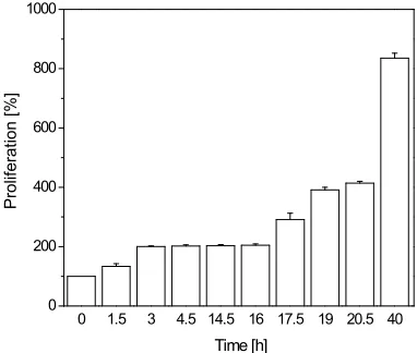
To analyse gap junctions during different phases of the cell cycle, granulosa cells were synchronised as describ- ed above. We found that mitotic cells harvested by mi-totic-shake-off completed mitosis within 3 h and contin-ued to undergo synchronised division for at least two ge- nerations every 16 h (Figure 1). Gap junction coupling
of the mitotic cells was analysed using a double whole cell patch-clamp beginning 2 h before the division and ending 1 h after the division. Correspondingly, the gap junction coupling of GFSHR-17 granulosa cells in inte- rphase was studied 4-14 h after the division. It was found that the macroscopic gap junction conductance (Gj)
in cells in mitotic phase was significantly reduced com-pared to the conductance of gap junctions in cells in the interphase (Figure 2(a)). At the molecular level, western
blotting revealed two forms of Cx43 (Figure 2(b)). A
0 1.5 3 4.5 14.5 16 17.5 19 20.5 40 0
200 400 600 800 1000
Time [h]
Prolif
erat
ion [
%
]
Figure 1. Synchronisation of GFSHR-17 granulosa
cells by the mitotis shake off method. It is shown that after selection, the mitotic cells finished mitosis wit- hin 3 h. Thereafter, they continued to divide in a syn-chronous manner for at least two generations. The re-sults were normalised to the population at the seeding time (105 cells) and are given as average SEM for at
least 10 experiments for each treatment.
0 5 10 15 20
25 Interphase
Gj [nS]
Mitosis
(a)
42 kDa 52 kDa
32 kDa
Mitosis Interphase
[image:4.595.349.498.80.370.2] [image:4.595.75.266.467.629.2](b)
Figure 2. (a) Absolute amplitudes of the
gap junction conductance in cells in inter- phase, in comparison with cells in mitotic phase. The amplitudes are significantly different (p < 0.01 student’s t-test); (b) Western blot analysis showing expression of Cx43, a representative of three indepe- ndent experiments. Two bands at 41-42 kDa and 44-46 kDa can be distinguished in both mitotic cells and in cells in interp- hase. The bands probably correlate to non- phosphorylated and phosphorylated Cx43, respectively. It is noteworthy that the non- phosphoprylated ba- nd is more intense in cells in interphase as compared to mitotic cells.
band at 41-42 kDa and a band to 44-46 kDa (Figure 2 (b), arrows). The Western blotting blot result showed in Figure 2(b) is a representative of three independent
Recently, it was shown that inhibition of gap junction coupling correlated with induction of apoptotic process- ses such as chromatin condensation or DNA strand br- eaks [10]. Therefore, we hypothesised that the sensitivity to gap junction inhibition-dependent stimulation of the apoptotic process would be increased in mitotic cells, which present low gap junction coupling (Figure 2(a)).
After applying the guanylyl cyclase inhibitor ODQ, which has been previously shown to suppress gap junc-tion coupling [10], the following results were obtained. 1) In agreement with our previous results, ODQ inhibits gap junction coupling as shown by dye transfer experi-ment (Figure 3). Quantitative analysis showed that, 10
min after the establishment of a whole-cell configuration on one cell within a monolayer (Figure 3), Lucifer
yel-low diffused in only 5.7 1.5 ODQ treated cells (n = 6 experiments), whereas under control conditions, 19.5
4.2 cells (n = 6 experiments) were achieved. In inter-phase cells a dye coupling and an ODQ-related reduction of coupling comparable to that observed in non synchro-nised cells (Figure 3) was found. In mitotic cells the dye
coupling was strongly reduced and ODQ treatment could not reinforce the reduction of coupling (results not sho- wn). 2) Analysis of chromatin structure and comet ass-
50 μm
50 μm
(a)
20 μm 20 μm
(b)
Figure 3.ODQ reduced gap junction coupling in GFSHR-17
granulosa cells. (a) Control experiment with cells not treated or treated (b) with 20µM ODQ for 6 h. The images were taken 10 min after establishment of the whole-cell patch-clamp conf- iguration on one cell in a monolayer with a pipette filling solu-tion containing 0.5% Lucifer yellow. Quantitative evaluasolu-tion showed that, in a monolayer, Lucifer yellow could diffuse into 19.5 4.2 cells (n = 6 experiments) under control conditions, whereas the treatment with ODQ reduced the coupled cells to 5.7 1.5 (n = 6 experiments).
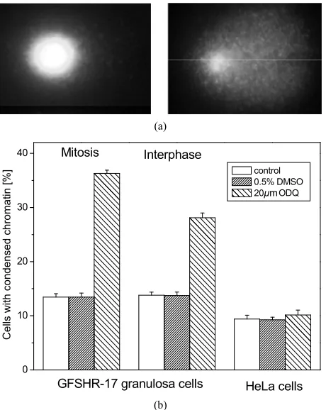
ays respectively revealed that the ODQ-dependent indu- ction of apoptotic reactions such as chromatin condensa-tion or DNA strand breaks was significantly increased in cells in mitotic phase, compared to cells in interphase (Figure 4 and Figure 5). ODQ dependent induction of
apoptotic reactions was only observed in cells which no- rmally express gap junction such as GFSHR-17 granul- osa cells. Cells which never relay on gap junctions such as HeLa cells were not affected (Figure 4(b) and Figure 5(b)).
4. DISCUSSION
The present report shows that gap junction coupling in the GFSHR-17 granulosa cells, the in vitro model for gr- anulosa cell in the maturing ovular follicle [12], is regu-lated in a cell cycle-dependent manner. We observed that cells in mitotic phase had a reduced gap junction condu- ctance (Gj) compared to cells in interphase (Figure 1,
Figure 2(a)). Gap junction communication serves as a
pathway for synchronisation of cells in tissues, allowing the formation of physiological units within an organ. Ho- wever, the cells must be able to evade the community of the cells for individual division. It is therefore hypothe-sised that mitotic cells reduce their gap junction coupling with neighbouring cells in order to undergo individual division. This hypothesis is in agreement with the obse- rvation that cellular division in tissues such as the ovular
0 2 4 6 8 10 12 14
HeLa cells Mitosis
Ta
ilmo
me
nt
control 0.5% DMSO 20礛 ODQ Interphase
GFSHR-17 granulosa cells
μm
Figure 4.Induction of chromatin condensation by ODQ. (a)
(a)
0 10 20 30
40 Interphase
HeLa cells
Ce
lls with
co
nd
en
sed
c
h
ro
ma
tin [
%
] control
0.5% DMSO 20礛 ODQ
GFSHR-17 granulosa cells Mitosis
μm
[image:6.595.58.288.79.368.2](b)
Figure 5. Induction of DNA strand breaking in GFSHR-17 gra-
llicle is not concomitant [22]. The closure of gap
junc-nction co
ulated that gap junction coupling re
ations presented in this report
nulosa cells by ODQ. (a) An example of non-affected cells (left) and affected cells (right) as revealed by comet assay experim- ents; (b) The tailmoment as a marker of DNA damage induced by treatment with ODQ is increased in cells in mitotic phase than in cells in interphase (p < 0.01 student’s t-test). The results represent the average SEM for n = 4 experiments. At least 1,000 cells were counted for each treatment.
fo
tion stops the exchange of metabolites such as second messengers between the cells [23] and thereby allows in- dividual mitotic division. Two forms of Cx43 were fou- nd in the GFSHR-17 granulosa cells by western blot an- alysis. These proteins had apparent molecular weights of 41-42 kDa and 44-46 kDa (Figure 2(b)). According to
Musil et al. [24], the revealed proteins correspond to the non-phosphorylated and phosphorylated forms of Cx43. A mitosis-dependent reduction of expression of connex-ins such as Cx43 was observed in different cell systems such as murine neocortical precursors [25]. However, our western blot analysis does not show a significant re- duction of the expression of Cx43, it only revealed that cells a dislevel of the non-phosphorylated form of Cx43 compared to cells in interphase (Figure 2(b)). We
hy-pothesise, therefore, that the observed reduction of the conductance of gap junction coupling is related to post-translational modifications such as phosphorylation. This hypothesis is in agreement with experiments that have
shown that phosphorylation reduced gap junction cou-pling [26] and that the phosphorylation of Cx- 43 was increased during the G2/M phase transition [11].
Recently, we showed that inhibition of gap ju upling in GFSHR-17 granulasa cells correlated with hallmarks of apoptosis, including induction of chromatin condensation, DNA strand breaks, and formation of ble- bs [10]. Since we observed that the cells in mitotic phase present a reduced Gj compared to cells in interphase and
non-synchronised cells (Figure 2(a)), we postulated that
the cells in mitotic phase should be more sensitive to the induction of apoptosis by ODQ, which inhibits gap junc-tion coupling [10] (Figure 3). This hypothesis was
con-firmed by the observation that ODQ induced an increase in the portion of cells with condensed chromatin (Figure 4), as well as an increased tailmoment in cells in mitotic
phase, compared to cells in interphase and non-synchr- onised cells (Figure 5). The importance of the gap
junc-tion coupling in the system is also shown by the experi-ments with HeLa cells which do not express gap juncti- ons. In the HeLa cells which do not relay on gap junc-tions the ODQ which acts by reducing gap junction did not induce the apoptotic reactions (Figure 4 and Figure 5). Furthermore the results with HeLa cells show that the
activation of apoptotic reactions is not a non specific effect of ODQ
Evidence has accum
gulates highly complex cellular functions. It was sho- wn that a reduction of gap junction coupling reduced the differentiation of cultured chick leg bud mesenchymal cells [27], the proliferation and fusion of the myoblast [6, 7] and the differentiation of progenitor cells of the retina [28,29]. Additionally, it was observed that inhibition of gap junction coupling was involved in stimulation of ap- optosis in granulosa cells [10,20]. Diverse molecular mechanisms that could link gap junction coupling to the cellular functions have been described. It was shown that gap junction coupling was involved in regulation of DNA synthesis [5] and in the expression and activation of cyclin-dependent kinase inhibitors (CDK-inhibitors) such as p21waf1/Cip1 and p27kip1[8,9]. Furthermore, it is
known that expression and activation of p21waf1/Cip1 and
p27kip1 is involved in regulation of the cell cycle and
apoptosis in granulosa cells [17,30].
5. CONCLUSIONS
Taken together, the observ
LEDGEMENTS
excel-y supported bexcel-y the project NANOTOME and th
uropean Graduate College; I fer
1) Emerging issues of connexin
chan-2003)Astrocytic
, Dang, X., Ping, P., Fandrich, R.R., Nickel,
, Becker, D.L., Dux, L., Stelkovics, E., Kren-
, J E. and Becker, D.L
a, I., Ikeda. M., Ma, K.W. and Mu-
eng,
.
204,137-144.
in43 phosphorylation at S368 is acute
Establishment of steroidegenic granulosa cell li-
43
(2001) Intercellular communication via
3 gap junction messenger ribonucleic acid and
and differential express-
one receptor and the cell cycle modulate apoptosis
ular
tion.
onal coupling, ion fluxes and cell
ytosolic phosph-
l Oncology,21(6),
004) Complex changes in cellular inositol
tion
cortical
propose that connexins phosphorylation-dependent mod- ulation of gap junction coupling is a relevant mechanism to regulate apoptosis of granulosa cells during the folic- ular maturation.
6. ACKNOW
The authors thank Hans-Georg Hannibal and Frank Koepke for lent technical support.
The project was partl
e DFG project Transregio 37/Q1.
Sabrina Schlie was supported by E nter- and connexin 45 but absence of connexin 40 in granulosa
cell gap junctions of rat ovary. Journal Reprod Fertil,
107(2),255-264.
[14] Ackert, C.L., Gittens, J.E.I., O’Brien, M.J., Eppig, J.J. and Kidder, G.M.
ence and Quantum Applications.
REFERENCES
[1] Harris, A.L. (200nels: Biophysics fills the gap. Quarterly Review of Bio-physics, 34(3), 325-472.
[2] White, T.W. and Paul D.L. (1999) Genetic diseases and gene knockouts reveal diverse connexin functions. Ann- ual Review of Physiology, 61(1),283-310.
[3] Willecke, K., Eiberger, J., Degen, J., Eckardt, J.D., Ro- mualdi, A., Guldenagel, M., Deutsch, U. and Sohl, G. (2002) Structural and functional diversity of connexin genes in the mouse and human genome. The Journal of Biological Chemistry, 383(5),725-737.
[4] Nakase, T., Fushiki, S. and Naus, C.C. (
gap junctions composed of connexin 43 reduce apoptotic neuronal damage in cerebral ischemia. Stroke, 34(8),
1987-1993. [5] Doble, B.W.
B.E., Jin, Y., Cattini, P.A. and Kardami, E. (2003) Phos-phorylation of serine 262 in the gap junction protein connexin-43 regulates DNA synthesis in cell-cell contact forming cardiomyocytes. Journal of Cell Science, 117(3),
507-514. [6] Gorbe, A.
acs, L., Bagdi, E. and Krenacs, T. (2005) Transient upr- egulation of connexin43 gap junctions and synchronized cell cycle control precede myoblast fusion in regenerat-ing skeletal muscle in vivo. Histochemistry and Cell Bi-ology, 123(6),573-583.
[7] Gorbe, A., Krenacs, T., Cook . oli
(2007) Myoblast proliferation and syncytial fusion both depend on connexin43 function in transfected skeletal muscle primary cultures. Experimental Cell Research,
313(6),1135-1148. [8] Zhang, Y.W., Morit
rota, S. (2001) Connexin43 suppresses proliferation of osteosarcoma U2OS cells through post-transcriptional regulation of p27. Oncogene, 20(31),4138-4149.
[9] Zhang, Y.W., Chen, X., Wu, D., Liu, W., Wang, J., F Z., Cai, G., Fu, B., Hong, Q. and Du, J. (2006) Downreg- ulation of connexin 43 expression by high glucose indu- ces senescence in glomerular mesangial cells. Journal of the American Society of Nephrology, 17(6),1532-1542.
[10] Ngezahayo, A., Altmann, B., Steffens, M. and Kolb, H A. (2005) Gap junction coupling and apoptosis in GF- SHR-17 granulosa cells. Journal of Membrane Biology
[11] Solan, J.L., Fry, M.D., TenBroek, E.M. and Lampe, P.D. (2003) Connex
during S and G2/M and in response to protein kinase C activation. Journal of Cell Science, 116(pt11), 2203- 2211.
[12] Keren, T.I., Dantes, A., Sprengel, R. and Amsterdam, A. (1993)
nes expressing follicle stimulating hormone receptors.
Molecular and Cellular Endocrinology, 95,R1-R10. [13] Okuma, A., Kuraoka, A., Iida, H., Inai, T., Wasano, K.
and Shibata, Y. (1996) Colocalization of connexin
connexin43 gap junctions is required for ovarian folicu- logenesis in the mouse. Developmental Biology, 233(2), 248-270.
[15] Wiesen, J.F. and Midgley, R.A. (1994) Expression of connexin4
protein during follicular atresia. Biology of Reproduction,
50(2),336-348.
[16] Wright, C.S., Becker, D.L., Lin, J.S., Warner, A.E. and Hardy, K. (2001) Stage-specific
ion of gap junctions in the mouse ovary: Connexin-spe- cific roles in follicular regulation. Reproduction, 121,77-
88.
[17] Quirk, M., Cowan, R.G. and Harman, R.M. (2004) Prog- ester
in granulosa cells. Endocrinol, 145(11),5033-5043. [18] Sasson, R. and Amsterdam, A. (2003) Pleiotropic anti-
apoptotic activity of glucocorticoids in ovarian follic cells. Biochemical Pharmacology, 66(8),1393-401.
[19] Lampe, P.D. and Lau, A.F. (2004) The effects of con-nexin phosphorylation on gap junctional communica
The International Journal of Biochemistry & Cell Biol-ogy, 36(7),1171-1186.
[20] Ngezahayo, A., Altmann, B. and Kolb, H.A. (2003) Regulation of gap juncti
volume by cGMP in GFSHR-17 granulosa cells. Journal of Membrane Biology, 194(3),165-176.
[21] Van Rossum, G.S.A.T., Vlug, A.S., van den Bosch, H., Verkleij, A.J. and Boonstra, J. (2001) C
pase A2 activity during the ongoing cell cycle. Journal of Cellular Physiology, 188,321-328.
[22] Schumer, S.T. and Cannistra, S.A. (2003) Granulosa cell tumor of the ovary. Journal of Clinica
1180-1189.
[23] Barker, C.J., Wright, J., Hughes, P.J., Kirk, C.J. and Mi-chel, R.H. (2
phosphate complement accompany transit through the cell cycle. Biochemical Journal, 380(pt 2),465-473. [24] Musil, L.S., Cunningham, B.A., Edelmann, G.M. and
Goodenough, D.A. (1990) Differential phosphoryla of the gap junction protein connexin43 in junctional co- mmunication-competent and deficient cell lines. The Journal of Cell Biology, 111(5 pt 1),2077-2088.
[25] Bittman, K.S. and LoTurco, J.J. (1999) Differential regulation of connexin 26 and 43 in murine neo precursors. Cerebral cortex, 9(2),188-195.
ction life
cy-, Tsakiricy-, N. and Cookcy-, J.E. (2007)
Mul-.L., Becker, D.L. and Mobbs,
tzuk, M.M. (2007) phosphorylation events regulate the gap jun
cle. Journal of Membrane Biology, 217(1-3),35-41. [27] Jin, E.J., Lee, S.Y., Jung, J.C., Bang, O.S. and Kang, S.S.
(2008) TGF-β3 inhibits chondrogenesis of cultured chick P. (2005) Gap junctions modulate interkinetic nuclear movement in retinal progenitor cells. The Journal of Neuroscience, 25(46),10803-10814.
[30] Andreu-Vieyra, C., Chen, R. and Ma leg bud mesenchymal cells via downregulation of
con-nexin 43 and integrin β4. Journal of Cellular Physiology,
214,345-353.
[28] Becker, D.L., Webb, K.F., Thrasivoulou, C., Lin, C.C., Nadershahi, R.
tiphoton imaging of chick retinal development in relation
to gap junctional communication. The Journal of Physi-ology, 585(pt 3),711-719
[29] Pearson, R.A., Lüneborg, N
Effects of granulosa cell-specific deletion of Rb in Inha-α