RESEARCH ARTICLE
CLINICAL EVALUATION OF 6% FERRIC OXALATE SOLUTION FOR PREVENTION OF ROOT
SENSITIVITY AFTER PERIODONTAL SURGERY: A RANDOMIZED, DOUBLE BLIND,
SPLIT MOUTH, CONTROLLED CLINICAL STUDY
*Dr. Kirti Satish Dulani, Dr. Neeta Vijay Bhavsar, Dr. Sakshee Rahul Trivedi
and Dr. Aarti Mangla
Department of Periodontology and Implantology, Government Dental College & Hospital, Asarwa, Ahmedabad
ARTICLE INFO ABSTRACT
Purpose: This is randomized, double blind, split mouth clinical study to evaluate the effectiveness of 6% ferric oxalate solution to prevent Root Sensitivity (RS) after periodontal flap surgery.
Methods: 25 subjects requiring periodontal surgery in similar bilateral posteriors quadrants were evaluated for RS with tactile, hot and cold test using Visual Analogue Scale (VAS) at baseline and 1, 2, 4, 6 weeks after surgery. Randomization was done with coin flip method for test (6% ferric oxalate in 0.9% saline) or control (0.9% saline) solution for each patient. Solutions were applied to the exposed root surfaces for 1 minute during surgery. Data were analyzed by repeated measures analysis of variance (ANOVA) for inter-group and paired t-test for intra-group comparisons.
Results: The test solution significantly reduced RS to tactile, hot and cold stimuliand for more time period than control solution.Sensitivity reduced to 85%, 66% and 53% for tactile, cold and hot stimuli respectively with test solution.Subjects got maximum discomfort on control sites from cold stimulus followed by tactile and least to hot stimulus during 1 and 2 week following surgery. Both the sides showed a gradual reduction in mean VAS, reaching to baseline values at 6 weeks.
Conclusion: The application of 6% ferric oxalate during periodontal flap surgery is a rapid and effective means of reducing RS after surgery. It can provide immediate relief to patient in a period when other agents will take longer time to act, thereby preventing discomfort from post-surgical RS.
Copyright © 2017, Dr. Kirti satish dulani et al. This is an open access article distributed under the Creative Commons Attribution License, which permits
unrestricted use, distribution, and reproduction in any medium, provided the original work is properly cited.
INTRODUCTION
Dentine Hypersensitivity (DH) is characterized by short sharp pain arising from exposed dentine in response to stimuli typically thermal, evaporative, tactile, osmotic or chemical and which cannot be ascribed to any other form of dental defect or pathology (Holland, 1997). Canadian Advisory Board has modified the above definition and has replaced the term “pathology” with “disease” (Canadian Advisory Board on Dentin Hpyersensitivity, 2003). DH can be caused by chronic trauma from tooth brushing, acid erosion from environment, gastric regurgitation or dietary substances, anatomical factors, gingival recession caused by periodontitis or periodontal surgery (Von Troil, 2002). Treatment of periodontal disease requires effective removal of bacterial deposits from the tooth surface by scaling and root planning and often access to deeper root surfaces by elevating periodontal flap.
*Corresponding author: Dr. Kirti Satish Dulani,
Department of Periodontology and Implantology, Government Dental College & Hospital, Asarwa, Ahmedabad.
This can lead to iatrogenic denudation of root dentin due to removal of the cementum layer and gingival recession (Tammaro, 2000). This causes increased DH after periodontal non surgical and surgical treatment. The occurrence of sensitivity on denuded root surface following periodontal therapy may be a condition distinct from DH occurring after hydrodynamic stimulation. The term “Root Sensitivity” (RS) is often used in this context (Von Troil, 2002). A multitude of methods have been described since many years for the management of DH/RSand there are many reviews providing information on the efficacy of the products used in the management of DH (Gillam, 2006; Dowell, 1983; Orchardson, 2006; Porto, 2009 and Wang, 1993). According to Gillam and Orchardson (2006), the treatment of DH/RS can be achieved by either dentinal tubule occlusion or blocking nerve activity
through direct iconic diffusion (increased K+ ions
concentration acting on the pulpal sensory nerve activity (Gillam, 2006). The products used for treatment of DH/RS have been classified according to their (a) mode of action, (b) whether they are self administered by the patients/over the counter (OTC) for home application or in office treatments
ISSN: 0975-833X
International Journal of Current Research
Vol. 9, Issue, 11, pp.61603-61608, November, 2017
OF CURRENT RESEARCH
Article History:
Received 15th August, 2017
Received in revised form 26th September, 2017
Accepted 25th October, 2017
Published online 30th November, 2017
Citation: Dr. Kirti Satish Dulani, Dr. Neeta Vijay Bhavsar, Dr. Sakshee Rahul Trivedi and Dr. Aarti Mangla. “Clinical evaluation of 6% ferric oxalate
solution for prevention of root sensitivity after periodontal surgery: a randomized, double blind, split mouth, controlled clinical study”, International Journal
of Current Research, Vol. 9, Issue, 11, pp.61603-61608, November, 2017
Key words:
Dentinal hypersensitivity, Root sensitivity, Periodontal flap surgery, Ferric oxalate,
applied by the dentists, (c) on their chemical or physical properties (Gillam, 2006). There effect could be either reversible or irreversible. The home use measures may be in form of dentifrices, gels or mouth rinses with active compounds such as formaldehyde, sodium fluoride, potassium
nitrate, strontium chloride, stannous fluoride etc. However,
they do not provide immediate effect and must be continuously used for a period of at least two weeks (Gillam, 2006). In office measures include the uses of cavity varnishes, sodium fluoride, stannous fluoride, adhesive resins, potassium nitrate, calcium phosphatesetcas well as periodontal grafting procedures and laser application (Gillam, 2006 and Porto, 2009). One of desensitizing agents used in the dental office is 6% ferric oxalate (FO) solution. The mechanism of action of ferric oxalate (Wang, 1993), is based on hydrodynamic hypothesis by Brannstrom (1967) (Brannstrom, 1986). The theory states that open dentinal tubules have an increase potential for dentinal fluid flow and therefore dentinal sensitivity (Brannstrom, 1986; Dragolich, 1993). It has been found that sensitive teeth have more concentration of open tubules and are wider in diameter (Dragolich, 1993). So, to treat DH, aim of the therapy should be towards either reducing the number of exposed tubules or reducing their diameter. The mechanism of action of ferric oxalate is by the dual precipitation of calcium oxalate and ferric phosphate saltsthat occlude open dentinal tubules (Dragolich, 1993). Following the application of ferric oxalate, 65% to 97% decrease in DH was recorded (Dragolich, 1993; Salvato, 1990 and Pashley, 1988). This agent has the added advantage of relative insolubility in acid (Yeh, 1990), making them resistant to dissolution after treatment. All of the above-mentioned desensitizing agents have been used only for the treatment of established tooth hypersensitivity, but have not been used for the prevention or reduction of the same specifically post surgically. Since DH is a common occurrence after periodontal surgery, a method to reduce or prevent this problem would be most helpful. Hence, an attempt was made in the present study, to evaluate the effect of 6% ferric oxalate solution applied during periodontal surgery for the prevention of RS.
MATERIALS AND METHODS
This study was randomized, split mouth, double blind, controlled clinical study over a period of 6 weeks. Twenty five individual of both the sexes, age ranging from 18 to 60 years were selected from the Department of Periodontology and Oral Implantology, Government Dental College and Hospital, Ahmedabad, Gujarat, India. According to power analysis, a sample size of 25 patients suffering from chronic generalized periodontitiswith bilateral similar suprabonyperiodontal defects achieves 80% power with a known standard deviation of 0.5 and with a significance level (alpha) of 0.050 using a two sided
dependent t test. The institutional Review Board and ethical
committee approved (dated April 2013) the study protocol and written and verbal consents were obtained from all study participants.
Patient selection criteria (By BNV)
Systemically healthy adults with chronic generalized severe
periodontitis with probing pocket depth of ≥ 5 mm and
radiographic evidence of suprabony horizontal bone loss requiring same type and extent of periodontal flap surgery were included in the study. Patients currently under treatment for DH, medications for chronic systemic disease, pregnancy and breast feeding, eating disorders, gastrointestinal disturbances,
cracked tooth, root canal treated, nonvital teeth, chipped teeth, generalized attrition/abrasion/erosion, defective restorations, orthodontic appliances, bridge work, denture, deep carious lesions and periodontal surgery done within last 6 months patients with number of teeth less than 6 were excluded from the study. Patients were tested for any allergy to ferric oxalate#. #All patients were referred to Skin Department of Civil hospital, Ahmedabad.
Study design (Supervised by BNV) (According to Wang et al
1993) (Wang, 1993)
The patient was selected and a detailed case history was recorded. Scaling and root planing was done. Patients were explained oral hygiene procedures and recalled after 3 to 4 weeks.Patients were revaluated and only those cases were included that were indicated for periodontal flap surgery and having RS. Baseline values for dentinal hypersensitivity tests were recorded, prior to flap surgery (before giving local anesthesia) and at 1, 2, 4 and 6 weeks after flap surgery by the first investigator (DKS). The examiner had been calibrated for performing all the tests by the supervisor (BNV).
Test stimuli
Patients were evaluated for RS level of the selected teeth by applying the following stimuli to the labial or buccal surface of each tooth using armamentarium shown in Fig 1a.The test (6% ferric oxalate in 0.9% saline) or control (0.9% saline) solutions were stored in identical colored bottles – labeled as Solution A and Solution B (Fig. 1b). The code of the solution applied to the particular site was maintained till statistical analysis was carried out.The solutions after breaking the code were found to be: Solution A: 6% ferric oxalate solution and Solution B: 0.9% normal saline solution. Therefore, the side to which Solution A was applied was the test side and side to which Solution B was applied was the control side. Sensitivty/pain response was assessed by using the Numerical 0-10 VAS where 0 = no pain and 10= intolerably severe pain (Fig. 1c).
Tactile test: A sharp dental explorer no. 23 was passed lightly
across the affected area, perpendicular to long axis of tooth (Fig. 1d). The test was repeated three times before the score was recorded. 2) Hot water test: This was done by loading hot water in a syringe of 5 ml volume and applying it to the exposed root surface for 3 seconds, after complete isolation of
teeth (Fig. 1e). The water was preheated in a water bath§ and
temperature of 50°C was measured with thermometer. 3) Cold water test: This was done by loading fresh ice water at 0°C in a pre-cooled syringe of 5 ml volume and applying it to the exposed root surface for 3 seconds, after complete isolation of teeth (Fig. 1e).
§:Avishkar international private limited, Mumbai,
Mahaarashtra, India.
Throughout the study, the stimuli were applied in the same order, with minimum 5 minutes gap between the applications of different stimuli. In any case, when discomfort becomes intolerable the stimulus was immediately removed.
Procedure for periodontal flap surgery
Periodontal flap surgery in quadrant including atleast 6 teeth was performed with a Modified Widman flap design (Ramfjord, 1974) and teeth were scaled, root planed and
debrided by the first investigator (DKS) only to standardize the procedure (Fig 1g). No regenerative or mucogingival techniques were used. Surgeries of bilateral periodontal pockets were scheduled in two consecutive appointments. The test or control solutions were applied randomly by a coin flip method on the buccal surfaces of exposed root surface of selected teeth by the second investigator (MAA) (Fig. 1h). Head was assigned for Solution A and tails was assigned for Solution B.
Procedure for applying solution: (MAA)
After complete debridement of the surgical area, it was isolated with cotton roles.Precaution was taken to see that the surgical area was not contaminated with saliva or blood. First investigator was then asked to step out. A small brush applicator was dipped in the solution A or B and the excess solution on the applicator was removed by dry cotton pellet.The solutions were applied on the buccal surfaces of exposed root surfaces of the teeth and left undisturbed for 60 seconds (Fig. 1h). Thereafter the tooth surfaces were irrigated with sterile water for 10 seconds.The flaps were adapted properly and sutured. Routine postsurgical instructions were given to each patient. Antibiotics and analgesics were prescribed.
Patients follow up: Patients were recalled at intervals of 1, 2, 4 and 6 weeks after surgery and were subjected to the tactile, hot and cold tests at each appointment and the responses were recorded for further analysis. At every follow up after surgery patients were asked at which side they were more comfortable with their daily food intake to assess their clinical outcomes in reduction of RS.
Statistical analysis: (Supervised by BNV)
The data was evaluated as Mean Standard of VAS. Student paired t – test was used to evaluate changes in sensitivity levels intra-group baseline 1, 2, 4 and 6 weeks after surgery. Mean scores were compared among groups at baseline, 1, 2, 4 and 6 week using repeated measures Analysis of Variance (ANOVA) to find out difference between the test and control group from baseline scores with the significance level of 0.05. Specific computer program used in statistical analysis was Statistical package for social sciences (SPSS) Version: 12. After the results were obtained, the second investigator (MAA) disclosed the identity of Solution A and Solution B.
RESULTS
A total number of 25 subjects were followed up for a period of 6 weeks. No post-operative complications such as delayed wound healing or adverse side effects were seen in any of the patient participating in the study. Table 1 shows intragroup and intergroup comparison of baseline VAS value of RS and observations at each interval for control and test solution with all the three stimuli. The recorded observation depict that after
surgery there is increase in RS from baseline to 1st week and 1st
week to 2nd week, except in test site with hot stimuli. The VAS
values for RS then gradually reduces from 4th week to 6th week
reaching values similar to baseline by end of 6 weeks. When comparing within group, the difference between baseline and each interval VAS values for RS for all the three stimuli is statistically significant for control site, contrary to the test site
where the difference is significant upto 2nd week only for cold
and tactile stimuli. While for hot stimuli the difference is not
significant at each interval. The intergroup comparison between test and control sites at various interval shows that the baseline mean VAS values for sensitivity to all the three stimuli are similar, the difference not being statistically significant (p>0.05). For each stimulus, the difference is statistically
significant at 1st week, 2nd week and 4th week between control
and test sites. The test site showing lower VAS values of RS.
At 6th week, VAS values for RS reach the baseline values for
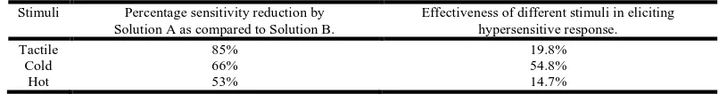
both the control and test site with no significant difference between the two. Table 2 shows percentage reduction of mean VAS values for sensitivity by test solution as compared to control. It was observed that tactile sensitivity reduced to 85%, cold sensitivity to 66% & hot sensitivity to 53% with test solution application. All the three stimuli were tested for their effectiveness to elicit hypersensitivity at baseline. It was found that patients got maximum discomfort from cold stimulus (54.7%) followed by tactile stimulus (19.8%), and least by hot stimulus (14.7%) (Table 2). Table 3 shows patients response to their daily food intake and oral hygiene procedures at different period intervals.
DISCUSSION
DH is frequently encountered and distinct clinical problem where patientsexperience considerable discomfort on eating hot, cold, acidic or sweet liquids and food (Brahmbhatt, 2012).Apart from attrition and abrasion, periodontal therapy appears to be a significant cause and several clinical studies and reviews have attempted to analyze the contribution of various clinical variables to the development of DH/RS after both non-surgical and surgical periodontal therapy (Nishida, 1976; Wallace, 1990; Chabanski, 2002 and Taani, 2002). Due to discomfort involved in brushing hypersensitive areas, patients tend to avoid these areas. Plaque and food debris are then allowed to remain on exposed surfaces, which often leads to increasing sensitivity which may create a vicious cycle. Therefore, DH resulting from periodontal surgery may influence plaque control measures and thus may compromise success of surgical therapy (Addy, 1987). So, it becomes important to prevent the post-surgical hypersensitivity, for the benefit of the patient. Different desensitizing agents available are used for the treatment of established tooth hypersensitivity, but none of them have been used for the prevention of the same. Thus present study evaluated the effect of 6% ferric oxalate solution applied during periodontal surgery for the prevention of DH/RS. Greenhill and Pashley 1981 (Greenhill, 1981), evaluated the ability of different desensitizing agents including oxalates on 133 dentin discs prepared from maxillary and mandibular unerupted third molars and reported that calcium oxalate crystals reduced the hydraulic conductance of dentin to approximately 98.4%. The crystals almost cover all dentinal tubules and appeared fairly regular. These crystals were connected to tubules with thread like structures.The authors concluded that the oxalate was most effective agent compared to fluoride, barium sulfate and silver nitrate. Yeh and Dangler (1990) conducted a study to measure the relative surface changes in dentin before and after application of ferric oxalate and the resistance of the effect to commonly experienced in vivo challenges such as tooth brushing and dietary acids. They concluded that 6% ferric oxalate solution is an effective dentin obturator which is also substantive when
evaluated in vitro. Dragolich et al. (1993) (Dragolich, 1993), in
of radicular dentin is indicated prior to the application of ferric oxalate in the treatment of root hypersensitivity. This agent has also got the ability to occlude dentinal tubules in the presence
or absence of smear layer. Gillam et al. (2001) (Gillam, 2001),
evaluated the effectiveness of four, in office oxalate products, in reducing dentine sensitivity including aluminium oxalate, ferric oxalate, oxalic acid and potassium oxalate. They concluded that professionally applied in-office products containing oxalate are capable of covering the dentine surface and/or occluding the tubules to varying degrees. The study was conducted as double blind where neither investigators nor patients were aware of solution’s name to avoid bias. Moreover for the advantage of same pain perception, oral hygiene habits, dietary habits and psychosomatic factors a split mouth study design was adopted (Brahmbhatt, 2012). According to Holland
et al. (1997) (Holland, 1997), DH most commonly presents on
buccal cervical surface of permanent teeth. So, sensitivity levels of the selected teeth were evaluated on the buccal surfaces of each tooth. Patients were evaluated for sensitivity level of the selected teeth by applying mechanical stimulation with a sharp dental explorer, hot water which was preheated to a temperature of 50°C, ice water with the temperature 0°C as these stimuli are both physiological and controllable. Scoring for hypersensitivity was done with VAS. It offers the advantages of being a continuous scale, thus providing quantitative measurements that are readily averaged and tested with parametric statistics (Holland, 1997).
Results from the study reveal that hypersensitivity scores for cold, hot and tactile stimuli were significantly lower in test group than control group at 1, 2 & 4 weeks after surgery. As
shown in Table 1 sensitivity level continues to increase from 1st
week to 4th week on control side, while it started reducing on
test side after 2nd week and for hot stimulus there was no
significant increase in RS on test side. This means that the post surgical sensitivity reached to its maximum in the control side at 2 weeks after surgery while at the same point of time, the mean VAS values of the test sides had already started
decreasing. The scores returned to baseline levels at 4th week
follow up for test side and at the 6th week for control side.
Baseline-4th week difference for test side is not statistically
significant (p>0.05), while on the control side difference is statistically significant (p<0.05) for all three stimuli. This indicates that the application of the test solution resulted in lower post-surgical sensitivity and the earlier reduction of sensitivity compared to the control side.On control side, there was 138% increase in sensitivity level to tactile stimulus, 162% to hot stimulus and 124% to cold stimulus whereas, there was increase of only 50% to tactile stimulus, 18% to hot stimulus
and 58% to cold stimulus on the test side at the end of 1st week
(Table 2). The results obtained in the present study for cold
stimulus are in agreement with Wang et al (Wang, 1993), and
Gillam et al (2004) (Gillam, 2004), who also reported
similar results. Wang et al. (1993) (Wang, 1993),demonstrated
statistically significant reduction in the responses of thermal stimuli, especially cold, between groups treated with ferric oxalate as compared to those treated with saline. It was concluded that 6% ferric oxalate was more effective in reducing post-surgical cold sensitivity when applied during
periodontal surgery. Gillam et al (2004) (Newman, 2004)
[image:4.595.46.549.87.169.2]demonstrated that a 1-min application of ferric oxalate is both rapid and effective in reducing DH although its long-term effectiveness still needs to be determined. However they used ferric oxalate during non surgical periodontal treatment. As
Table 1. Intra group and Intergroup comparison of sensitivity scores for tactile, hot and cold stimuli between test and control solutions
Stimuli Cold Hot Tactile
Time Interval Control(mean±sd) Test(mean±sd) ‘t’
value
Control(mean ±sd)
Test(mean ±sd)
‘t’ value
Control(mea n±sd)
Test(mean ±sd)
‘t’ value
Baseline 2.43±0.51 2.32±0.43 1.86 0.56±0.12 0.58±0.09 0.70 0.84±0.18 0.82±0.21 0.97
1 week 5.08±0.83 a 3.68±0.38 a 16.45* 1.32±0.45 a 0.69±0.38 4.74* 1.98±0.52 a 1.23±0.37 a 12.45*
2 week 5.45±0.79 a 3.15±0.38 a 17.82* 1.47±0.32 a 0.61±0.29 5.01* 1.54±0.57 a 1.00±0.38 a 10.75*
4 week 3.84±0.43 a 2.45±0.25 8.44* 0.97±0.25 a 0.56±0.17 3.39* 1.04±0.41 a 0.78±0.39 5.42*
6 week 2.56±0.19 2.18±0.23 2.25 0.55±0.12 0.48±0.09 1.20 0.79±0.14 0.76±0.12 1.24
[image:4.595.94.497.227.280.2]a: significant difference from baseline; p<0.05. (intragroup) *: significant difference; p<0.05.(intergroup).
Table 2. Percentage sensitivity reduction by Solution A as compared to Solution B and Effectiveness of different stimuli in eliciting hypersensitive response
Stimuli Percentage sensitivity reduction by
Solution A as compared to Solution B.
Effectiveness of different stimuli in eliciting hypersensitive response.
Tactile 85% 19.8%
Cold 66% 54.8%
Hot 53% 14.7%
Table 3. Response of patient’s for daily food intake at 1st, 2nd, 4th and 6th week interval
Patient’s satisfaction With cold food With hot food With regular food
Test control test control test control
1st week 0 0 5 satisfied
20 not satisfied
0 11 satisfied
14 not satisfied
2 satisfied 23 not satisfied
2nd week 11satisfied
14 not satisfied
0 13 satisfied
12 not satisfied
7 satisfied 18 not satisfied
20 satisfied 5 not satisfied
2 satisfied 23 not satisfied
4th week 20 satisfied
5 not satisfied
12 satisfied 13 not satisfied
25 satisfied 21 satisfied
4not satisfied
22 satisfied 3 not satisfied
15 satisfied 10 not satisfied
6th week 23 satisfied
2 not satisfied
15 satisfied 10 not satisfied
25 satisfied 22 satisfied
3 not satisfied
25 satisfied 25 satisfied
[image:4.595.57.539.310.410.2]were lower than that of cold test with lowest level for hot test.
Fig. 1. Surgical procedure, solutions application and testing stimuli for root sensitivity 1. Armamentarium for testing root sensitivityTwo
identical bottles containing test & control solution a.Visual Analog Scale
b.Application of Tactile Stimulus c.Application of hot and cold stimulus
d.Patient using Visual Analog Scale e.After flap reflection & debridement f.Application of Solution with a brush applicator
This is because most of the subjects did not have as strong a response to hot stimuli as they did to cold stimuli. Approximately, 75% of patients with DH complain of pain with application of cold stimuli (Chidchuangchai, 2007). In a
study by Gillam et al. in 2002 (Gillam, 2002), for evaluation of
frequency, distribution and severity of DH in subjects recruited for clinical trial of desensitizing agents, DH to cold was the main presenting symptom. Ong & Strahan (1989) (Ong, 1989), in their study to assess the effectiveness of a dentrifice containing 2% dibasic sodium citrate in poloxamer 407 for treatment of DH showed of all the stimuli used cold was the most effective in eliciting hypersensitivity response, followed by chemical stimulation and air, while heat and tooth brushing caused least discomfort (Gillam, 2002). As shown in Table 1, for the hot stimulus there was no statistically significance
difference between the Baseline-1st ,2nd ,3rd and4th in the test
side, while in the control side statistically significant differences (p<0.05) were observed between Baseline and 1,2 and 4 weeks post surgically. This indicates that on test side sensitivity level to hot stimulus was almost near to baseline values i.e. on test side patients experienced almost no sensitivity than on control sides. This finding is in agreement
1990). Salvato et al. in 1990 (Salvato, 1990), studied the
effectiveness of 6% aqueous ferric oxalate solution in relieving dentinal hypersensitivity in 38 patients. Sensitivity was recorded by using the Yeaple probe, air sensitivity and global subjective assessment utilizing the visual analog scale. Data was collected post application at 5 minutes and again at 1, 4 and 8 weeks. The data for ferric oxalate group showed 1) significant subject improvement from air sensitivity at all points over placebo 2) significance at week 1 and 8 for subjective response over placebo and 3) improved tactile sensitivity from baseline at all points. From the study they concluded that the ferric oxalate is a rapid and effective agent for the relief of dentinal hypersensitivity. In the present study, most of the patients experienced the highest level of sensitivity
during the 1st or 2nd week following surgery. This result is in
agreement with that reported byWang et al. (1993), Nishida et
al. (1976), Wallace et al. (1990), Uchida et al. (1980),
Al-Sabbagh et al. (2010) and Vaitkeviciene I et al. (2006). In all
the three stimuli tested, mean VAS values returned to almost near to baseline values at 6 weeks. This may be explained by the natural occlusion of dentinal tubules. Pashley (1996) stated that spontaneous remission of symptoms which is observed in most instances occur somewhere between 7-14 days after surgery but may require several weeks to fully resolve.
Tamminen et al. (1998) reported in a study that post-surgical
hypersensitivity reaches to its peak in 2 to 4 weeks after surgery and it may take several weeks to reach to its baseline values. This occurs due to natural occlusion of dentinal tubules which can occur through the formation of calculus, intratubular crystals from salivary minerals, peritubular dentin, collagen plugs, or the absorption of large plasma proteins leaking into the blood vessels and leaking into the tubules (Kerns, 1991). As shown in Table 3 more patients were satisfied to their daily food intake at different intervals on test side as compared to control side. This is in accordance to VAS values for RS to all stimuli. DH is the condition where clinician has to depend on subjective assessmentof the individual response and it is extremely difficult to evaluate DH objectively, thus there can be variability in response and lacks standardized measurability, VAS being the only practical method.
Conclusion
Thus study suggests that the application of 6% ferric oxalate during periodontal flap surgery is a rapid & effective means of reducing the post-surgical hypersensitivity when applied during surgery. This agent can provide immediate relief to the patient in a period when other agents will take time to act. So, it can help to reduce pain and discomfort of the patient. In future, it would be interesting to carry out studies monitoring effects of various desensitizing agents for the prevention of post-surgical hypersensitivity.
Conflict of Interest and Sources of funding
No conflict of interest declared by any of the authors.
This study has been self supported by the authors and
that has not been funded by any organization.
REFERENCES
Al-Sabbagh M, Beneduce C, Andreana S, Ciancio SG 2010. Incidence and time course of dentinal hypersensitivity after
periodontal surgery. J Gen Dent.; 58:e14-9.
Brahmbhatt N, Bhavsar N, Sahayata V, Acharya A, Kshatriya P 2012. Adouble blind controlled trial comparing three
treatment modalities for dentin hypersensitivity. Med Oral
Patol Oral Cir Bucal.; 17:e483-490. doi:10.4317/mwdoral.
17594
Brannstrom M 1986. The hydrodynamic theory of dentinal pain: sensation in preparations, caries, and the dentinal
crack syndrome. J Endod.; 12:453-457. doi:
http://dx.doi.org/10.1016/50009-2399(86)80198-4
Canadian Advisory Board on Dentin Hpyersensitivity 2003. Consensus-based recommendations for the diagnosis and
management of dentin hypersensitivity. J Can Dent Assoc.;
69:221-226.
Chabanski MB, Gillam DG, Bulman JS, Newman HN 1996. Prevalence of cervical dentine hypersensitivity in a
population of patients referred to a specialist
Periodontology Department. JClinPeriodontol.;23:989-992.
Chidchuangchai W, Vongsavan N, Mathews B 2007. Sensory transduction mechanisms responsible for pain caused by
cold stimulation of dentine in man. Arch Oral Biol.;
52:154-160. doi:10.1016/j.archoralbio.2006.09
Dowell P, Addy M 1983. Dentin hypersensitivity – A review
Aetiology, symptoms and theories of pain production. J
ClinPeriodontol.; 10:341-350. doi:
10.1111/j.1600-051x.1983.tb01284.x
Dragolich WE, Pashley DH, Beennan WA, O’ Neal RB, Horner JA, Van Dyke TE 1993. An in vitro srtudy of
dentinal tubule occlusion by ferric oxalate. J Periodontol.;
64:1045-1051.doi: 10.1902/jop.1993:64.11.1045
Gillam DG, Aris A, Bulman JS, Newman HN, Ley F 2002. Dentine hypersensitivity in subjects recruited for clinical trials: clinical evaluation, prevalence and intra-oral
distribution. J Oral Rehabil.; 29:226-231.
Gillam DG, Mordan NJ, Sinodinou AD, Tang JY, Knowles JC, Gibson IR 2001. The effects of oxalate-containing products
on the exposed dentine surface: an SEM investigation. J
Oral Rehabil.; 28:1037-44.doi: 10.1111/j.1365-2842.
2001.00775.x
Gillam, D.G., Newman, H.N., Davies, E.H., Bulman, J.S., Troullos, E.S. & Curro, F.A. 2004. Clinical evaluation of
ferric oxalate in relieving dentine hypersensitivity. J Oral
Rehabil., 31:245–252. doi:10.1046/j.0305-182x.2003.012
30.x
Gillam, D.G.,Orchardson, R 2006. Advances in the treatment of root dentine sensitivity: mechanisms and treatment
principles. Endod Topics.;13:13–33. doi:
10.1111/j.1601-1546.2006.00209.x
Greenhill JD, Pashley DH 1981. The effects of desensitizing agents on the hydraulic conductance of human dentin in
vitro. J Dent Res.; 60:686-98. . doi: 10.1177/ 00220345810
600030401
Holland GR, Nahri MN, Addy M, Gangarosa L, Orchardson R 1997. Guidelines for the design and conduct of clinical
trials on dentine hypersensitivity. J ClinPeriodontol.;
24:808-813. doi: 10.1111/j.1600-051x.1997.tb01194.x Kerns DG, Scheidt MJ, Pashley DH, Horner JA, Strong SL,
Van Dyke TE 1991. Dentinal tubule occlusion and root
hypersensitivity. JPeriodontol.; 62:421-428. doi: 10.1902/
jop1991.62.7.421
Lin YH, Gillam DG 2012. The prevalence of root sensitivity
following periodontal therapy: A systemic review. Int J
Dent.; 1-12. doi: 10.1155/2012/407023
Nishida M et al 1976. Hypersensitivity of the exposed root
surfaces after surgical periodontal treatment. J Osaka Univ Dent Sch.;16:73-85. doi: 10.2329/ perio.18.502. 197618 (4) 502.510
Ong G, Strahan JD 1989. Efffect of a desensitizing dentrifice
on dentinal hypersensitivity. Endod Dent Traumatol
.;5:213-218.
Orchardson R, Gillam DG 2006. Managing dentin
hypersensitivity. J Am dent Assoc.; 137:990-998.
Pashley, D.H., 1996. Dynamics of the pulpodentin complex.
Crit Rev Oral Biol M.;7:104-133.doi: 10.1177/ 104544119
60070020101
Pashley, D.H., Derkson, G.D. Tao, L., Derkson, M., Kalathoor, S. 1988. The effects of a muti-step dentin bonding system on dentin permeability. Dent Mater.;4:60-63. doi: http://dx. doi.org/10.1016/s0109-5641(88)80091-5
Porto IC, Andrade AK, Montes MA 2009. Diagnosis and
treatment of dentinal hypersensitivity. J Oral Sci.;
51:323-332.
Ramfjord SP, Nissle RR 1974. The modified widman flap. J Periodontol.;43:601-607. doi: 10.1902/jop.1974.45.8.2.601 Salvato A, Troullos ES, Curro FA, Merola MR, Gingold J
1990. The effectiveness of ferric oxalate (FO) in relieving
dentinal hypersensitivity. J Dent Res 69(Spec. Issue):169
(Abstr no. 482). doi:10.1177/0022034590069s101
Tammaro S, Wennstrom JL, Bergenholtz G 2000. Root–dentin
sensitivity following non-surgical periodontal treatment. J
ClinPeriodontol.; 27:690-697. doi: 10.1034/j.1600-051x
2000.027009690.x
Tamminen U, Konturri-Narhi V, Narhi M 1998. Development
of pulp and dentin densitivity in flap operated teeth. J Dent
Res 77(Special Issue): 672 (abstract no. 322). doi:
10.1177/0022034598077s201
Taani Q, Awartani F 2002. Clinical evaluation of cervical dentin sensitivity (CDS) in patients attending general dental
clinics (GDC) and periodontal specialty clinics (PSC). J
ClinPeriodontol.; 29:118-122.
Uchida A, Wakano Y, Fukuyama O, Miki T, Iwayama Y, Okada H 1980. Controlled clinical evaluation of a 10% strontium chloride dentifrice in treatment of dentin
hypersensitivity following periodontal surgery. J
Periodontol.; 51:578-581. doi: 10.1902/jop.1980.51.10.578
Vaitkeviciene I, Vaitkevicius R, Paipaliene P, and Zekonis G 2006. Morphometric analysis of pulpal myelinated nerve fibres in human teeth with chonic periodontitis and root
sensitivity. Medicina.;42:914-922.
Von Troil B, Needleman I, Sanz M 2002. A systematic review of the prevalence of root sensitivity following periodontal
therapy. J ClinPeriodontol.; 29(Suppl 3):173-177. doi:
10.1034/j.1600-051x.29.s3.19x
Wallace JA, Bissada NF 1990. Pulpal and root sensitivity rated to periodontal therapy. OralSurg Oral Med Oral Pathol.;69:743-747. doi: http://dx.doi.org/10.1016/0030-4220(90) 90360-5
Wang HL, Yeh CT, Smith F, Burgett FG, Richards P, Shyr Y, O’Neal R 1993. Evaluation of ferric oxalate as an agent for use during surgery to prevent post-operative root
hypersensitivity. J Periodontol.; 64:1040-1044. doi: 10.
1902/jop.1933.64.11.1040
Yeh K, Dangler L Sena F, Zientek L, Denooyer M, Weedle L
1990. Use of ferric oxalate to reduce dentin permeability. J
Dent Res.; 69 (Spec. Issue):168 (Abstr no. 479). doi:
10.1177/0022034590069s101