Original Article
Effects of sevoflurane and propofol on right ventricular
function and pulmonary circulation in patients
undergone esophagectomy
Wen-Yun Xu*, Na Wang*, Hai-Tao Xu, Hong-Bin Yuan, Hai-Jing Sun, Chun-Li Dun, Shuang-Qiong Zhou, Zui
Zou, Xue-Yin Shi
Department of Anesthesiology, Changzheng Hospital, Second Military Medical University, 415 Fengyang Road, Shanghai, People’s Republic of China. *Equal contributors.
Received September 11, 2013; Accepted October 23, 2013; Epub December 15, 2013; Published January 1, 2014
Abstract: Object: Sevoflurane and propofol are both widely used in clinical anesthesia. The aim of this study is to compare the effects of sevoflurane and propofol on right ventricular function and pulmonary circulation in pa
-tients receiving esophagectomy. Methods: Forty adult pa-tients undergoing an elective open-chest thoracotomy for esophagectomy were randomized to receive either propofol (n=20) or sevoflurane (n=20) as the main anesthetic agent. The study was performed in Changzheng Hospital. Hemodynamic data were recorded at specific intervals: before the surgery (T0), BIS values reaching 40 after anesthesia induction (T1), two-lung ventilation (T2), ten min
-utes after one-lung ventilation (T3), the end of the operation (T4) using PiCCO and Swan-Ganz catheter. Results: CI, RVEF, RVSWI and RVEDVI were significantly smaller in propofol group than those in sevoflurane group through
-out the surgery (P<0.05). However, SVRI was significantly greater in propofol group than that in sevoflurane group (P<0.05). Compared with the patients in propofol group, the patients who received sevoflurane had a greater reduc
-tion in OI and increase in Os/Ot (P<0.05). And, PVRI was significantly smaller in sevoflurane group than in propofol group (P<0.05). Conclusion: Anesthesia with sevoflurane preserved better right ventricular function than propofol in patients receiving esophagectomy. However, propofol improved oxygenation and shunt fraction during one-lung ventilation compared with sevoflurane anesthesia. To have the best effect, anesthesiologists can choose the two anesthetics flexibly according to the monitoring results.
Keywords: Sevoflurane, propofol, right ventricular function, pulmonary circulation, esophagectomy
Introduction
Mortality from esophagectomy has declined
over the past 30 years [1]. Anastomotic and cardiopulmonary complications contribute to
the majority of perioperative morbidity and mortality. How to reduce the incidence of these
complications has become more and more con-cerned. Some studies have demonstrated that
the choice of an appropriate anesthesia meth -od may achieve a go-od outcome. So we are
intended to investigate the effects of different
anesthetics on patients who are undergoing esophagectomy.
Esophagectomy has obvious effects on hemo -dynamics. One-lung ventilation can result in increased pulmonary vascular resistance (PVR)
induced by hypoxic pulmonary vasoconstriction (HPV), increased ventilation pressure, hypox-emia or hypercarbia [2]. These consequences can lead to hemodynamic instability. A great
number of disputes have risen about volatile
anesthetics and intravenous anesthetics in the open chest surgery. Researches showed that
fluoroalkenes and sevoflurane could reduce pulmonary inflammatory reaction when com
-pared with propofol. However, other studies found that sevoflurane would cause more seri
-ous inflammation reactions than propofol in
thoracic surgery [3].
useful to optimize hemodynamic management of patients undergoing esophagectomy.
However, there are few researches on the impact of sevoflurane and propofol on RVF in
esophageal surgery. This study is aimed to
investigate the effects of sevoflurane and pro
-pofol on the right ventricular function and the pulmonary circulation of patients receiving
esophagectomy. We hypothesized that
oxygen-ation and right ventricular function might become better during propofol and during sevo
-flurane respectively. There are many methods to monitor the right heart function. The Swan-Ganz catheter methods as the golden standard of hemodynamic monitoring have been used for many years [5]. In addition, this study adopt -ed PiCCO as an advanc-ed hemodynamic moni-toring indicator which allowed the
anesthesiol-ogists to improve fluid management,
hemodynamics and pulmonary gas exchange.
Methods
Patient population
The study was approved by the Institutional Ethical Committee (Second Military Medical
University) and written informed consent was
obtained. 40 patients (between 50 and 75 years old) undergoing an elective open-chest
thoracotomy for esophagectomy were prospec -tively enrolled whose ASA physical status were
I-II. Patients with cardiac disease, heart failure, arrhythmia, bronchial inflammation, coagula
-tion disorders, hepatic and renal insufficiency
were excluded. Patients were randomly (by
opening of an envelope) allocated to receive either propofol (group P) or sevoflurane (group
S).
Anesthesia and surgery
In the operating room, the patients received routine monitoring, including electro-cardio-gram, pulse oximetry, capnoelectro-cardio-gram, arterial pres-sure, and bispectral index (BIS). A Swan-ganz catheter was placed into the right external jugu-lar vein. A thermistor-tipped catheter (4 F,
PiCCO Catheter, Pulsion, Munich, Germany) was placed into the right femoral artery in the purpose of detecting transcardiopulmonary
thermodilution. In group P, anesthesia was
induced with a target-controlled infusion of pro
-pofol at a target plasma concentration of 6 μg/
ml, adjusting BIS value between 40-60. In
group S, sevoflurane was initially given at the concentration of 8% (6 L/min) so as to prime the anesthesia machine pipeline for 3 minutes.
As soon as the patients lost consciousness and
BIS values dropped to 40, remifentanil was given at a target plasma concentration of 4 ng/ ml. The patients were intubated with a left-sid -ed double-lumen tube (DLT) (Smiths, Hythe,
Kent, UK: 39 F for males and 37 F for females) after cisatracurium (0.15 mg kg-1) was given.
Fiberoptic bronchoscopy was used to confirm the correct positioning of the DLT immediately after its blind insertion. The lungs were venti
-lated with a Primus Ventilator (Drager, Lubeck, Germany). VT was set at 8 ml kg-1 and FiO2 was
100%. Respiratory frequency was adjusted to
12-14/min to maintain end-tidal-carbon-diox-ide tension (EtCO2) between 35-45 cmH2O.
Hemodynamic data analysis
The following parameters were recorded: mean
arterial pressure (MAP), central venous pres-sure (CVP), mean pulmonary artery prespres-sure (PMAP), pulmonary artery wedge pressure
(PAWP), stroke volume index (SVI), cardiac index
(CI), and systemic vascular resistance index (SVRI), RV hemodynamic data (right ventricular
ejection fraction [RVEF], RV end-diastolic vol
-ume index [RVEDVI], RV stroke work index
[RVSWI]), pulmonary vascular resistance index (PVRI), transmembrane pressure, mixed-venous and arterial blood gas analysis.
Pulmonary shunt fraction (Qs/Qt) and oxygen
-ation index (OI) were calculated by the following formula:
Qs/Qt = (PAa-DO2×0.0331)/PA-aDO2×0.0331+
(CaO2-CvO2)
OI = PaO2/FiO2
transmembrane pressure = PADP (pulmonary
artery diastolic pressure)-PAWP
These parameters were all recorded at specific intervals: before the surgery (T0), BIS values reaching 40 after anesthesia induction (T1), two-lung ventilation (T2), 10 minutes after one-lung ventilation (T3) and the end of the opera
-tion (T4). Each interval lasted for 10 minutes after stability, and we took the average value in three times as the final data for all the patients.
Statistical analysis
and variance analysis of repeated measure
-ment were performed. For count data, constitu
-ent ratio and chi-square analysis were used. Wilcoxon
rank-sum test was used in comparison of non-normal
distribution and variance heterologous measurement data. For all data analyzed, a
P value≤0.05 was consid
-ered statistically significant.
Statistical tests were
per-formed using SPSS version
17.0 (SPSS Inc., Chicago, IL, USA).
Results
There were no significant dif
-ferences in the characteris
-tics of the patients (Table 1).
And there were no differenc -es between th-ese two groups
in the duration of operation,
anesthesia and one-lung
ventilation, the amount of bleeding, the total amount of fluid infusion and urine vol -ume. Compared with group P,
the incident of hypoxemia in
the operation was higher in group S. One patient in group
P developed and died of anastomotic leakage after
operation. Another three patients in group S also died,
in which one died of massive
hemorrhage in the digestive
tract, and two died of anasto
-motic leakage. There were no differences in the number of
days in hospital and the time
of removing gastric tube. But the number of days staying
at intensive care unit in group P was much more than that in group S (Table 2). The patients were
compara-ble for all measured and
derived variables at baseline (Table 3). The values for MAP,
SVI, PMAP, CVP and PAWP
did not significantly differ
[image:3.612.92.383.84.150.2]between two groups (Figure
Table 1. Patient characteristics and baseline findings
Propofol (n=20) Sevoflurane (n=20)
Age (yr) 59.0±7.8 60.6±6.6
BMI (kg/m2) 22.1±2.2 23.1±2.2
Sex (M/F) 15/5 14/6 ASA (I/II) 16/4 17/3
[image:3.612.92.384.196.435.2]Data were expressed as mean ± SD. There were no differences between two groups.
Table 2. Other data in perioperative
Propofol (n=20) Sevoflurane (n=20)
Operation time (min) 281.15±90.01 226.77±58.84
Anesthesia time (min) 329.77±67.65 298.00±54.83
One lung ventilation time (min) 143.23±34.02 147.08±28.62
hypoxemia (n) 2 8*
Mount of bleeding (ml) 337.5±102.52 250.00±67.7
Mount of fluid infusion (ml) 2269.23±655.92 1929.85±585.15
Urine volume (ml) 784.62±256.89 789.23±404.67
Intensive care unit stay (d) 4.69±3.66 2.23±1.36* Hospital stay (d) 19.92±14.22 26.60±24.87 Time of removing gastric tube (d) 10.31±8.42 14.90±17.55 Complication:
ARDS 1 0
Pulmonary infection 2 0
Anastomotic leakage 3 6
arrhythmia 1 0
Cardiac insufficiency 0 1
death 1 3
Data were expressed as mean ± SD. *P<0.05 sevoflurane group vs propofol group.
Table 3. Hemodynamic findings at baseline
Propofol (n=20) Sevoflurane (n=20)
MAP (mmHg) 109.5±13.9 107.7±12.3 CVP (mmHg) 5.8±2.8 5.4±2.0 PMAP (mmHg) 19.2±5.6 15.8±2.5
PAWP (mmHg) 9.8±3.5 8.5±2.5
CI (L/min·m2) 3.9±0.8 3.9±0.6
RVEF (%) 38.4±7.3 37.3±7.8
RVSWI (g·m/m2) 7.8±2.8 7.0±2.4 RVEDVI (ml/m2) 122.6±22.3 135.1±23.0
GEDVI (ml/m2) 755.5±132.8 832.8±152.1
EVLWI (ml/kg) 6.9±2.0 8.2±4.0
OI (mmHg) 425.7±41.5 427.8±74.0
PVRI (dyn·s-1·cm-5) 185.1±62.2 157.5±60.6
Qs/Qt (%) 12.8±3.2 13.6±3.6
Data were expressed as mean ± SD. There were no differences between two groups.
1). Both groups exhibited a significant reduc
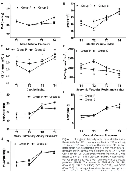
[image:3.612.92.380.480.667.2]-Figure 1. Changes in hemodynamic data at after anes -thesia induction (T1), two lung ventilation (T2), one lung
ventilation (T3) and the end of the operation (T4) in pro
-pofol group and sevoflurane group. A was mean arterial pressure (MAP), B was stroke volume index (SVI), C was Cardiac index (CI), D was stroke volume index (SVI), E was
mean pulmonary artery pressure (PMAP), F was central
venous pressure (CVP), G was pulmonary artery wedge pressure (PAWP). The values for MAP (P=0.140), SVI (P=0.369), PMAP (P=0.790), CVP (P=0.899), and PAWP (P=0.203) did not significant differ between two groups. CI was significantly smaller in propofol group than in sevo
-flurane group throughout the surgery (P=0.007). SVRI was significantly greater in propofol group than in sevoflu
-rane group (P=0.022). *P<0.05 between groups.
[image:4.612.88.525.65.654.2]sevoflurane group (Figure 1). Similarly, RVEF,
RVSWI and RVEDVI of the patients who received
sevoflurane were significantly greater all the time after baseline than those of the patients who received propofol (Figure 2).
Conversion from TLV to OLV caused a signifi
-cant decrease in OI (p=0.000) and an increase in Qs/Qt (p=0.000) in both groups. Compared with the patients who received propofol, those who received sevoflurane got even greater
reduction in OI and increase in Os/Ot (Figure 3).
However, PVRI was significantly smaller in the sevoflurane group than in the propofol group. And there were no significant differences in
transmembrane pressure between the two groups (Figure 3).
Discussion
In order to reduce the postoperative
complica-tion, anesthesiologists have adopted plenty of approaches, including the choice of appropri
-ate way of anesthesia, dose of anesthetics,
respiratory parameters, ventilation mode,
goal-directed fluid therapy, oxygenation improve -ment with drugs, ventilated-lung protection and
etc. Propofol and sevoflurane are common regi
-men in clinical anesthesia. This study employed
PiCCO and Swan-Ganz catheter methods to compare the influence of sevoflurane and pro
-pofol on the right heart function and pulmonary
circulation.
Patients’ characteristics, the duration of opera -tion, anesthesia and one-lung ventilation were similar in both groups. The intraoperative
bleeding loss, the total amount of fluid infusion,
and urine volume were also similar among all
the patients, which suggested that the differ
-ences in cardiac function between the groups were not caused by the differences in patients’
characteristics and intraoperative events but
were related to the choice of anesthetic agent.
The results of the present study indicated that the choice of the anesthetic regimen influenced cardiac function and pulmonary circulation.
Indeed, the patients who were anesthetized
with sevoflurane obtained significantly better cardiac function, but lower oxygenation than
the patients who were anesthetized with
propo-fol. And sevoflurane had greater influence on
the peripheral vascular resistance than
[image:5.612.94.321.73.370.2]propofol.
Figure 2. Changes in right ventricular function data at after anesthesia induction (T1), two lung ventilation (T2), one lung ventilation (T3) and the end of the op
-eration (T4) in propofol group and sevoflurane group. A was right ventricular ejection fraction (RVEF), B was RV stroke work index (RVSWI), C was RV end-diastol
-ic volume index (RVEDVI). RVEF (P=0.008), RVEDVI (P=0.019), RVSWI (P=0.003) were significantly smaller in propofol group than in sevoflurane group throughout
In this study, we had shown that although there
were no significant differences in MAP, SVI, CVP
and PMAP between the two groups, the patients
who received propofol experienced a greater
and more sustained reduction in CI, RVEDVI, RVSWI and RVEF when compared with those
who received sevoflurane. Some studies have found that sevoflurane preserved myocardial function better than propofol in myocardial ischemia-reperfusion [6, 7]. These results may be explained by the vasodilatory effects of pro
-pofol and a reduction in cardiac baroreflex sen
-sitivity by propofol. Immunoregulatory effects of volatile anesthetic might contribute to our results. Recent studies have identified an immunoregulatory role of sevoflurane that had significant reduction of inflammatory mediators and a remarkably better clinical outcome (defined by postoperative adverse events) dur -ing anesthesia in patients undergo-ing thoracic
surgery with OLV [8, 9]. But some scholars have an opposite opinion. Sevoflurane was found to
cause a greater proinflammatory response than propofol during thoracic surgery [10]. Therefore, the contribution of the anesthetic type to the inflammatory response during
esophagectomy and clinical outcome remains
unknown. Along with the progress of monitoring
methods, researchers have been paying more
attention to the right ventricular function. But data about the effects of sevoflurane and pro
-pofol on it were limited. In one study, investiga
-tors found the patients who received propofol obtaining significantly smaller CI and RVEF, but
greater end-systolic volume index (ESVI) than
those who received isoflurane [11]. Although the type of volatile anesthetics was different,
two studies demonstrated that patients who
received isoflurane and sevoflurane made smaller influences on RVF than those who received propofol.
The patients who received propofol had higher
[image:6.612.93.523.73.367.2]SVRI and PVRI than the patients who received Figure 3. Changes in pulmonary circulation data at two lung ventilation (T2), one lung ventilation (T3) and the end of the operation (T4) in propofol group and sevoflurane group. A was pulmonary shunt fraction (Qs/Qt), B was oxygen -ation index (OI), C was pulmonary vascular resistance index (PVRI), D was transmembrane pressure. OI decreased
sevoflurane, in consistent with some other
studies. One study examined the dose-related
effects of sevoflurane and isoflurane on sys -temic vascular resistance during
cardiopulmo-nary bypass in patients suffering elective coro -nary artery surgery. The result showed that
there were no significant changes of SVRI in patients receiving 1.0 and 2.0 vol% sevoflu
-rane. However, 3 vol% sevoflurane decreased SVRI significantly [12]. De Blasi compared the effects of remifentanil-based general anaes
-thesia with propofol or sevoflurane on muscle microcirculation. He found that in patients who received general anesthesia with remifentanil– propofol, muscle blood flow increased greater than in those who received remifentanil– sevoflurane [13].
Another finding of this study was that the OI in the propofol group was greater than that in the sevoflurane group, and Qs/Qt in the propofol group was lower than that in the sevoflurane
group. Kazuo Abe observed the same conse-quence as our study. He demonstrated that
propofol improved oxygenation and shunt frac -tion during one-lung ventila-tion compared with
sevoflurane and isoflurane [14]. The reason
was intravenous anesthetic agents had little
effect on HPV. However, some studies showed different outcomes. D.H. Beck found the admin
-istration of sevoflurane (1 MAC) resulted in smaller increase in shunt fraction during tho -racic surgery with OLV, when compared with
propofol [15]. And the reason was the inhibition of HPV by sevoflurane. The low concentration of sevoflurane resulted in small changes in shunt fraction as mechanism of propofol.
In conclusion, anesthesia with sevoflurane pre
-served the right ventricular function, when compared with propofol in esophagectomy. However, propofol improved better oxygenation and shunt fraction during one-lung ventilation sevoflurane. Two anesthetics take their own
advantages and disadvantages. Accordingly, anesthesiologists should choose them
accord-ing to the different requirements of operation. However, further studies are required to verify and optimize these beneficial effects as well as
the underlying mechanism.
Acknowledgements
The study was supported by the project of Health Department of General Logistics
Department of Chinese People’s Liberation
Army (13QNP097).
Disclosure of conflict of interest
None.
Address correspondence to: Dr. Xue-Yin Shi or Dr.
Zui Zou, Department of Anesthesiology, Changzheng
Hospital, Second Military Medical University, 415 Fengyang Road, Shanghai 200003, People’s
Republic of China. Tel: 86-21-55231805; Fax: 86-21-63520020; E-mail: shixueyin1128@163.com (Xue-Yin Shi); zouzui1980@163.com (Zui Zou)
References
[1] Chang AC, Ji H, Birkmeyer NJ, Orringer MB and Birkmeyer JD. Outcomes after transhiatal and transthoracic esophagectomy for cancer. Ann Thorac Surg 2008; 85: 424-9.
[2] Trepte C, Haas S, Meyer N, Gebhardt M, Goep
-fert MS, Goetz AE and Reuter DA. Effects of
one-lung ventilation on thermodilution-derived
assessment of cardiac output. Br J Anaesth 2012 Jun; 108: 922-8.
[3] Ng JM. Update on anesthetic management for
esophagectomy. Curr Opin Anaesthesiol 2011 Feb; 24: 37-43.
[4] Redington AN. Right ventricular function. Car -diol Clin 2002; 20: 341-9, v.
[5] Munro HM, Wood CE, Taylor BL, Smith GB. Con -tinuous invasive cardiac output
monitoring--the Baxter/Edwards Critical-Care Swan Ganz
IntelliCath and Viligance system. Clin Intensive Care 1994; 5: 52-5.
[6] Bein B, Renner J, Caliebe D, Scholz J, Paris A,
Fraund S, Zaehle W, Tonner PH. Sevoflurane but not propofol preserves myocardial function
during minimally invasive direct coronary ar-tery bypass surgery. Anesth Analg 2005 Mar; 100: 610-6.
[7] Nader ND, Li CM, Khadra WZ, Reedy R, Panos AL. Anesthetic myocardial protection with
sevo-flurane. J Cardiothorac Vasc Anesth 2004 Jun; 18: 269-74.
[8] De Conno E, Steurer MP, Wittlinger M, Zalu-nardo MP, Weder W, Schneiter D, Schimmer
RC, Klaghofer R, Neff TA, Schmid ER, Spahn DR, Z’graggen BR, Urner M, Beck-Schimmer B. Anesthetic-induced improvement of the inflam -matory response to one-lung ventilation. Anes-thesiology 2009 Jun; 110: 1316-26.
[9] Sugasawa Y, Yamaguchi K, Kumakura S, Mu
-rakami T, Suzuki K, Nagaoka I, Inada E. Effects of sevoflurane and propofol on pulmonary in
-flammatory responses during lung resection. J
[10] Abou-Elenain K. Study of the systemic and pul -monary oxidative stress status during
expo-sure to propofol and sevoflurane anaesthesia
during thoracic surgery. Eur J Anaesthesiol 2010 Jun; 27: 566-71.
[11] Ewalenko P, Brimioulle S, Delcroix M, Lejeune P, Naeije R. Comparison of the effects of isoflu
-rane with those of propofol on pulmonary vas -cular impedance in experimental embolic pul-monary hypertension. Br J Anaesth 1997 Nov; 79: 625-30.
[12] Rödig G, Keyl C, Wiesner G, Philipp A, Hobbhahn J. Effects of sevoflurane and isoflu
-rane on systemic vascular resistance: use of
cardiopulmonary bypass as a study model. Br J Anaesth 1996 Jan; 76: 9-12.
[13] De Blasi RA, Palmisani S, Boezi M, Arcioni R,
Collini S, Troisi F, Pinto G. Effects of remifent
-anil-based general anaesthesia with propofol or sevoflurane on muscle microcirculation as assessed by near-infrared spectroscopy. Br J Anaesth 2008 Aug; 101: 171-7.
[14] Abe K, Shimizu T, Takashina M, Shiozaki H, Yo
-shiya I. The effects of propofol, isoflurane, and sevoflurane on oxygenation and shunt fraction
during one-lung ventilation. Anesth Analg
1998 Nov; 87: 1164-9.