Original Article
Rhesus
monkey is a new model of secondary
lymphedema in the upper limb
Guojun Wu1*, Hao Xu1*, Wenhong Zhou2*, Xianshun Yuan3, Zhe Yang4, Qing Yang1, Feng Ding1, Zhigang
Meng1, Weili Liang1, Chong Geng1, Ling Gao5, Xingsong Tian1
1Department of Breast and Thyroid Surgery, 2Department of Nursing, 3Radiology Center, 4Radiotherapy Center, 5Central Laboratory, Provincial Hospital Affiliated to Shandong University, Jinan 250021, Shandong, P. R. China. *Equal contributors.
Received July 15, 2014; Accepted August 21, 2014; Epub August 15, 2014; Published September 1, 2014
Abstract: Objective: This study is to establish the rhesus monkey model of lymphedema in the upper limbs, and assess the suitability of this model. Methods: An animal model of lymphedema was established by the combined irradiation and surgical techniques in the upper limbs of these rhesus monkeys. Physical examination, high-reso-lution MR lymphangiography, bioelectrical impedance analysis (BIA), and immunohistochemical staining were per-formed to determine the severity of the edema in the upper limbs of the animal model. Results: Our results from physical examination indicated that the rhesus monkey model present with typical appearance and features of lymphedema. MR lymphangiography further demonstrated pathologically modified lymphatic vessels in our rhesus monkey model. BIA revealed increased water content in the upper limb in these rhesus monkeys, which was in line with the pathology of lymphedema. Immunohistochemical staining showed the curvature of the lymphatic vessels in the rhesus monkey model, typical pathological changes in lymphedema. Conclusion: Rhesus monkey lymphedema model provides a more consistent background to elucidate the pathophysiology of the disease. This new model would help to increase our understanding of acquired upper limb lymphedema, and promote the development of new treatments for this intractable disorder.
Keywords: Secondary lymphedema, rhesus monkeys, upper extremity, breast cancer, animal model
Introduction
Lymphedema is a chronic, progressive disease resulting from the abnormality of the lymphatic system. It is mainly caused by the interstitial
accumulation of lymph fluid, which leads to inflammation, adipose tissue hypertrophy and fibrosis [1]. Lymphedema can be categorized as
primary and secondary concerning congenital and acquired etiologies. Generally, secondary lymphedema is more common, and has been associated with malignancy, particularly the
breast cancer, and its treatment [2]. The patho
-logical course induces significant swelling of
the upper limbs after the lymph node dissec-tion in the axilla. Although major advances have been made in the diagnosis and therapeutic treatments in recent years, lymphedema still represents one of the leading causes of physi-cal and psychologiphysi-cal morbidity in patients with breast cancer. Researchers are continuing with
the aim to improve the lifestyle and care of these patients.
An important aspect in researches on arm lymphedema after breast cancer treatment is about the animal model. So far, various animal models for clinical lymphedema have been described, including rodent and carnivore spe-cies. For examples, Liu et al. produced a practi-cal model of secondary lymphedema in rats, and found that when treated with local intrader-mal VEGF-C transfection, a reduction of lymph-edema would be observed in the therapy group
[3]. Moreover, Kinjo and co-workers showed
that lymphatic vessel-to-vein anastomosis would be an effective therapeutic method for the management of secondary lymphedema, in
dog models [4]. Although encouraging results
disad-vantages in these models. The non-primate
models of lymphedema are often difficult to
create, with relatively low achievement ratio. In
addition, significant differences exist in physiol -ogy and anatomy between the upper and the lower extremities in humans, which could not
be reflected in these non-primate species. Due
to the above reasons, pathophysiological pro-cesses in lymphedema in the upper limbs have not been easily replicated in these convention-al animconvention-al models.
In the present study, a rhesus monkey model of lymphedema in the upper limbs was estab-lished, and high-resolution MR lymphangiogra-phy, bioelectrical impedance analysis (BIA), and immunohistochemisty were performed to assess the suitability of the model. Our results showed that the rhesus monkey model could achieve a similar pathophysiological environ-ment to humans following the treatenviron-ment of breast cancer, in physiology, neurobiology, sus-ceptibility to infection, and metabolic diseases. This study provided a preferable model to study lymphedema in the upper extremities after the treatment of breast cancer.
Materials and methods
Animals
[image:2.612.92.527.72.216.2]Female rhesus monkeys of 4-5 years old, approximately 5.5-6.2 kg, were obtained from The Central for New Drug Evaluation of Shandong University (CNDE; Jinan, Shandong, China). From 7 days before experiments, all ani-mals started to be kept in standard laboratory
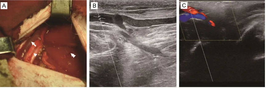
Figure 1. ALN dissection and ultrasound detection of axillary vein in rhesus monkeys. A: A transverse straight inci-sion was made at the right axillary region and the subcutaneous fat and the deep lymphatic tissues around the axil-lary vessels were excised. Axilaxil-lary blood vessels (arrow), nerves (arrowhead), and pectoralis major muscle (double arrowhead) were indicated. B, C: Ultrasound detection of axillary vein in rhesus monkeys after surgery. After wound healing, B-mode ultrasonic diagnostic equipment was used to confirm whether the blood flow in the axillary vein was pulsatile. B: Axillary vein; C: Ultrasound signal of axillary vein.
with an artificial 12-hour light/dark cycle. All
ani-mal expe-riments were conducted accoring to the ethical guidelines of The Ins-titutional Animal Care and Use Committee of Shandong University (Protocol number 2012).
Animal model establishment
Animal model of lymphedema was established by the combined irradiation and surgical tech-niques in the upper limbs of rhesus monkeys. The right upper limbs were chosen for the inter-vention, and the left ones were used as control. Axillary fossa was irradiated two weeks before and four weeks after the surgery, with a single dose of 30 Gy emitted from an X-ray machine (MBR-1520R-3; Hitachi, Tokyo, Japan), at a dose rate of 4.12 Gy/min (150 kVp, 20 mA). Axillary lymph node (ALN) and fat dissection were performed to facilitate lymphedema, just duplicating the ALN dissection in humans. Before the surgery, the animals were
anesthe-tizes with ketamine (4 mg/kg i.m.; Jiangsu
Hengrui Medicine Co., Ltd., Lianyungang,
Jiangsu, China) combined with diazepam (2
mg/kg s.c.; Jiangsu Hengrui Medicine Co., Ltd.). First, 5 ml of 2% patent methylene blue (Beijing Double-crane pharmaceutical Co., Ltd., Beijing, China) was injected into the subcutaneous tis-sue layer adjacent to the right mammary gland to identify drainage lymphatic vessels. ALNs
were readily identified by blue staining within 5
min. A transverse straight incision was made at the right axillary region, and then the subcuta-neous fat and the deep lymphatic tissues around the axillary vessels were excised to
-min. Four doses of gadodiamide (0.1 mmol/kg) were injected cutaneously into the dorsal aspect of hand, at the region of the four intedig-ital webs, with a 25 G needle (BD Biosciences, Franklin Lakes, NJ, USA). After the injection, the injection sites were massaged for 60 s. The massage was repeated during data acquisition.
The four stations were first imaged without
gadodiamide, and subsequently repeated 5, 25, and 45 minutes after intracutaneous
appli-cation of gadodiamide. To emphasize the gado -linium-containing structures, baseline images were subtracted, and 3D MIP reconstructions were performed.
Bioelectrical impedance analysis (BIA)
The extracellular fluid volume changes were
detected by BIA with a multiple-frequency
bio-electrical impedance analyzer (Inbody 3.0; Biospace, Seoul, Korea). During impedance
measurements, the animals were seated at nonmetal desks, with their feet resting on the plate electrode of the equipment. The animals rested their arms with palms facing down on the cushion at shoulder level. The skin areas
where electrodes were applied were first wiped
with 75% alcohol. Then, lightly adhesive resting electrodes were placed on each hand at the dorsal surface of the wrist between the radial and ulnar bones, and at the dorsal surface of the hand, 1 cm proximal from the peak of the
knuckle of the middle finger. The foot electrode
was placed between the 2 bony processes on the ankle in the front of the foot. Color-coded alligator clips were placed on the targeted
elec-trodes as previously described [6]. Histology and immunohistochemistry
Upper arm subcutaneous tissue was fixed in
4% formaldehyde in PBS overnight, and then ways (Figure 1A). After wound healing, B-mode
ultrasonic diagnostic equipment was used to
confirm whether the blood flow in the axillary
vein was pulsatile (Figure 1B, 1C). Postoperative analgesia was obtained by application of tram-adol hydrochloride (2.5 mg/kg i.m.; Hexal
GmbH, Holzkirchen, Germany) for three days
after surgery.
Limb circumference assessment
For the circumference measurement of the extremity, water displacement method was
used as previously described [5]. Tape rule was used to analyze the circumference of forearm,
elbow, and upper arm.
Magnetic resonance imaging (MRI)
The animals were scanned by MRI with a 3.0-T scanner, Magnetom Symphony (Siemens Medical Systems, Erlangen, Germany) equipped with high-performance gradients. Four stations were examined: the palm, the forearm, the upper arm, and the shoulder region. The phased-array body coil was used to examine the upper extremity. Before MR lymphangiogra-phy, the extent and distribution of the lymph-edema were evaluated using a heavily T2-weighted turbo spin-echo sequence (TR/TE, 2,400/705; matrix, 256 × 256; bandwidth, 247
Hz/pixel; slices, 66; FoV, 350 × 350; acquisition time, 5 min). To highlight the edema, maximum-intensity-projection (MIP) reconstructions were performed.
For HR MR lymphangiography, a spoiled gradi-ent-echo sequence (volumetric interpolated
breath-hold examination [VIBE]) was used, with
the following parameters: TR/TE, 2400/724; matrix, 448 × 448; bandwidth, 490 Hz/pixel;
[image:3.612.89.522.72.181.2]slices, 79; FoV, 350 × 350; acquisition time, 5
decalcified in 10% formic acid in 10% formalin/
PBS overnight. The tissues were embedded in
paraffin after tissue processing of dehydration,
clearance, and impregnation. Each tissue block was cut into 5 mm serial sections, and every
fifth section was stained with rabbit anti-human
vascular endothelial growth factor receptor-3/ fms-related tyrosine kinase 4 (VEGFR-3/FLT-4) IgG polyclonal antibody (dilution 1:500; Santa
Cruz Biotech, Santa Cruz, CA, USA) for immuno -histochemical staining of lymphatic vessels. Slides were imaged with an upright Olympus BX-50 microscope equipped with a Moticam 2300 color camera (Cole-Parmer; Vernon Hills, IL, USA).
Statistical analysis
Data were expressed as mean ± SD. SPSS 22.0 software was used to perform statistical
analy-sis. Differences of the extracellular fluid volume
in the upper limb of rhesus monkeys before and after operation were compared by paired t-test.
P < 0.05 was considered as statistically
significant.
Results
Macroscopic observation of the upper limb in rhesus monkeys
There are various diagnostic tests that could be counted to detect and assess lymphedema. In this study, physical examination, soft tissue imaging, bioelectrical impedance analysis (BIA), and immunohistochemical staining were performed to determine the severity of the edema in the upper limbs of the animal model. Firstly, macroscopic observation indicated that,
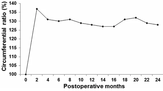
showed that the circumferential ratios of the affected limb to the contralateral control were between 100% and 137% over the 24 months after modeling (Figure 3), and the thickness of the palm increased about 15-40%. Accordingly, physical examination indicates that the rhesus monkey model present with typical appearance and features of lymphedema.
Magnetic resonance imaging (MRI) in the up-per limb in rhesus monkeys
Soft tissue imaging, like MRIs and CTs, detects
excess fluid in the tissues. Since lymphedema
mainly results from the accumulation of
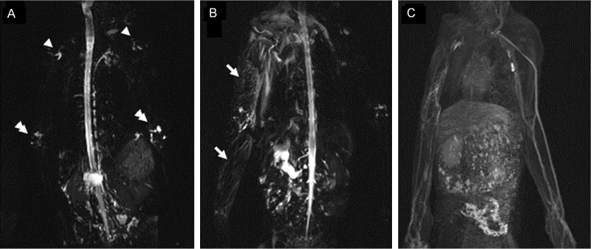
inter-stitial fluid, these imaging technologies are usu -ally used to assess lymphedema conditions. In this study, MR lymphography was performed, to evaluate the lymphedema in the upper limb in these rhesus monkeys. Our results showed
sig-nificant changes in lymphatic vessels, at 3 m
after the surgery. The major lymphatic trunks disappeared on the treated arm, compared with the normal side. The vessel structure was
replaced by a bright, punctate fluorescence
pattern against a foggy background (Figure 4A,
4B). In all animals, the lymphedema showed an epifascial distribution with high signal intensity on T2-weighted images. In frontal 3D spoiled gradient-echo high-resolution MRI and digital subtraction angiography (DSA)
lymphangiogra-phy image, delayed lymphatic flow with reticular
pattern of dilated lymphatic vessels was
observed, indicating neovascularization associ -ated with obstruction (Figure 4C). These MRI results further demonstrate the pathologically
[image:4.612.92.373.73.230.2]modified lymphatic vessels in our rhesus mon -key model.
Figure 3. The circumferential ratios in rhesus monkeys after intervention.
to the accumulation of fluid
and/or fat deposition in the extremity. On the other hand, the changes in the limb cir-cumference were measured
and recorded, from zero
Bioelectrical impedance analysis (BIA) in the upper limb in rhesus monkeys
BIA detected water content in the tissue by inducing a small, unharmful electrical current through the limb, and the impedance to current
flow was measured. Higher water content in tis -sues was linked with lower resistance. The lymphedema condition was assessed by
com-paring the resistance of electrical flow in the intracellular and extracellular fluid [7]. The rhe -sus monkey model of lymphedema was
sub-jected to BIA to determine the extracellular fluid
volume changes. As shown in Table 1, the
extracellular fluid volume was significantly ele -vated after the operation, compared with the volume before the surgery. These results indi-cate increased water content in the upper limb in these rhesus monkeys, which is in line with the pathology of lymphedema.
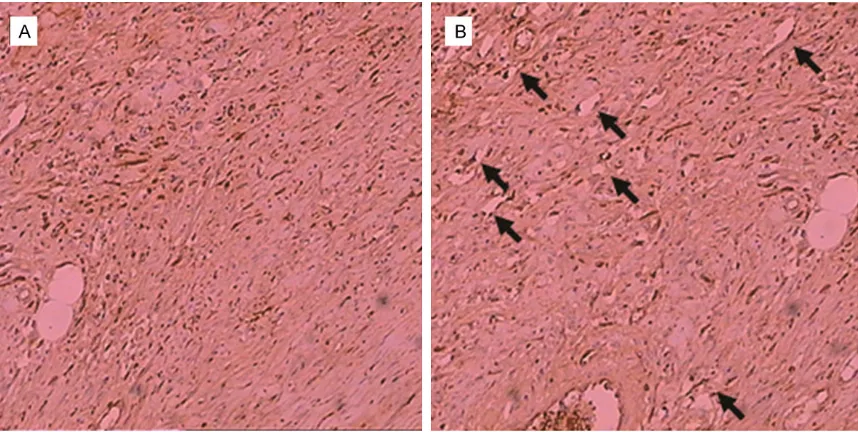
Immunohistochemical staining of the subcu-taneous tissue in the upper limb in rhesus monkeys
The lymphatic vessels in the upper limbs in the rhesus monkeys were next detected with
immu-These results suggest the curvature of the lym-phatic vessels in the rhesus monkeys models, in accordance with the pathological changes in lymphedema.
Discussion
Nowadays, there are approximately 45 million people suffering from the secondary lymphede-ma of limbs worldwide. A recent meta-analysis has assessed the incidence of arm lymphoe-dema after breast cancer and explored the risk
factors. The findings suggest that more than
20% women who survive breast cancer will develop arm lymphoedema, and it is very important to improve the understanding of risk factors and management strategies to reduce the individual and public health burden of this
disorder [8].
Although breast cancer mortality rates have
been significantly declined throughout recent
years due to the adequate management and
treatment, side-effects are still a significant
issue, which have a severe impact on the
qual-ity of life [9]. Lymphedema is a long-term disor -der, which lacks effective therapeutic
strate-Table 1. Extracellular fluid volume changes in the upper limb in rhesus
monkeys
[image:5.612.91.528.72.254.2]Preoperative (L) Postoperative (L) P Extracellular fluid volume (n) 0.326 ± 0.022 (5) 0.430 ± 0.047 (5) 0.0220
Figure 4. Magnetic resonance imaging (MRI) of lymphedema in the upper limb in rhesus monkeys. A: MR lymphog-raphy of bilateral upper limbs in rhesus monkeys at 1 m after surgery. Only elbow (arrowheads) and axillary lymph nodes (double arrowheads) were visible, without delayed lymphatic flow. B: MR lymphography of bilateral upper limbs in rhesus monkeys at 3 m after surgery. In the right limb, delayed lymphatic flow with reticular pattern of dilated lymphatic, and dermal backflow (arrows) was detected. C: Frontal 3D spoiled gradient-echo high-resolution MRI and digital subtraction angiography (DSA) lymphangiography image. In the right limb, delayed lymphatic flow with reticular pattern of dilated lymphatic vessels was observed.
nohistochemical staining. Our results showed that the lymphatic vessels were
significantly increased and
permanent lymphedema is inconsistent with humans. In addition, there are considerable dif-ferences in physiology and anatomy between the upper and lower extremities in humans and factors responsible for lymphatic transport in
the upper extremity, including capillary filtra -tion, muscular activity, breathing moments, body position or gravity, etc., are far more com-plex than those in the lower limb. Conventional
animal models, like rodents and carnivore [1, 27-30] cannot fully replicate the pathophysio -logical conditions of lymphedema in the upper limb. Furthermore, there are few studies con-cerning the animal models with lymphedema associated with axillary lymph node dissection, and none of these studies have been
conclu-sive [31]. Actually, an ideal animal model should
closely simulate the biology and pathogenesis of the particular disease in human, including the natural history and the temporal patterns of the clinical expression of the disease. One might then expect to make valid predictions about the applicability of the therapy in humans, by extrapolating the observations from the
ani-mal models [27].
In this study, we performed the combined surgi-cal and irradiation techniques in the upper limbs of rhesus monkeys. All animals survived, and developed progressive lymphedema as expected, demonstrating the reproducibility
gies [10]. The disease causes functional impair -ment and psychological burden to the affected
ones [11-17], and poses a complicated thera -peutic challenge for the physicians. Traditional treatments, such as elevating the swollen limb, compression bandages, massage and remedial
exercises, etc., can only promote the fluid circu -lation, reduce edema volume, and result in
par-tial relief in the patients [10, 11, 18, 19].
Moreover, these conservative measures can-not guarantee the long-term freedom from
pathophysiological alterations [10]. Based on
these reasons, there has been substantial interest on the studies of disease treatments concerning stem cells and growth factor-medi-ated lymphangiogenesis. It has been suggest-ed that the administration of growth factors and/or regulatory genes, and stem cell trans-plantation may provide new therapeutic options
for lymphedema [20-26]. Cell biology and
molecular biology approaches may provide a therapeutic method to reverse the
pathophysi-ological changes in lymphatic insufficiency.
Such approaches will require the documenta-tion of appropriate treatment responses, and the assessment of safety, in suitable preclinical
animal models [27].
[image:6.612.93.522.71.287.2]Various animal models of lymphedema have been described, however their achievement ratios are relatively low, and the formation of
Hospital Affiliated to Shandong University, No. 324, Jing 5 Road, Jinan 250021, Shandong, China. Tel: 86-053168776940; E-mail: txs0509@hotmail.com; Ling Gao, Central Laboratory, Provincial Hospital Affiliated to Shandong University, No. 324, Jing 5 Road, Jinan 250021, Shandong, China. Tel: 86- 053168776910; E-mail: gaoling1@medmail.com.cn
References
[1] Hadamitzky C and Pabst R. Acquired lymph -edema: an urgent need for adequate animal models. Cancer Res 2008; 68: 343-345. [2] Paskett ED, Dean JA, Oliveri JM and Harrop JP.
Cancer-related lymphedema risk factors, diag-nosis, treatment, and impact: a review. J Clin Oncol 2012; 30: 3726-3733.
[3] Liu Y, Fang Y, Dong P, Gao J, Liu R, Tian H, Ding Z, Bi Y and Liu Z. Effect of vascular endothelial growth factor C (VEGF-C) gene transfer in rat model of secondary lymphedema. Vascul Pharmacol 2008; 49: 44-50.
[4] Kinjo O and Kusaba A. Lymphatic vessel-to-isolated-vein anastomosis for secondary lymphedema in a canine model. Surg Today 1995; 25: 633-639.
[5] Gjorup C, Zerahn B and Hendel HW. Assess-ment of volume measureAssess-ment of breast can-cer-related lymphedema by three methods: circumference measurement, water displace-ment, and dual energy X-ray absorptiometry. Lymphat Res Biol 2010; 8: 111-119.
[6] Frank V, Chirag S, Maureen L and Pat W. Bio-electrical Impedance for Detecting and Moni-toring Patients for the Development of Upper Limb Lymphedema in the Clinic. Clin Breast Cancer 2012; 12: 133-137.
[7] Stout Gergich NL, Pfalzer LA, McGarvey C, Springer B, Gerber LH and Soballe P. Preopera-tive assessment enables the early diagnosis and successful treatment of lymphedema. Cancer 2008; 112: 2809-2819.
[8] Disipio T, Rye S, Newman B and Hayes S. Inci-dence of unilateral arm lymphoedema after breast cancer: a systematic review and meta-analysis. Lancet Oncol 2013; 14: 500-515. [9] Berlit S, Brade J, Tuschy B, Földi E,
Walz-Eschenlohr U, Leweling H and Sütterlin M. Whole-body versus Segmental Bioelectrical Impedance Analysis in Patients with Edema of the Upper Limb After Breast Cancer Treatment. Anticancer Res 2013; 33: 3403-3406. [10] Szuba A and Rockson SG. Lymphedema: Clas
-sification, diagnosis and therapy. Vasc Med 1998; 3: 145-156.
[11] Rockson SG. Lymphedema. Am J Med 2001; 110: 288-295.
[12] Velanovich V and Szymanski W. Quality of life of breast cancer patients with lymphedema. Am J Surg 1999; 177: 184-187.
and consistency of the models and methods. Compared with rodents, which are separated from humans by more than 70 million years
[32, 33], monkeys are closely related to humans
and share a last common ancestor from about
25 million years ago [32]. This species shares
about 93% of the DNA sequence with humans
[34]. In addition, compared with other previ -ously described models, rhesus monkeys exhibit greater similarity to humans in terms of physiology, neurobiology, and susceptibility to infectious and metabolic diseases, and they have a greater range of research tools available such as antibodies and various databases. Therefore, our rhesus monkey model of upper limb lymphedema is more suitable for the com-parison with humans. To the best of our
knowl-edge, this is the first report of an experimental
model with acquired lymphedema in the pri-mate upper extremity to mimic the disease pro-cess in humans. However, due to the limited duration of the study, the late effects such as delayed reappearance of lymphedema could not be assessed.
In conclusion, we have successfully established a novel model of upper limb lymphedema in rhesus monkeys. Our model provides a more consistent background to elucidate the patho-physiology of the disease in humans. This new model would help to increase our understand-ing of acquired upper limb lymphedema, and promote the development of new treatments for this intractable disorder.
Acknowledgements
This study was funded by Shandong Province Science and Technology Plan Project (Grant No. 2009GG10002060) and Shandong Medical and Health Science and Technology Deve- lopment Plan Project (Grant No. 2011HZ071). We thank Baoqiu Li at The Central for New Drug Evaluation (CNDE) of Shandong University, for providing animal experimental technique and equipment support.
Disclosure of conflict of interest
All authors declare no financial competing inter
-ests. All authors declare no non-financial com -peting interests.
models for the study of lymphatic insufficiency. Lymphat Res Biol 2003; 1: 159-169.
[28] Kanter MA, Slavin SA and Kaplan W. An experi -mental model for chronic lymphedema. Plast Reconstr Surg 1990; 85: 573-580.
[29] Casley-Smith JR, Clodius L and Földi M. Experi-mental blood vascular and lymphatic occlu-sion in the rabbit ear and the effect of benzo -pyrones. Arzneimittelforschung 1977; 27: 379-382.
[30] Park HS, Jung IM, Choi GH, Hahn S, Yoo YS and Lee T. Modification of a Rodent Hindlimb Mod -el of Secondary Lymphedema: Surgical Radi-cality versus Radiotherapeutic Ablation. Biomed Res Int 2013; 2013: 208912.
[31] Huang P, Li S, Han M, Xiao Z, Yang R and Han ZC. Autologous transplantation of granulocyte colony-stimulating factor-mobilized peripheral blood mononuclear cells improves critical limb ischemia in diabetes. Diabetes Care 2005; 28: 2155-2160.
[32] Kumar S and Hedges SB. A molecular times -cale for vertebrate evolution. Nature 1998; 392: 917-920.
[33] Gibbs RA, Weinstock GM, Metzker ML, Muzny DM, Sodergren EJ, Scherer S, Scott G, Steffen D, Worley KC, Burch PE, Okwuonu G, Hines S, Lewis L, DeRamo C, Delgado O, Dugan-Rocha S, Miner G, Morgan M, Hawes A, Gill R, Celera, Holt RA, Adams MD, Amanatides PG, Baden-Tillson H, Barnstead M, Chin S, Evans CA, Fer-riera S, Fosler C, Glodek A, Gu Z, Jennings D, Kraft CL, Nguyen T, Pfannkoch CM, Sitter C, Sutton GG, Venter JC, Woodage T, Smith D, Lee HM, Gustafson E, Cahill P, Kana A, Doucette-Stamm L, Weinstock K, Fechtel K, Weiss RB, Dunn DM, Green ED, Blakesley RW, Bouffard GG, De Jong PJ, Osoegawa K, Zhu B, Marra M, Schein J, Bosdet I, Fjell C, Jones S, Krzywinski M, Mathewson C, Siddiqui A, Wye N, McPher-son J, Zhao S, Fraser CM, Shetty J, Shatsman S, Geer K, Chen Y, Abramzon S, Nierman WC, Havlak PH, Chen R, Durbin KJ, Egan A, Ren Y, Song XZ, Li B, Liu Y, Qin X, Cawley S, Worley KC, Cooney AJ, D’Souza LM, Martin K, Wu JQ, Gon -zalez-Garay ML, Jackson AR, Kalafus KJ, McLeod MP, Milosavljevic A, Virk D, Volkov A, Wheeler DA, Zhang Z, Bailey JA, Eichler EE, Tu-zun E, Birney E, Mongin E, Ureta-Vidal A, Wood -R. Predictors of psychological distress, sexual
dysfunction and physical functioning among women with upper extremity lymphedema re-lated to breast cancer. Psycho Oncology 1995; 4: 255-263.
[16] Rockson SG. Lymphedema after surgery for cancer: The role of patient support groups in patient therapy. Dis Manag Health Outcomes 2002; 10: 345-347.
[17] Ozcinar B, Guler SA, Kocaman N, Ozkan M, Gulluoglu BM and Ozmen V. Breast cancer re -lated lymphedema in patients with different loco-regional treatments. Breast 2012; 21: 361-365.
[18] Rockson SG, Miller LT, Senie R, Brennan MJ, Casley Smith JR, Földi E, Földi M, Gamble GL, Kasseroller RG, Leduc A, Lerner R, Mortimer PS, Norman SA, Plotkin CL, Rinehart-Ayres ME and Walder AL. American Cancer Society Lymphedema Workshop. Work group III: Diag-nosis and management of lymphedema. Can-cer 1998; 83: 2882-2885.
[19] Földi E, Földi M and Weissleder H. Conserva-tive treatment of lymphoedema of the limbs. Angiology 1985 Mar; 36: 171-180.
[20] Asahara T, Murohara T, Sullivan A, Silver M, van der Zee R, Li T, Witzenbichler B, Schatte -man G, Isner JM. Isolation of putative progeni-tor endothelial cells for angiogenesis. Science 1997; 275: 964-967.
[21] Conrad C, Niess H, Huss R, Huber S, von Lu-ettichau I, Nelson PJ, Ott HC, Jauch KW and Bruns CJ. Multipotent mesenchymal stem cells acquire a lymphendothelial phenotype and en-hance lymphatic regeneration in vivo. Circula-tion 2009; 119: 281-289.
[22] Religa P, Cao R, Bjorndahl M, Zhou Z, Zhu Z and Cao Y. Presence of bone marrow- derived circulating progenitor endothelial cells in the newly formed lymphatic vessels. Blood 2005; 106: 4184-4190.
[23] Rutkowski JM, Boardman KC and Swartz MA. Characterization of lymphangiogenesis in a model of adult skin regeneration. Am J Physiol Heart Circ Physiol 2006; 291: 1402-1410. [24] Kim H and Dumont DJ. Molecular mechanisms
EE, Hahn MW, Hardison RC, Makova KD, Miller W, Milosavljevic A, Palermo RE, Siepel A, Sikela JM, Attaway T, Bell S, Bernard KE, Buhay CJ, Chandrabose MN, Dao M, Davis C, Delehaunty KD, Ding Y, Dinh HH, Dugan-Rocha S, Fulton LA, Gabisi RA, Garner TT, Godfrey J, Hawes AC, Hernandez J, Hines S, Holder M, Hume J, Jhan -giani SN, Joshi V, Khan ZM, Kirkness EF, Cree A, Fowler RG, Lee S, Lewis LR, Li Z, Liu YS, Moore SM, Muzny D, Nazareth LV, Ngo DN, Ok -wuonu GO, Pai G, Parker D, Paul HA, Pfannkoch C, Pohl CS, Rogers YH, Ruiz SJ, Sabo A, San -tibanez J, Schneider BW, Smith SM, Sodergren E, Svatek AF, Utterback TR, Vattathil S, Warren W, White CS, Chinwalla AT, Feng Y, Halpern AL, Hillier LW, Huang X, Minx P, Nelson JO, Pepin KH, Qin X, Sutton GG, Venter E, Walenz BP, Wallis JW, Worley KC, Yang SP, Jones SM, Mar -ra MA, Rocchi M, Schein JE, Baertsch R, Clarke L, Csürös M, Glasscock J, Harris RA, Havlak P, Jackson AR, Jiang H, Liu Y, Messina DN, Shen Y, Song HX, Wylie T, Zhang L, Birney E, Han K, Konkel MK, Lee J, Smit AF, Ullmer B, Wang H, Xing J, Burhans R, Cheng Z, Karro JE, Ma J, Raney B, She X, Cox MJ, Demuth JP, Dumas LJ, Han SG, Hopkins J, Karimpour-Fard A, Kim YH, Pollack JR, Vinar T, Addo-Quaye C, Degenhardt J, Denby A, Hubisz MJ, Indap A, Kosiol C, Lahn BT, Lawson HA, Marklein A, Nielsen R, Vallen-der EJ, Clark AG, Ferguson B, Hernandez RD, Hirani K, Kehrer-Sawatzki H, Kolb J, Patil S, Pu LL, Ren Y, Smith DG, Wheeler DA, Schenck I, Ball EV, Chen R, Cooper DN, Giardine B, Hsu F, Kent WJ, Lesk A, Nelson DL, O’brien WE, Prüfer K, Stenson PD, Wallace JC, Ke H, Liu XM, Wang P, Xiang AP, Yang F, Barber GP, Haussler D, Kar -olchik D, Kern AD, Kuhn RM, Smith KE and Zwieg AS. Evolutionary and biomedical insights from the rhesus macaque genome. Science 2007; 316: 222-234.
wark C, Zdobnov E, Bork P, Suyama M, Torrents D, Alexandersson M, Trask BJ, Young JM, Huang H, Wang H, Xing H, Daniels S, Gietzen D, Schmidt J, Stevens K, Vitt U, Wingrove J, Ca -mara F, Mar Albà M, Abril JF, Guigo R, Smit A, Dubchak I, Rubin EM, Couronne O, Poliakov A, Hübner N, Ganten D, Goesele C, Hummel O, Kreitler T, Lee YA, Monti J, Schulz H, Zimdahl H, Himmelbauer H, Lehrach H, Jacob HJ, Brom-berg S, Gullings-Handley J, Jensen-Seaman MI, Kwitek AE, Lazar J, Pasko D, Tonellato PJ, Twig -ger S, Ponting CP, Duarte JM, Rice S, Good-stadt L, Beatson SA, Emes RD, Winter EE, Web-ber C, Brandt P, Nyakatura G, Adetobi M, Chiaromonte F, Elnitski L, Eswara P, Hardison RC, Hou M, Kolbe D, Makova K, Miller W, Nek -rutenko A, Riemer C, Schwartz S, Taylor J, Yang S, Zhang Y, Lindpaintner K, Andrews TD, Cac -camo M, Clamp M, Clarke L, Curwen V, Durbin R, Eyras E, Searle SM, Cooper GM, Batzoglou S, Brudno M, Sidow A, Stone EA, Venter JC, Payseur BA, Bourque G, López-Otín C, Puente XS, Chakrabarti K, Chatterji S, Dewey C, Pachter L, Bray N, Yap VB, Caspi A, Tesler G, Pevzner PA, Haussler D, Roskin KM, Baertsch R, Clawson H, Furey TS, Hinrichs AS, Karolchik D, Kent WJ, Rosenbloom KR, Trumbower H, Weirauch M, Cooper DN, Stenson PD, Ma B, Brent M, Arumugam M, Shteynberg D, Copley RR, Taylor MS, Riethman H, Mudunuri U, Peter-son J, Guyer M, Felsenfeld A, Old S, Mockrin S, Collins F; Rat Genome Sequencing Project Consortium. Genome sequence of the Brown Norway rat yields insights into mammalian evolution. Nature 2004; 428: 493-521. [34] Rhesus Macaque Genome Sequencing and