Original Article
Differential expression of MST4, STK25 and PDCD10
between benign prostatic hyperplasia
and prostate cancer
Heyu Zhang1,3,4, Xi Ma5, Saihui Peng1,3, Xu Nan2,3, Hongshan Zhao2,3
1Department of Immunology, School of Basic Medical Sciences, Peking University, No. 38 Xueyuan Road, Beijing, PR China; 2Department of Medical Genetics, School of Basic Medical Sciences, Peking University, 38 Xueyuan Road, Beijing, PR China; 3Human Disease Genomics Center, Peking University, 38 Xueyuan Road, Beijing, PR China; 4Central Laboratory, Peking University School of Stomatology, 22 South Zhongguancun Road, Beijing, PR China; 5State Key Lab of Animal Nutrition, China Agricultural University, 2 Yuanmingyuan West Road, Beijing 100193, PR China
Received September 22, 2014; Accepted November 8, 2014; Epub October 15, 2014; Published November 1, 2014
Abstract: Both benign prostatic hyperplasia (BPH) and prostate cancer (PC) are common diseases for men around the world. Both serine/threonine protein kinase MST4 (MST4) and serine/threonine kinase 25 (STK25) belong to the Ste20-like kinases and interact with programmed cell death 10 (PDCD10) which is closely linked to cancer diseases. To clarify the roles of MST4, STK25 and PDCD10 in prostate carcinogenesis, we examined MST4, STK25 and PDCD10 expression in tissue microarray blocks containing 110 cores of BPH and 160 cores of PC
immunohisto-chemically and evaluated their correlation with clinicopathological findings. MST4 was not expressed in all the BPH
cases and expressed in 38.7% of PC cases (P < 0.0001). STK25 expression was found in 77.3% of BPH cases and 93.1% of PC cases (P < 0.0001). PDCD10 staining was considered weak in 82 (74.5%) and strong in 28 (25.5%) of BPH cases. However, in prostate cancer cases, PDCD10 staining was weak in 95 (59.4%) and strong in 65 (40.6%)
(P < 0.05). PDCD10 and STK25 immunostaining were associated with age in prostatic hyperplasia cases (P < 0.05).
The staining intensity for STK25 was significantly greater in Gleason grades 3-5 (47.1% of such cases staining
strongly) compared with other grades of prostate cancer (only 26.5% of these cases staining strongly; P < 0.05). Our results suggest that MST4, STK25 and PDCD10 are unregulated in prostate cancer and may play roles in prostate tumorigenesis. MST4 may be a helpful marker for identifying prostate cancer.
Keywords: MST4, STK25, PDCD10, benign prostatic hyperplasia, prostate cancer
Introduction
Prostate cancer is the most frequently diag-nosed cancer in males [1]. The incidence of prostate cancer is increasing steadily, yet our knowledge of its causes is very limited [2-4]. The previous studies which showed elevated levels of active mitogen-activated protein kinase (MAPK) in high-grade and advanced stage prostate and specimens from the andro-gen-insensitive tumors implicated that MAPK pathway might be involved in the development of prostate cancers and its androgen-indepen-dent growth [5].
Sterile 20 (STE20) serine/threonine protein
cerevisiae as a mitogen-activated protein kinase (MAP4K) involved in the mating pathway [6]. In mammals, more than 30 members of the STE20 superfamily of kinases have been described. MST4 (serine/threonine protein kinase MST4) and STK25 (serine/threonine
pro-tein kinase 25) are members of the GCK group
III family of kinases, which are a subset of the Ste20-like kinases. Both of MST4 and STK25
are localized to the Golgi apparatus and specifi
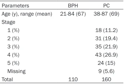
Table 1. Clinicopathologic characteristics of patients in the TMA dataset
Parameters BPH PC
Age (y), range (mean) 21-84 (67) 38-87 (69) Stage
1 (%) 18 (11.2)
2 (%) 31 (19.4)
3 (%) 35 (21.9)
4 (%) 43 (26.9)
5 (%) 24 (15)
Missing 9 (5.6)
Total 110 160
way [9]. STK25 translocates from the Golgi to
the nucleus upon chemical anoxia and induces apoptotic cell death [10]. STK25 also plays a role in cell migration [11, 12].
Programmed cell death 10 (PDCD10) gene,
also named TFAR15 (TF-1 cell apoptosis-relat
-ed gene 15) and CCM3 (cerebral cavernous malformation 3) was originally cloned in our laboratory [13]. Mutations of PDCD10 are one cause of cerebral cavernous malformations, which are vascular malformations that cause seizures and cerebral hemorrhages. Several reports have indicated that PDCD10 interacts with MST4 and STK25 [14-18]. PDCD10 forms
a ternary complex with GCKIII (MST4, STK24,
STK25) and Golgi matrix protein GM130 to reg
-ulate the Golgi morphology [12, 16, 19].
PDCD10 could advance cell proliferation and transformation by activating the MST4 activity and involve itself in the ERK pathway [17]. PDCD10 stabilizes STK25 to accelerate cell apoptosis under oxidative stress [14]. Increa- sing evidence indicates that PDCD10 may play a part in the tumor signaling because it is up-regulated in many cancer tissues, such as pan-creatic adenocarcinomas and colorectal can-cer [20-22]. Our previous studies have showed that MST4, STK25 and PDCD10 were expressed in prostate cancer cell line PC-3 and could pro-mote cell growth or apoptosis. However, the expression patterns of the three molecules in human prostate cancer tissues are not clear. In the present study, we evaluated the expres-sion of MST4, STK25 and PDCD10 in prostatic hyperplasia and prostate cancer by using tis-sue microarray (TMA) technology. Then, we explored their association with clinicopatholog-ic parameters.
Materials and methods
Experimental specimens
Tissue microarrays were obtained from Chao- ying Biotechnology Co. (Shanxi, China).
Antibodies
Monoclonal mouse-antibody against PDCD10 was constructed in our lab [23]; polyclonal rab-bit-antibody against MST4 was obtained from Cell Signaling Technology Inc. (Danvers, USA); and monoclonal mouse-antibody against STK-
25 (clone 1G6) from Abnova Co. (Taipei, Taiwan).
Immunohistochemistry
Sections were deparaffinized and rehydrated
with xylene and a series of grades of alcohol. Epitopes were retrieved by heating in a micro-wave oven with 10 mM citrate buffer (PH 6.0) or 1 Mm EDTA buffer (PH 8.0) for 10 min, followed by cooling for 1 hr at room temperature. Endogenous peroxidase activity was inhibited
by 3% hydrogen peroxide. Unspecific binding
was blocked with 10% normal serum (for goat) for 30 min. Sections were incubated with anti-PDCD10, MST4 and STK25 antibodies at 4°C over night. Staining procedures were performed in an automated immunostainer (TechMate 1000; Dako) in accordance with the ChemMate protocol using the biotin ± streptavidin detec-tion system (ChemMate-HRP/DAB; Dako). After- wards, sections were counterstained in haema-toxylin. Primary anti-human antibodies were anti-PDCD10, mouse monoclonal diluted 1: 100, anti-MST4, rabbit polyclonal diluted 1:50 and anti-STK25 mouse monoclonal diluted
1:50. The unrelated rabbit IgG and mouse IgG
at the same concentration were used as nega-tive controls.
Scores of antibody staining
Cytoplasmic immunoreactivity was scored sep-arately according to staining intensity and grad-ed semi-quantitatively as negative (-), weekly positive (+) and strongly positive (++). Each anti-body was evaluated in double-blind fashion. Statistical analyses
Statistical analyses were performed using
data. Statistical significance was defined as a
P-value of < 0.05.
Results
Clinical features of prostatic hyperplasia and prostate cancer
Table 1 demonstrates the clinical and patho-logic characteristics of all TMA samples. There were 9 cases of prostate cancer missing the
Gleason grade records.
Tissue localization of PDCD10, MST4 and STK25 in the prostate tissue
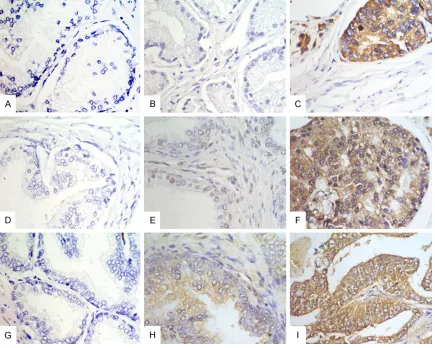
Representative examples of reactivity for MST4, STK25 and PDCD10 are shown in Figure
1. The immunoexpression of them in the benign and the cancerous prostatic epithelium was cytoplasmic. Some cases showed nuclear staining of STK25.
Expression patterns of MST4, STK25 and PDCD10 in prostate specimens by using im-munohistochemistry
[image:3.612.90.525.72.420.2]We did not detect MST4 expression in 110 benign prostatic hyperplasias. MST4 analysis showed positive staining in 38.7% of prostate cancer (Table 2). A statistically significant dif -ference in MST4 staining between hyperplasia and cancer tissues was found (P < 0.0001). STK25 was found positive in 77.3% of prostatic hyperplasia and 93.1% in malignant prostate
Figure 1. Immunohistochemical analysis of MST4, STK25 and PDCD10 antigen expression in benign prostatic hy-perplasia tissues and prostate cancer tissues. A. Negative staining of MST4 in benign prostatic hyhy-perplasia. B. Negative staining of MST4 in prostate cancer. C. Positive staining of MST4 in prostate cancer. D. Negative staining
of STK25 in benign prostatic hyperplasia. E. Positive staining of STK25 in prostatic hyperplasia (+). F. Positive stain
-ing of STK25 in prostate cancer (++). G. Negative stain-ing of PDCD10 in benign prostatic hyperplasia. H. Positive
staining of PDCD10 in prostatic hyperplasia (+). I. Positive staining of PDCD10 in prostate cancer (++). Original
found the three molecules were upregulated in prostate cancer than in benign prostatic hyper-plasia, implying that they may play a role in prostate carcinoma progression.
The results presented here suggest that the MST4 is expressed in a higher level in prostate cancer than in benign prostatic hyperplasia, which is consistent with previous reports focused on cell level. Sung, et al detected high-er expression levels of MST4 in prostate canchigh-er cell lines DU145 and PC-3 than in normal cell lines [9]. The experiments demonstrated that the over expression of MST4 could promote cell
growth by specifically activating the ERK path -way [8, 9, 17]. The ERK path-way functions in cellular proliferation, differentiation and surviv-al. Its inappropriate activation is a common occurrence in human cancers. It may be the role that MST4 plays in prostate cancer pro-gression [9]. Our data indicate MST4 as a potential marker or prospective target for the most aggressive forms of prostate carcinoma. The mammalian kinase STK25 is another
mem-ber of GCKIII kinases. STK25 can be activated
by oxidative stress and induce apoptotic cell death, suggesting STK25 is an important medi-ator of oxidant-mediated signal transduction [10, 24]. In prostate cancer, oxidative stress, an innate key event characterized by
supraphysio-logical ROS concentrations, has been identified
as one of the hallmarks of the aggressive
[image:4.612.91.384.97.177.2]dis-ease phenotype [25]. Specifically, oxidative
Table 2. MST4, STK25 and PDCD10 protein expression in benign prostatic hyperplasia and prostate cancer
MST4 STK25 PDCD10
BPH PC BPH PC BPH PC
- (%) 110 (100) 98 (61.3) 25 (22.7) 11 (6.9) 0 0 + (%) 0 62 (38.7) 78 (70.9) 88 (55) 82 (74.5) 95 (59.4) ++ (%) 0 0 7 (6.4) 61 (38.1) 28 (25.5) 65 (40.6)
P value < .0001 < .0001 .010
cancer (Table 2). The frequency of positive
cores was significantly higher in cancer tissues
than in hyperplasia (P < 0.0001).
PDCD10 was expressed in all BPH and PC cases in our study (Table 2). PDCD10 staining was considered weak in 82 (74.5%) and strong in 28 (25.5%) of the prostatic hyperplasia cases. In prostate cancer cases, PDCD10 stain-ing was weak in 95 (59.4%) and strong in 65 (40.6%). Hence, the expression of PDCD10 pro-tein was stronger in cancer than hyperplasia (P = 0.01).
Clinicopathologic characteristics in associa-tion with the intensity of MST4, STK25 and PDCD10 stainings
The correlation between MST4, STK25 and PDCD10 immunoreactivity and several clinico-pathologic characteristics was investigated. The TMA had been validated as representative for traditional prognostic variables of prostatic hyperplasia and cancer. These protein expres-sion and clinicopathologic data of the patients are summarized in Tables 3 and 4. According to
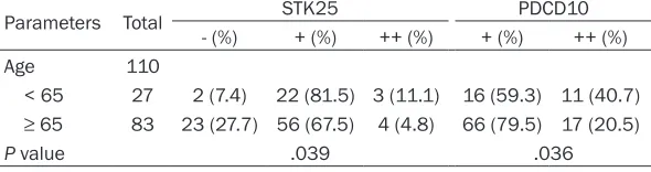
our predefined criteria, both PDCD10 and
STK25 immunostaining were associated with age in prostatic hyperplasia cases (P < 0.05) (Table 3). However, MST4 immunostaining was not associated with age in prostatic hyperpla-sia. The association between patients’ age and MST4, STK25 and PDCD10 expression did not
exceed the threshold for statistical significance
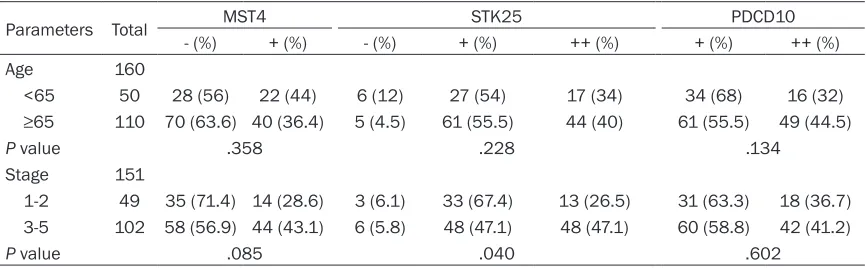
in prostate cancer (P > 0.05) (Table 4). In prostate cancer cases, the staining intensity
for STK25 was significantly greater in Gleason grades
3-5 (47.1% of such cases staining strongly) compared with other grades of prostate cancer (only 26.5% of these cases staining strongly; P = 0.04). The positive MST4 and PDCD10 staining was not
associated with Gleason
grade in prostate cancer (Table 4).
Discussion
In this paper, we examined the expression of MST4, STK25 and PDCD10 and
Table 3. Correlation of STK25 and PDCD10 expression with clinico-pathologic factor of benign prostatic hyperplasia
Parameters Total STK25 PDCD10
- (%) + (%) ++ (%) + (%) ++ (%)
Age 110
< 65 27 2 (7.4) 22 (81.5) 3 (11.1) 16 (59.3) 11 (40.7)
≥ 65 83 23 (27.7) 56 (67.5) 4 (4.8) 66 (79.5) 17 (20.5)
[image:4.612.91.386.226.304.2]stress is associated with prostate cancer devel-opment, progression and response to therapy [26]. Nevertheless, a thorough understanding of the relationships between oxidative stress, redox homeostasis and the activation of prolif-eration and survival pathways in healthy and malignant prostate remains elusive [25]. Our
date showed a significant difference of STK25
expression level between prostate hyperplasia and cancer, while STK25 expression was asso-ciated with age in prostate cancer, implying STK25 may play a role in prostate cancer pro-gression. More studies need to be conducted in
order to confirm the function of STK25 in
tumorigenesis.
Our study provides a new insight into the clini-cal relevance of PDCD10 in prostate cancer. The mutations of CCM1, CCM2 and PDCD10 gene occur in familial cerebral cavernous mal-formations, a condition associated with sei-zures and strokes [27, 28]. Both CCM1 and CCM2 are in a complex which involves in p38 MAPK and integrin signaling pathways [29]. PDCD10 is demonstrated to interact with CCM2 in vitro and may function as a stabilizing molec-ular link between CCM1 and CCM2 [18]. The PP2A phosphatase high-density interaction
network defined a large molecular assembly
(STRIPAK) that links the PDCD10 and associ-
ated GCK-III kinases to a PP2A•striatin•
Mob3•STRIP complex [30]. Katrin, et al
identi-fied that STK25 could phosphorylate PDCD10
[18]. Our previous studies showed that PDCD10 could promote PC-3 cell growth and transfor-mation and exerted its effect through interac-tion with MST4, via modulainterac-tion of the ERK path-way, by increasing the activated form of ERK [17]. PDCD10 also interacted with and
stabi-lized STK25 to accelerate cell apoptosis under oxidative stress [14]. Programmed cell death 10 enhances proliferation and protects malig-nant T cells from apoptosis [31]. Based on all
these findings we hypothesized that PDCD10
may be involved in prostate cancer progres-sion. In our experiment, we did observe that dif-ference of PDCD10 expression levels between benign and malignant prostate tissues. PDCD10 is expressed in almost all tissues of which the expression in normal level may play an important role in preserving health. When PDCD10 is expressed in an abnormally high level, it may contribute to tumorigenesis. In summary, our study showed increased pro-tein expressions of MST4, STK25 and PDCD10
in prostate cancer for the first time, indicating
that they may play a role in prostate cancer
pro-gression or at least reflect a stage of carcinoma
progression. Our data suggest that MST4 may be a novel biomarker for identifying prostate cancer from prostatic hyperplasia.
Acknowledgements
This work was supported the National Natural
Science Foundation of China (grant numbers
30872940 and 81300894). The authors thank Ms. Zhang Zirong for assistance with immuno-histochemical staining.
Disclosure of conflict of interest
None.
Address correspondence to: Dr. Hongshan Zhao,
Department of Medical Genetics, School of Basic
[image:5.612.90.523.96.230.2]Medical Sciences, Peking University, 38 Xueyuan Road, Beijing, PR China. Tel: +86-10-82802846;
Table 4. Correlation of MST4, STK25 and PDCD10 expression with clinicopathologic factors of pros-tate cancer
Parameters Total MST4 STK25 PDCD10
- (%) + (%) - (%) + (%) ++ (%) + (%) ++ (%)
Age 160
<65 50 28 (56) 22 (44) 6 (12) 27 (54) 17 (34) 34 (68) 16 (32)
≥65 110 70 (63.6) 40 (36.4) 5 (4.5) 61 (55.5) 44 (40) 61 (55.5) 49 (44.5)
P value .358 .228 .134
Stage 151
1-2 49 35 (71.4) 14 (28.6) 3 (6.1) 33 (67.4) 13 (26.5) 31 (63.3) 18 (36.7) 3-5 102 58 (56.9) 44 (43.1) 6 (5.8) 48 (47.1) 48 (47.1) 60 (58.8) 42 (41.2)
Fax: +86-10 -82801149; E-mail: hongshan@bjmu.
edu.cn
References
[1] Siegel R, Naishadham D, Jemal A. Cancer sta-tistics, 2013. CA Cancer J Clin 2013; 63: 11-30.
[2] Grönberg H. Prostate cancer epidemilolgy.
Lancet 2003; 361: 859-64.
[3] Schaid DJ. The complex genetic epidemiology
of prostate cancer. Hum Mol Genet 2004; 13:
R103-21.
[4] Xia SJ, Cui D, Jiang Q. An overview of prostate
diseases and their characteristics specific to
Asian men. Asian J Androl 2012; 14: 458-64. [5] Gioeli D, Mandell JW, Petroni GR, Frierson HF
Jr, Weber MJ. Activation of mitogen-activated protein kinase associated with prostate cancer progression. Cancer Res 1999; 59: 279-84. [6] Wu C, Whiteway M, Thomas DY, Leberer E.
Mo-lecular characterization of Ste20p, a potential mitogen-activated protein or extracellular sig-nal-regulated kinase kinase (MEK) kinase ki-nase from Saccharomyces cerevisiae. J Biol Chem 1995; 270: 15984-92.
[7] Dan I, Ong SE, Watanabe NM, Blagoev B, Nielsen MM, Kajikawa E, Kristiansen TZ, Mann M, Pandey A. Cloning of MASK, a novel mem-ber of the mammalian germinal center kinase III subfamily, with apoptosis-inducing proper-ties. J Biol Chem 2002; 277: 5929-39. [8] Lin JL, Chen HC, Fang HI, Robinson D, Kung HJ,
Shih HM. MST4, a new Ste20-related kinase that mediates cell growth and transformation via modulating ERK pathway. Oncogene 2001; 20: 6559-69.
[9] Sung V, Luo W, Qian D, Lee I, Jallal B, Gishizky
M. The Ste20 kinase MST4 plays a role in pros-tate cancer progression. Cancer Res 2003; 63: 3356-63.
[10] Nogueira E, Fidalgo M, Molnar A, Kyriakis J, Force T, Zalvide J, Pombo CM. SOK1 translo
-cates from the Golgi to the nucleus upon
chemical anoxia and induces apoptotic cell death. J Biol Chem 2008; 283: 16248-58. [11] Chen XD, Cho CY. Downregulation of SOK1
pro-motes the migration of MCF-7 cells. Biochem
Biophys Res Commun 2012; 407: 389-92. [12] Preisinger C, Short B, De Corte V, Bruyneel E,
Haas A, Kopajtich R, Gettemans J, Barr FA. YSK1 is activated by the Golgi matrix protein GM130 and plays a role in cell migration
through its substrate 14-3-3zeta. J Cell Biol 2004; 164: 1009-20.
[13] Wang Y, Liu H, Zhang Y, Ma D. cDNA cloning and expression of an apoptosis-related gene,
humanTFAR15 gene. Sci China C Life Sci
1999; 42: 323-9.
[14] Zhang H, Ma X, Deng X, Chen Y, Mo X, Zhang Y, Zhao H, Ma D. PDCD10 interacts with STK25 to accelerate cell apoptosis under oxidative
stress. Front Biosci (Landmark Ed) 2012; 17:
2295-305.
[15] Ceccarelli DF, Laister RC, Mulligan VK, Kean MJ, Goudreault M, Scott IC, Derry WB, Chakrab
-artty A, Gingras AC, Sicheri F. CCM3/PDCD10
heterodimerizes with germinal center kinase III
(GCKIII) proteins using a mechanism analo -gous to CCM3 homodimerization. J Biol Chem 2011; 286: 25056-64.
[16] Fidalgo M, Fraile M, Pires A, Force T, Pombo C, Zalvide J. CCM3/PDCD10 stabilizes GCKIII pro
-teins to promote Golgi assembly and cell orien -tation. J Cell Sci 2010; 123: 1274-84.
[17] Ma X, Zhao H, Shan J, Long F, Chen Y, Chen Y,
Zhang Y, Han X, Ma D. PDCD10 interacts with Ste20-related kinase MST4 to promote cell growth and transformation via modulation of the ERK pathway. Mol Biol Cell 2007; 18: 1965-78.
[18] Voss K, Stahl S, Schleider E, Ullrich S, Nickel J,
Mueller TD, Felbor U. CCM3 interacts with
CCM2 indicating common pathogenesis for ce-rebral cavernous malformations. Neurogenet-ics 2007; 8: 249-56.
[19] Zhang M, Dong L, Shi Z, Jiao S, Zhang Z, Zhang
W, Liu G, Chen C, Feng M, Hao Q, Wang W, Yin
M, Zhao Y, Zhang L, Zhou Z. Structural
mecha-nism of CCM3 heterodimerization with GCKIII
kinases. Structure 2013; 21: 680-8.
[20] Gibson S, Shillitoe EJ. Analysis of
apoptosis-associated genes and pathways in oral cancer cells. J Oral Pathol Med 2006; 35: 146-54. [21] Aguirre AJ, Brennan C, Bailey G, Sinha R, Feng
B, Leo C, Zhang Y, Zhang J, Gans JD, Bardeesy
N, Cauwels C, Cordon-Cardo C, Redston MS, DePinho RA, Chin L. High-resolution character-ization of the pancreatic adenocarcinoma ge-nome. Proc Natl Acad Sci U S A 2004; 101: 9067-72.
[22] Cardoso J, Boer J, Morreau H, Fodde R. Expres
-sion and genomic profiling of colorectal can -cer. Biochim Biophys Acta 2007; 1775: 103-37.
[23] Chen Y, Zhao Y, Zhang T, Xu L, Ma X, Zhao H,
Chen Y. Preparation and identification of
monoclonal antibodies against human pro-grammed cell death 10 (PDCD10). Beijing Da Xue Xue Bao 2006; 38: 586-91.
[24] Pombo CM, Bonventre JV, Molnar A, Kyriakis J,
Force T. Activation of a human Ste20-like ki
-nase by oxidant stress defines a novel stress
response pathway. Embo J 1996; 15: 4537-46.
paradigm for prevention and therapeutics. Prostate Cancer Prostatic Dis 2013; 16: 217-25.
[26] Khandrika L, Kumar B, Koul S, Maroni P, Koul HK. Oxidative stress in prostate cancer. Cancer Lett 2009; 282: 125-36.
[27] Bergametti F, Denier C, Labauge P, Arnoult M,
Boetto S, Clanet M, Coubes P, Echenne B,
Ibra-him R, Irthum B, Jacquet G, Lonjon M, Moreau JJ, Neau JP, Parker F, Tremoulet M,
Tournier-Lasserve E. Mutations within the programmed cell death 10 gene cause cerebral cavernous
malformations. Am J Hum Genet 2005; 76:
42-51.
[28] Guclu B, Ozturk AK, Pricola KL, Bilguvar K, Shin D, O’Roak BJ, Gunel M. Mutations in apopto -sis-related gene, PDCD10, cause cerebral cav-ernous malformation 3. Neurosurgery 2005; 57: 1008-13.
[29] Zawistowski JS, Stalheim L, Uhlik MT, Abell AN,
Ancrile BB, Johnson GL, Marchuk DA. CCM1
and CCM2 protein interactions in cell signal-ing: implications for cerebral cavernous
mal-formations pathogenesis. Hum Mol Genet
2005; 14: 2521-31.
[30] Goudreault M, D’Ambrosio LM, Kean MJ, Mul
-lin MJ, Larsen BG, Sanchez A, Chaudhry S, Chen GI, Sicheri F, Nesvizhskii AI, Aebersold R, Raught B, Gingras AC. A PP2A phosphatase high density interaction network identifies a
novel striatin-interacting phosphatase and ki-nase complex linked to the cerebral cavernous malformation 3 (CCM3) protein. Mol Cell Pro-teomics 2009; 8: 157-71.
[31] Lauenborg B, Kopp K, Krejsgaard T, Eriksen
KW, Geisler C, Dabelsteen S, Gniadecki R,