REVIEW
Effects of skeletal muscle energy availability on protein turnover
responses to exercise
William J. Smiles1, John A. Hawley1,2,* and Donny M. Camera1
ABSTRACT
Skeletal muscle adaptation to exercise training is a consequence of repeated contraction-induced increases in gene expression that lead to the accumulation of functional proteins whose role is to blunt the homeostatic perturbations generated by escalations in energetic demand and substrate turnover. The development of a specific ‘exercise phenotype’ is the result of new, augmented steady-state mRNA and protein levels that stem from the training stimulus (i.e. endurance or resistance based). Maintaining appropriate skeletal muscle integrity to meet the demands of training (i.e. increases in myofibrillar and/or mitochondrial protein) is regulated by cyclic phases of synthesis and breakdown, the rate and turnover largely determined by the protein’s half-life. Cross-talk among several intracellular systems regulating protein synthesis, breakdown and folding is required to ensure protein equilibrium is maintained. These pathways include both proteasomal and lysosomal degradation systems (ubiquitin-mediated and autophagy, respectively) and the protein translational and folding machinery. The activities of these cellular pathways are bioenergetically expensive and are modified by intracellular energy availability (i.e. macronutrient intake) and the ‘training impulse’(i.e. summation of the volume, intensity and frequency). As such, exercise– nutrient interactions can modulate signal transduction cascades that converge on these protein regulatory systems, especially in the early post-exercise recovery period. This review focuses on the regulation of muscle protein synthetic response-adaptation processes to divergent exercise stimuli and how intracellular energy availability interacts with contractile activity to impact on muscle remodelling.
KEY WORDS: Carbohydrate, Endurance exercise, Fat, Resistance exercise, Muscle bioenergetics, Protein synthesis, Training adaptation
Introduction and background
Human skeletal muscle is characterized by a high degree of plasticity in its adaptive response to external stimuli such as habitual level of contractile activity, substrate availability and the prevailing environmental conditions (Flück and Hoppeler, 2003; Zierath and Hawley, 2004). Individually or in combination, these stimuli perturb cellular homeostasis and alter the metabolic demands of the tissue, leading to numerous structural and functional changes that ultimately impact on the tissue phenotype. With regard to contractile activity, the processes underlying any exercise-induced training adaptation centre on the accumulation of specific proteins, with gene expression promoting an increase in protein concentration fundamental to any training-induced response (Hansen et al., 2005; Hawley et al., 2011).
The four fundamental cellular processes involved in gene expression are transcription, mRNA degradation, translation and protein degradation, with each step of this cascade controlled by gene-regulatory events. Recent evidence also suggests that acute gene activation is associated with a dynamic change in DNA methylation in skeletal muscle and that DNA hypomethylation is an early event in contraction-induced gene activation (Barrès et al., 2012). While the responsiveness of individual mRNA species to contractile loading is variable between individuals, any increase in steady-state mRNA levels is ultimately the result of synthesis (transcription) and degradation (mRNA stability), with the relative contribution of these processes playing an important role in phenotypic adaptation (Coffey and Hawley, 2007). In this regard, up to 40% of the variance in protein levels can be explained by changes in mRNA levels, with this variability largely due to different transcription rates rather than mRNA stability (Schwanhäusser et al., 2011).
The functional consequences of contraction-induced adaptations are largely determined by the training stimulus (e.g. endurance versus resistance based), with the‘training impulse’(the product of the volume, intensity and frequency of this stimulus)‘fine-tuning’ this response-adaptation process. In the final analyses, however, the ability of a given muscle cell to alter the type and quantity of protein is a function of the protein’s half-life; proteins that turn over rapidly and have high rates of synthesis are capable of attaining a new steady-state level faster than those that turn over slowly during adaptation to contractile and other stimuli.
Over the past two decades, it has become evident that nutrient availability can serve as a potent modulator of many of the acute responses and chronic adaptations to both endurance- and resistance-based training (Hawley et al., 2011). Changes in macronutrient intake and availability alter the concentration of circulating metabolites and hormones, triggering large perturbations in the storage profile of skeletal muscle and other insulin-sensitive tissues. In turn, the pre-exercise energy status of muscle (and other organs) alters patterns of fuel utilization during contractile activity and directly impacts on many of the acute regulatory processes underlying gene expression (Yang et al., 2005) and cell signalling (Creer et al., 2005; Wojtaszewski et al., 2003). Of note is that the time courses of many of these intracellular events typically peak in the 3–4 h period post-exercise and have returned to pre-exercise baseline values within 24 h (Creer et al., 2005). This‘window’of heightened cellular activity also coincides with sports nutrition recommendations for aggressive post-exercise refuelling (Beelen et al., 2010). Accordingly, these nutrient–exercise interactions have the potential to either activate or inhibit many of the signalling pathways with roles in training adaptation during this period. A comprehensive review of the intracellular signalling events involved in both endurance- and resistance-based training adaptation is provided in an accompanying paper in this issue (Hoppeler, 2016). Here, our principal focus is on how these contraction-stimulated
1
Centre for Exercise and Nutrition, Mary MacKillop Institute for Health Research, Australian Catholic University, Melbourne, VIC 3065, Australia.2Research Institute for Sport and Exercise Sciences, Liverpool John Moores University, Liverpool L3 3AF, UK.
*Author for correspondence ( john.hawley@acu.edu.au)
Journal
of
Experimental
signalling pathways can be either amplified or attenuated by the prevailing skeletal muscle energy status before, during and after exercise. As we have recently published several reviews on the role of carbohydrate availability on training adaptation (Bartlett et al., 2015; Hawley and Morton, 2014), our emphasis here will be on how energy availability from protein and fat interacts with contractile activity to impact on skeletal muscle protein synthetic responses.
Cellular regulation of exercise training adaptation: a primer There are several potential sites of regulation that impact on exercise-induced training adaptation (Fig. 1), including, but not limited to, alterations in mRNA processing, degradation and stability, as well as altered protein kinetics (i.e. processing, transport and degradation/stability). Many of the intracellular pathways involved in these events comprise complex networks with a high degree of crosstalk, feedback regulation and transient activation by both contractile activity and the prevailing cellular environment (i.e. energy availability). However, it is worth noting that, in many cases, normal responses to a single bout of exercise and the adaptations to chronic exercise training can be seen when one or more key pathways are absent, are blocked with drugs, or are otherwise disturbed by low cellular energy availability. Such biological redundancy suggests that perhaps the only obligatory response to contractile activity, independent of energy availability, is the defence of cellular homeostasis itself (Hawley et al., 2014).
The maintenance of cellular mRNA and protein levels by recycling from their building blocks (nucleotide monophosphates and amino acids, respectively) is a bioenergetically expensive process. Degradation of proteins is also necessary in many cellular processes including cell cycle progression, signal transduction and apoptosis. In this regard, skeletal muscle mass is highly regulated by coordinate rates of muscle protein synthesis (MPS) and muscle protein breakdown (MPB). Whole-body protein homeostasis is largely achieved by the consumption of protein-rich meals, which results in a systemic hyperaminoacidemia stimulating the synthesis of new muscle
proteins (Bohé et al., 2001). Feeding-induced changes in MPS are severalfold higher than in MPB and account for the greatest fluxes in turnover (Biolo et al., 1997). In order for the expression of a given polypeptide to increase, the specific genetic sequence of amino acids encoded in DNA must be transcribed and acquired by mRNA. These amino acids are then translated into a functional protein by sequential reactions of translation initiation, elongation and eventually termination of the peptide chain.
Translation initiation is the most well-characterized level of MPS with predominant focus on the mechanistic target of rapamycin (mTOR) protein kinase (Goodman, 2014). mTOR exists in two multi-protein complexes (mTORC1 and mTORC2) with mTORC1 established as the principal regulator of protein translation. Activation of mTORC1 has been linked to a myriad of energetic and hormonal signals (e.g. growth factors, amino acids and mechanical stimuli) that converge on the lysosome to augment protein synthesis by enhancing its interaction with two direct activators, a small GTPase called Rheb (Inoki et al., 2003a,b) and the glycerophospholipid phosphatidic acid (PA) (Sun et al., 2008; Yoon et al., 2011; You et al., 2014).
As noted previously, protein synthesis is an energy-consuming process, requiring four high-energy phosphates per peptide bond throughout translation (Merrick, 1992). With perturbations to cellular energy homeostasis, the AMP-activated protein kinase (AMPK) propagates signal transduction to conserve ATP and restore energy balance by repressing protein synthesis and upregulating systems implicated in the restoration of cellular homeostasis (Browne et al., 2004; Gwinn et al., 2008; Inoki et al., 2003b, 2006; Zhang et al., 2014). In this regard, the regulation of protein balance is largely an intrinsic process with several intracellular systems synergistically impacting synthesis, breakdown and folding of proteins. These intracellular systems (in addition to the protein translational machinery), which include autophagy, the ubiquitin proteasome system (UPS) and the unfolded protein response (UPR), are inextricably linked to energy availability and can thus mediate exercise-induced adaptation responses. Autophagy is an intracellular lysosomal recycling system that regulates endogenous amino acid availability through the bulk non-selective and selective turnover of superfluous organelles and macromolecules (Mizushima and Klionsky, 2007). Briefly, autophagy commences with the formation of a double-membrane cup-like structure termed the autophagosome, which sequesters and delivers a fraction of cytoplasm for degradation by the lysosome (Mizushima and Klionsky, 2007). AMPK and mTORC1 are rate limiting for autophagy as both kinases phosphorylate (at different residues) the unc-51-like kinase 1 (ULK1) to promote and inhibit autophagy induction, respectively (Kim et al., 2011b). Similar to protein synthesis, autophagy is temporally sensitive to energy availability and utilizes ATP in proportion to its rate of protein degradation (Plomp et al., 1987; Schellens et al., 1988).
In contrast to autophagy, the UPS is an ATP-dependent process that targets the majority of short-lived proteins for ubiquitination and subsequent degradation by the proteasome (Rock et al., 1994). In skeletal muscle, the ubiquitin ligases muscle RING finger protein 1 (MuRF1) and muscle atrophy F-box or Atrogin-1 (atrogenes) are critical mediators of this process (Bodine and Baehr, 2014) and are mitigated by contractile activity and energy availability. Lastly, the UPR also mediates protein turnover in response to perturbations to intracellular protein metabolism. The endoplasmic reticulum (ER) is a small, dynamic organelle that folds, processes and traffics newly synthesized polypeptides. Misfolded proteins in the ER are List of symbols and abbreviations
4E-BP1 eukaryotic initiation factor 4E binding protein 1 AMPK AMP-activated protein kinase
EAA essential amino acid eEF eukaryotic elongation factor eIF eukaryotic initiation factor ER endoplasmic reticulum FFA free fatty acid
LAT L-type amino acid transporter MPB muscle protein breakdown MPS muscle protein synthesis mTOR mechanistic target of rapamycin MuRF1 muscle RING finger protein 1 p70S6K p70 ribosomal protein S6 kinase PA phosphatidic acid
PAT proton-assisted transporters PERK PKR-like ER protein kinase
PGC-1α peroxisome proliferator-activated receptor-γ (PPAR-γ)-coactivator-1α
REX resistance exercise rpS6 p70 ribosomal protein S6
SNAT sodium-coupled neutral amino acid transporters ULK1 unc-51-like kinase 1
UPR unfolded protein response UPS ubiquitin proteasome system
Journal
of
Experimental
targeted for degradation by the proteasome or autophagy (called ER-associated degradation) (Travers et al., 2000). However, if the rate of protein synthesis exceeds that of the ER’s handling capacity, unfolded proteins accumulate in the ER lumen and prompt the UPR. The UPR can be induced by fluctuations in energy availability (Kaufman et al., 2002) and substrate utilization, with skeletal muscle ER (sarcoplasmic reticulum) emerging as being sensitive to such changes, particularly in the context of elevated contractile activity (Deldicque et al., 2012). The following sections of this review focus on the regulation of these cellular systems in response to both resistance- and endurance-based exercise and how the prevailing cellular energy availability interacts with contractile activity to impact on protein synthetic responses.
Effects of energy availability on resistance exercise-induced muscle protein turnover
Increased protein availability
The ingestion of high-quality protein (i.e. lean, intact sources) increases MPS and decreases MPB following resistance exercise (REX) in humans, thereby promoting net muscle protein accretion (Biolo et al., 1995; Chesley et al., 1992; Tipton et al., 1999). A recent meta-analysis examining the efficacy of protein supplementation to increase lean body mass and strength after REX training revealed that protein supplementation significantly increased muscle mass and strength compared with placebo (Cermak et al., 2012). While the anabolic capacity of protein
availability in human skeletal muscle is primarily attributable to the ingestion of essential amino acids (EAAs) and in particular the branched chain amino acid leucine, factors such as the dose (Moore et al., 2009a; Witard et al., 2013), source (Reidy et al., 2013; Tang et al., 2009; Tipton et al., 2004) and the timing/distribution of intake (Areta et al., 2013; Esmarck et al., 2001) also modulate the effectiveness for protein supplementation to augment muscle mass with REX. The‘anabolic’signalling pathways activated by REX and protein ingestion centre on intramuscular mTORC1 signalling (Kimball and Jefferson, 2010). This is largely based on the capacity for leucine to stimulate mRNA translation initiation through mTORC1 activation, although the precise mechanism(s) in humans are not clear (Fig. 2). Regardless, increases in MPS with leucine/EAA ingestion are blocked by the mTORC1 inhibitor rapamycin, confirming the required role for mTORC1 activation as a key regulator of human MPS (Dickinson et al., 2011). Moreover, unlike other amino acids, leucine exclusively stimulates mTOR phosphorylation (Atherton et al., 2010). Increases in signalling markers downstream of mTORC1, including p70 ribosomal protein S6 (rpS6) kinase ( p70S6K), rpS6 and the eukaryotic initiation factor
[image:3.612.128.478.56.328.2]4E (eIF4E) binding protein 1 (4E-BP1), are routinely observed during early recovery (i.e. up to ∼30 h post-exercise) following combined REX and protein ingestion in human skeletal muscle (Burd et al., 2010; Dreyer et al., 2008; Karlsson et al., 2004; Koopman et al., 2007). This response may be dose dependent as evidenced by greater mTOR (Kakigi et al., 2014) and p70S6K(Areta
Fig. 1. Mechanical and amino acid signals are sensed and converge on the lysosome.Combined resistance exercise (REX) and protein ingestion optimize
protein translation initiation via the mammalian target of rapamycin complex 1 (mTORC1) by potentially enhancing the activity of its direct regulators phosphatidic acid (PA) and Ras homologue enriched in brain (Rheb) that are modulated upstream by diacylglycerol kinaseζ(DGKζ) and tuberous sclerosis complex catalytic subunit 2 (TSC2), respectively. The mTORC1 kinase promotes protein synthesis by phosphorylating its substrates eIF4E binding protein 1 (4E-BP1) and p70 rpS6 kinase ( p70S6K). Ultimate peptide formation depends on energy availability and this is potentially governed by the lysosome, which, in addition to being the site of protein translation initiation, is the terminal site of autophagy. Autophagy regulates energy balance and is stimulated by proximal energy sensors, most notably AMP-activated protein kinase (AMPK) that phosphorylates and activates unc-51-like kinase 1 (ULK1), whereas mTOR phosphorylation of ULK1 is inhibitory. The proposed‘inside-out’model by Zoncu et al.(2011) in which lysosomes sequester amino acids for mTORC1 signalling, raises the possibility that some of these amino acids originate from an initial autophagy-mediated breakdown of protein macromolecules. Under this context, the relationship between REX, energy and protein availability, and the temporal balance of systems controlling anabolism (synthesis) and catabolism (autophagy and proteasome mediated) is in need of investigationin vivoin human skeletal muscle.
Journal
of
Experimental
et al., 2013; D’Souza et al., 2014; Kakigi et al., 2014) phosphorylation when greater amounts (i.e. 20–40 g) of protein are ingested. However, not all studies have demonstrated such a dose dependency for mTORC1 signalling with increasing amounts of protein following REX (Moore et al., 2009a; West et al., 2011). This may be a result of differences in the selection of muscle biopsy sampling points or induced aminoacidaemia from manipulating feeding patterns between studies. Indeed, no differences in mTOR phosphorylation were observed at 1, 3 or 5 h post-exercise when whey protein was consumed as either a single 25 g bolus dose or smaller, regular‘pulsed’feedings (10×2.5 g amounts; West et al., 2011), suggesting any dose response may only be apparent when bolus protein amounts greater than 20 g are consumed.
The type of protein ingested may also regulate cell signalling responses following REX. Previous studies investigating the feeding-induced stimulation of MPS with whey compared with soy or casein protein have shown greater (Burd et al., 2012; Pennings et al., 2011; Tang et al., 2009) or similar (in the case of casein) (Dideriksen et al., 2011; Reitelseder et al., 2011; Soop et al., 2012; Tipton et al., 2004) MPS responses in humans, although the cellular basis for these divergent responses is not clear and may be
attributable to differences in the type of an individual protein ingested. For example, in some studies where no differences in MPS induced by protein ingestion (i.e. casein versus whey) post-REX were observed, caseinate was the type of casein ingested (Dideriksen et al., 2011; Reitelseder et al., 2011) compared with the superior effect of whey when miceller casein was the protein source (Burd et al., 2012; Tang et al., 2009). These differences may be ascribed to the window of biopsy sampling time points not capturing the optimal time frame (i.e. >5 h) for casein to elicit maximum rates of MPS compared with the earlier and more rapid effects of whey consumption. However, cell signalling and MPS responses to a‘blend’of soy, whey and casein proteins following REX in humans recently showed similar phosphorylation responses in mTOR, 4E-BP1, p70S6Kand rpS6 compared with a whey protein
[image:4.612.131.483.59.330.2]beverage (Reidy et al., 2013). This signalling response closely matched changes in post-exercise rates of MPS, indicating a soy– dairy-based mixture of proteins can equally activate mTOR signalling and promote muscle growth responses to whey protein. Intuitively, this indicates that the ingestion and subsequent digestive kinetics of these different protein sources are responsible for the increased mTOR and MPS response when whey, soy and casein are
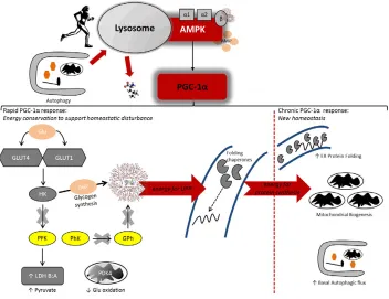
Fig. 2. The peroxisome proliferator-activated receptorγ-coactivator-1α(PGC-1α) regulates acute and chronic adaptations to endurance exercise
training.Endurance exercise contraction-induced metabolic stress promotes an AMPK-mediated autophagic response which could partition amino acids and
other substrates for prevailing anabolic events (i.e. protein synthesis). AMPK also has phosphotransferase activity toward PGC-1α, which may promote its nuclear localization and the elicitation of a stress-responsive gene transcription programme. Initially in this endurance exercise adaptation paradigm, PGC-1αhas been shown to promote energy conservation, in particular glycogen, by upregulating genes involved in glucose (Glu) uptake (GLUT1 and GLUT4 transporters), repression of Glu oxidation ( pyruvate dehydrogenase kinase 4, PDK4) glycogen synthesis [hexokinase (HK), which catalyses Glu 6 phosphate (G6P) formation], lactate buffering (lactate dehydrogenase isoforms LDH B/A, to favour pyruvate accumulation) and downregulation (as indicated by crosses over arrows) of glycolytic genes ( phosphofructokinase, PFK; phosphorylase kinase, PhK; glycogen phosphorylase, GPh), which collectively would enhance glycogen storage and preservation during exercise. Such rapid energy conservation following acute exercise bouts in the untrained state is conceivably necessary to support adaptation to the contraction-invoked challenges to homeostasis, in particular disturbances to protein-folding reactions in the endoplasmic reticulum (ER) which, themselves, are energy dependent. Indeed, rodent data reveal that PGC-1αis implicated in the adaptive ER unfolded protein response (UPR) that reacts to, and ameliorates exercise-induced perturbations to protein folding and trafficking. Furthermore, a role for PGC-1αin transcriptionally regulating autophagy, including that of select mitochondrial turnover termed mitophagy, has been identified. With chronic training (as indicated by the dashed line), PGC-1αappears to promote a new, augmented homeostasis that engenders greater intrinsic energy availability via substrate conservation, an augmented autophagic flux and enhanced ER protein folding that would synergistically support mitochondrial biogenesis; hence, mitochondrial biogenesis would require synthesis of nascent mitochondrial protein and, therefore, autophagy would partition requisite energy substrate (i.e. amino acids) to support these processes, while an equilibrated ER lumen would mitigate the secretory demands arising from repeated endurance exercise bouts.
Journal
of
Experimental
consumed as a blend as opposed to separately. Whether repeated feeding of soy or a soy–dairy-based mixture versus whey alone across prolonged REX recovery (i.e. 24–48 h) results in differential MPS and translational signalling responses is unknown. Furthermore, the effects of variable compositions of soy, whey and casein within this mixture and the ‘threshold’dose of dairy protein (i.e. to reach a certain leucine concentration) relative to the amount of soy to maximize post-REX anabolism represents another area for future research.
In regard to the potential for the type and amount of protein ingested to modulate signalling markers of translation initiation, it should also be noted that other systemic factors, or those released by the muscle itself (e.g. insulin-like growth factor 1 and interleukin-6) may propagate intracellular messages that augment the rate of synthesis of sarcomeric proteins. Furthermore, the rate of peptide elongation, the most energetically expensive step of translation (Merrick, 1992) that depends on eukaryotic elongation factor (eEF2) activities (most notably phosphorylation of eEF2) has not been well characterized with respect to the time course of post-translational modifications and their temporal relationship with MPS following REX and protein ingestion. The effects of novel, important regulators of skeletal muscle mass including, but not limited to, Wnt/beta catenin (Armstrong and Esser, 2005), Hippo/Yes-Associated Protein (Goodman et al., 2015), bone morphogenetic protein (Sartori et al., 2013) and regulators of ribosome mass (for overall translational capacity) (Chaillou et al., 2014) should also be coupled to the analysis of mTORC1 signalling and MPS in future investigations of REX–nutrient interactions in human skeletal muscle.
Recent evidence suggests a role for amino acid transporters in the post-exercise adaptation responses with protein ingestion following REX. Amino acid transporters regulate amino acid flux (both influx and efflux) across the muscle cell membrane and therefore, in part, may mediate mTORC1 activation. The plasma membrane system L transporters regulate the import of large neutral amino acids (such as leucine) and include theL-type amino acid transporter (LAT) 1 and 2
[or solute-linked carrier 7A5 and 7A8 (SLC7A5 or SLC7A8, respectively)] (Hundal and Taylor, 2009). Other types of transporters include the sodium-coupled neutral amino acid transporters (SNAT) and proton-assisted transporters (PAT1, SLC36). Changes in amino acid availability selectively increase the mRNA expression of these transporters post-exercise in humans (Areta et al., 2013; Churchward-Venne et al., 2012; Dickinson et al., 2013, 2014; Drummond et al., 2011, 2010; Reidy et al., 2014). This response occurs concomitantly with elevated mTORC1 signalling and MPS, indicating a precursory role for these transporters to promote MPS. While emerging evidence suggests the expression of select transporters may be dependent on the type and dose of protein ingested (Reidy et al., 2014), further work is required to ascertain the exact cellular location and functional capacity of these transporters.
Protein ingestion following REX can also regulate markers of skeletal muscle proteolysis and degradation. The UPS, autophagy– lysosomal, calpain and caspase systems all contribute to muscle proteolysis with the acute response derived predominantly from the UPS. The atrogenes Atrogin-1 and MuRF1 are central mediators of the UPS through their ability to ubiquitinate cleaved myofibril segments for subsequent degradation by the proteasome. Previous studies have reported reduced MuRF1 and Atrogin-1 expression post-REX with EAA ingestion compared with a placebo beverage (Borgenvik et al., 2012). Moreover, this reduction may be dose dependent (Areta et al., 2013), suggesting larger boluses (>30 g) of
protein, in this case whey, may be more advantageous for suppressing the muscle catabolic response compared with smaller to moderate quantities (10–20 g) during the early post-REX recovery period. We have also reported reduced MuRF1 and Atrogin-1 mRNA abundance when 25 g of whey protein was consumed compared with a placebo following a bout of concurrent resistance and endurance exercise (Camera et al., 2015). In this regard, it is possible this coordinated attenuation in MuRF1 and Atrogin-1 expression represents a mechanism by which exogenous amino acid ingestion can ameliorate the classical endurance exercise-induced ‘interference’ toward muscle hypertrophy adaptations with concurrent training, although this hypothesis requires further investigation. However, not all studies report reduced expression of UPS markers with post-exercise protein ingestion (Dalbo et al., 2013; Hulmi et al., 2009; Reitelseder et al., 2014; Stefanetti et al., 2014) and it is yet to be established whether increased MuRF1 and/or Atrogin-1 responses following REX are indicative of general tissue remodelling or a greater induction of protein degradation, as there is evidence demonstrating that Atrogin-1 mRNA, but not MuRFAtrogin-1 mRNA, increases intramuscularly after 10 weeks of REX training (Stefanetti et al., 2015). Thus, elucidating the specific roles of individual ubiquitin ligases in human skeletal muscle with REX is important for understanding the impact of protein availability on these molecules.
Increased fat availability
Little is known regarding the effects of excess fat availability, whether through consumption of a high-fat diet (i.e. ∼45–70% energy from fat) or by direct intralipid infusion (to rapidly increase circulating free fatty acids, FFAs), on MPS responses following REX. Increased intracellular accumulation of lipid within skeletal muscle with human ageing is implicated in so-called ‘anabolic resistance’through direct inhibition of anabolic pathways and/or activation of proinflammatory cytokines (Rivas et al., 2012; Savage et al., 2005). Results from animal studies initially demonstrated that a short-term 7-fold elevation of plasma FFA (via intralipid infusion) decreased basal rates of MPS (Lang, 2006). This attenuation was associated with decreased eIF4G but not mTOR, p70S6Kor 4E-BP1
phosphorylation, supporting recent findings showing 2 weeks of a high-fat diet preceding exercise to have little effect on mTORC1 signalling in rat skeletal muscle (Castorena et al., 2015). As eIF4G is inhibited by rapamycin (Raught et al., 2000), it is possible that short-term or transient excess fat availability causes select defects in mTORC1 substrate targeting. To add complexity, immunoprecipitation data from Lang (2006) demonstrated that an intralipid infusion over 5 h increased the association of 4E-BP1 with eIF4E in rodent muscle, thus reducing its affinity for eIF4G. In contrast, 14 weeks of a high-fat, low-carbohydrate diet reduced rates of MPS, concomitant with attenuated Akt ( protein kinase B) and p70S6K phosphorylation, following a 14 day functional
overload stimulus in mice (Sitnick et al., 2009). The disparity in signalling responses from these studies could be a result of a longer high-fat diet, and suggests that chronic excess lipid availability impairs the ability of muscle to hypertrophy through attenuated protein translation in response to mechanical loading.
The effects of increasing FFA availability on direct (Katsanos et al., 2009; Stephens et al., 2014) or indirect (Ferrannini et al., 1986; Gormsen et al., 2008) measures of MPS have also been studied in humans. Katsanos and colleagues (2009) reported little attenuation of MPS in response to the ingestion of 7 g of EAAs after the initiation of a 10 h intralipid infusion (20% Liposyn, 90 ml h−1).
It should be noted, however, that the Liposyn infusion produced a
Journal
of
Experimental
4-fold higher circulating insulin concentration before and after amino acid ingestion compared with a control saline infusion, which probably dampened any potential negative effect of insulin resistance on MPS responses. These findings are in contrast to a recent study showing that a 10% intralipid infusion prevented the 2.2-fold increase in MPS after ingestion of a 21 g bolus of amino acids compared with a saline control (Stephens et al., 2014). This intralipid-induced attenuated MPS response was associated with reduced 4E-BP1 but not Akt or mTOR phosphorylation, providing further evidence that this decrease in MPS is mediated by distal regulators of translation initiation responses. Whether the responses of these downstream regulators are conserved from rodents to humans is unknown. Furthermore, future studies are required to ascertain whether excess fat availability can attenuate increases in MPS and associated translational signalling induced by REX and/or protein ingestion in human skeletal muscle and whether training status, or exercise performed during an intralipid infusion can overcome this fat-induced attenuation of MPS.
Excess lipid availability has also been implicated in the activation of the ER UPR. C2C12 cells exposed for 17 h to palmitic acid, a saturated FA, increased UPR markers including the ER protein chaperone BiP and PKR-like ER protein kinase (PERK), while concomitantly reducing p70S6K phosphorylation and protein
synthesis (Deldicque et al., 2010). These findings suggest that the increased intracellular presence of specific FAs can repress protein synthesis responses, via the UPR, as a result of increased inflammation and/or as a potential compensatory means to restrain the accumulation of newly synthesized proteins waiting proper folding in the ER. Moreover, as the ER is a site of triglyceride formation (Wolins et al., 2006), UPR-mediated repression of protein synthesis by FA infiltration may be required to prevent ER overcrowding. Contrasting the effects of palmitic acid, mice fed a high-fat diet for 6 weeks showed simultaneous increases in p70S6K
phosphorylation and UPR markers (Deldicque et al., 2010). This disparity in findings may have been the result of negative feedback of the mTORC1/p70S6K axis toward the insulin receptor substrate-1
during periods of high circulating FAs. The same group (Deldicque et al., 2011) also investigated UPR responses to high-fat feeding in human skeletal muscle. Despite increases in body mass and subcutaneous fat deposits following a 6 week high-fat diet, there were no changes in intramuscular BiP, PERK or inositol-requiring enzyme 1αexpression (Deldicque et al., 2011). Given the limited characterization of the time course of UPR expression in human skeletal muscle, it is possible that the biopsy sampling time points and/or incision site (vastus lateralis) missed the activation of these markers, or a longer period of high-fat feeding and greater impairment in muscle insulin sensitivity may have been required to activate the UPR. In addition, as 8 weeks of supplemental long chain
n-3 polyunsaturated FAs has been shown to augment skeletal muscle protein anabolism in young, middle-aged and older individuals in response to insulin and amino acid infusions (Smith et al., 2011a,b), the differential effects of saturated versus polyunsaturated FAs on muscle protein turnover in humans need to be considered.
Reduced energy availability
As MPS is energetically expensive, it is hardly surprising that periods of energy deficit attenuate resting-, fasted- (Areta et al., 2014; Murphy et al., 2015; Pasiakos et al., 2010) and feeding-induced MPS (Murphy et al., 2015; Pasiakos et al., 2013). However, the magnitude to which energy deficit affects post-REX MPS responses has received little attention. Recently, we showed that REX undertaken subsequent to a 5 day, moderate
(∼500 kcal day−1) energy deficit, which lowered resting MPS by
27% compared with resting energy balance, only increased MPS to the rate originally observed at rest when subjects were in energy balance (Areta et al., 2014). Post-exercise whey protein provision in graded amounts of either 15 or 30 g augmented MPS and prolonged mTOR phosphorylation in a dose-responsive manner, demonstrating that the energy deficit-induced repression of MPS is relieved with a larger supply of exogenous amino acids. However, it remains to be determined whether protein ingestion at amounts greater than 30 g saturates the anabolic response to REX in energy deficit. A recent study by Murphy and colleagues (2015) found that MPS responses (specifically the myofibrillar fraction) of older, overweight men to acute protein feeding in energy deficit (4 weeks of energy restriction,∼300 kcal day−1) could be partly rescued by
consuming a balanced daily distribution of protein (25% per meal) during energy deficit as opposed to consuming the majority of protein in an evening meal. A complete restoration of MPS was mitigated by REX training in weeks 3 and 4 of the energy deficit period, but these MPS values still failed to exceed that of resting energy balance ( presumably from intrinsic factors of anabolic resistance in this population) (Murphy et al., 2015). These data support the notion that prior REX sensitizes the protein synthetic machinery to amino acid ingestion (Burd et al., 2011; Moore et al., 2009b).
Selectively restricting nutrients, specifically glycogen, can attenuate Akt phosphorylation immediately after REX in humans (Creer et al., 2005). We (Camera et al., 2012) have reported that MPS responses to a single bout of REX, in which subjects ingested either a placebo or protein/carbohydrate beverage (20 g whey/40 g maltodextrin) immediately post-exercise and 2 h later, was unaffected when exercise was commenced with low intramuscular glycogen stores. Although MPS was higher in the nutrient-fed group, no differences between legs in the two groups were observable, suggesting low endogenous glycogen does not suppress acute anabolic responses to REX. In fact, resting Akt phosphorylation was higher in the low glycogen leg, conceivably as a compensatory means to increase glucose uptake and promote glycogen resynthesis. Furthermore, despite a higher post-exercise nutrient-dependent increase in mTOR phosphorylation with normal glycogen, no significant differences in downstream effectors ( p70S6K/rps6) were
observed between legs (Camera et al., 2012), indicating a threshold level of mTOR activity is sufficient to saturate substrate phosphorylation for peak MPS (myofibrillar) responses. The fact that low energy availability, but not glycogen per se, elicits divergent effects on MPS at rest and following acute REX is potentially a consequence of energy deficit attenuating intracellular amino acid availability. The effects of commencing endurance exercise with low carbohydrate availability have been extensively reviewed elsewhere (Bartlett et al., 2015; Hawley, 2013; Hawley and Morton, 2014).
How MPS is modulated by short-term energy deficit at rest and in response to REX is poorly understood. We have recently demonstrated that commensurate with MPS, the abundance of the autophagy-related gene (Atg) protein Atg5 was attenuated by a 5 day energy deficit (Smiles et al., 2015), whereas its conjugated form (cAtg5) along with several additional proteins implicated in autophagy were increased when protein was ingested post-REX. As autophagy has been shown to provide an amino acid source in myotubes when amino acids are restricted in the culture media (Yu and Long, 2014), and autophagy itself is an energy-consuming process (Plomp et al., 1987; Schellens et al., 1988), our data suggest that energy deficit-induced perturbations to MPS could be due to a lower autophagic flux, at least in the short-term. Consequently,
Journal
of
Experimental
repressed autophagy may diminish the available pool of endogenous amino acids that otherwise become available in response to high-resistance contractions (MacKenzie et al., 2009). As post-exercise protein ingestion augmented the protein expression of members of the autophagic machinery in energy deficit (Smiles et al., 2015), it is possible that increasing protein availability repressed the ATP-dependent UPS (Areta et al., 2013) and, thus, potential ubiquitination of autophagy signalling proteins (Platta et al., 2012). Such mechanisms could therefore enable autophagy to support the anabolic demands of REX, particularly when overall energy is limiting.
Effects of energy availability on endurance exercise-induced muscle protein turnover
Increased protein availability
Endurance exercise of an intermittent or prolonged nature reduces muscle glycogen levels, body fluids and electrolytes. Research on the nutritional requirements of endurance athletes has predominantly focused on carbohydrate ingestion protocols designed to replace glycogen stores and rehydration (Burke et al., 2006). However, endurance exercise can also induce skeletal muscle damage caused by muscle contraction and subsequent MPB. The ingestion of high-quality protein sources is an emerging nutritional strategy to provide amino acids for enhanced muscle repair and regeneration as well as to stimulate myofibrillar and mitochondrial protein synthesis. Previous studies in humans have shown that acute and/or chronic endurance exercise increases mixed MPS independent of protein ingestion (Harber et al., 2009, 2010; Mascher et al., 2011; Pikosky et al., 2006). While it has been assumed such increases in mixed MPS are mitochondrial-related proteins/enzymes, recent studies have instead shown increases in myofibrillar protein synthesis with endurance-based exercise (Breen et al., 2011; Coffey et al., 2011; Rowlands et al., 2015). Such increases in myofibrillar protein synthesis may increase muscle hypertrophy and enhance power output at the expense of increased body weight. Alternatively, the increased synthesis of this protein fraction could represent a greater overall protein turnover arising from the endurance exercise-induced increase in protein degradation (Breen et al., 2011). Another consideration is the extent to which increased protein availability accompanying endurance exercise bouts and the resultant MPS response augments the translation and ultimate accruement of myosin heavy chain type I fibres, thereby promoting, over time, a more fatigue-resistant trained musculature (Harber et al., 2002; Kohn et al., 2007).
Several investigations have examined the effects of protein– carbohydrate feeding on MPS and cell signalling responses with endurance exercise. Increases in mixed muscle (Howarth et al., 2009; Levenhagen et al., 2002) and myofibrillar (Breen et al., 2011) rates of MPS have been reported with protein–carbohydrate compared with carbohydrate-only beverages following endurance exercise. We have also reported increases in rates of myofibrillar, but not mitochondrial, protein synthesis when protein was ingested following sprint cycling (Coffey et al., 2011). This protein-mediated increase in myofibrillar protein synthesis following endurance exercise does not appear to be dose dependent, with similar increases in rates of myofibrillar protein synthesis recently observed between 15 and 5 g leucine doses following high-intensity cycling (Rowlands et al., 2015). The select increases in rates of myofibrillar but not mitochondrial protein synthesis may seem paradoxical, but may serve as a counter-measure to the seemingly inevitable increased protein degradation that forms part of the training-induced transition to oxidative metabolism (van Wessel et al., 2010). It is also noteworthy that the increased myofibrillar
protein synthetic response to endurance exercise and amino acid ingestion may be the result of a delayed temporal response (i.e. 24– 28 h post-exercise) of mitochondrial proteins to endurance exercise (Di Donato et al., 2014). Moreover, in addition to the previously mentioned increase in myosin heavy chain I, protein ingestion with chronic endurance programmes may be required to cause a shift toward mitochondrial-specific responses (Wilkinson et al., 2008). Regardless, protein ingestion appears to acutely facilitate muscle remodelling and adaptive processes with endurance exercise through select increases in myofibrillar protein synthesis that may subsequently enhance power output capacity. Future work should be aimed at delineating the effects of high-protein availability following divergent forms of endurance-based exercise including high-intensity intervals, sprints and classical low-intensity endurance exercise, and whether any differential intramuscular remodelling responses manifest after a specific training intervention. The signalling pathways mediating increases in MPS with protein ingestion following endurance exercise have largely centred on the mTORC1 pathway. This is unsurprising given the necessity to initiate and increase gene translation and protein expression responses that ultimately form the basis of adaptation and muscle remodelling to exercise (Camera et al., 2010). Increases in mTOR, rpS6 and 4E-BP1 phosphorylation have been reported when protein was ingested following endurance exercise compared with a carbohydrate–fat beverage (Rowlands et al., 2015) or a non-energetic placebo (Ivy et al., 2008). Breen and co-workers (2011) also showed increases in mTOR and p70S6Kphosphorylation that
matched myofibrillar protein synthesis responses when a protein– carbohydrate beverage was consumed after 90 min cycling. These results are in contrast to a recent investigation showing a disparity between mTOR, p70S6K and rpS6 phosphorylation and rates of
myofibrillar protein synthesis in response to different amounts of leucine ingestion after endurance exercise (Rowlands et al., 2015). However, changes in the phosphorylation status of these proteins only represent a small snapshot of their activity throughout recovery and it is likely that changes in these markers may have matched protein synthesis responses at other time points during recovery.
To investigate the effect of protein ingestion following endurance exercise, Rowlands and co-workers determined acute (3 h post-exercise) and late (48 h post-post-exercise) transcriptome and regulatory signalling responses using microarray analyses following 100 min of submaximal cycling (Rowlands et al., 2011). Protein ingestion was shown to downregulate gene expression from the muscle contractile protein (e.g. genes encoding myosin isoforms and associated proteins) and proteolytic ontologies at the 3 h post-exercise time point. This was also associated with increased mTORC1 substrate and reduced AMPK phosphorylation, indicating that protein ingestion may acutely enhance muscle remodelling processes with endurance exercise through increased MPS and targeted muscle–protein breakdown responses (Rowlands et al., 2011). Indeed, later (48 h post-exercise) transcriptome responses presented upregulated ontology responses for muscle contraction, mitochondrial FA transporter and mitochondrial electron transporter expression, along with downregulated glycolytic enzyme expression (Rowlands et al., 2011). These ‘delayed’ responses suggest protein ingestion following endurance exercise promotes a metabolic–mitochondrial transcriptome characteristic of muscle remodelling processes toward a slow-fibre phenotype. Moreover, this response was also associated with increased peroxisome proliferator-activated receptor-γ (PPAR-γ) activation and expression of PPARγ-coactivator-1α (PGC-1α), putative transcriptional regulators of mitochondrial proteins (Rowlands
Journal
of
Experimental
et al., 2011). Importantly, these findings support recent work demonstrating that 2 weeks of protein–carbohydrate supplementation with endurance training increased PGC-1αmRNA compared with a carbohydrate-only intervention (Hill et al., 2013). We have also shown that protein ingestion increases PGC-1αmRNA following a bout of concurrent resistance and endurance exercise, although this response occurred despite there being no change in mitochondrial protein synthesis rates (Camera et al., 2015). While no study to date has directly correlated PGC-1αand mitochondrial protein synthesis in human skeletal muscle following endurance exercise and protein ingestion, such a relationship is likely to be dependent on the apparent temporal differences in their post-exercise response patterns. Furthermore, the biopsy sampling site would presumably influence these adaptation-response processes, as acute increases in mRNA species of genes regulating protein turnover have been shown to be more sensitive to endurance exercise in the predominantly slow fibre soleus than that of the vastus lateralis (Harber et al., 2009). Thus, future research is required to elucidate the molecular mechanisms and/or surrogate markers that may mediate increases in mitochondrial protein synthesis with protein following endurance exercise in different fibre types, and the temporal relationship between changes in these cellular markers and mitochondrial protein synthesis. Additionally, further work should also target the optimal dose, type and timing of protein to enhance these adaptation responses.
Reduced energy availability
The precise energetic requirements of mitochondrial protein synthesis following endurance exercise are unknown. Furthermore, as endurance exercise invokes signals that can repress protein translation (e.g. AMPK toward mTORC1), the time course and mechanisms by which nascent mitochondrial protein is incorporated into existing and/or new mitochondria under these circumstances is also unknown. It seems reasonable to suggest that a level of homeostasis must be acquired prior to any meaningful changes in mitochondrial function. In that regard, autophagy, the intracellular recycling system that mitigates energy balance (Singh and Cuervo, 2011), is well positioned to direct the requisite substrate flux to support anabolic adaptive events.
Endurance exercise training can induce autophagy, and, appropriately, intracellular energetic stress elicited by exercise seems to be the nexus for autophagy induction, as ultra-endurance exercise (Jamart et al., 2012a,b), running to exhaustion (Pagano et al., 2014; Vainshtein et al., 2015b) and exercise commenced when fasted (Jamart et al., 2013; Møller et al., 2015) all upregulate autophagic signalling processes. However, in well-trained human skeletal muscle there is no synergistic effect of prior fasting on autophagy signalling compared with commencing the exercise bout in the fed state, with exercise intensity mitigating the largest autophagic response to contraction (Schwalm et al., 2015). Regardless, specific autophagosomal targeting of mitochondria for degradation, termed mitophagy, constitutes a form of mitochondrial remodelling provoked by endurance exercise. Early work by Salminen and Vihko (1984) using electron micrography of rodent skeletal muscle revealed autophagic vacuoles, presumably autophagosomes, engulfing partly degraded mitochondria after strenuous exercise (9 h uphill running). More recent evidence demonstrates that Parkin, an E3 ubiquitin ligase that tags mitochondrial substrates for autophagosomal sequestration, and light chain 3b-II (LC3b-II), a marker of autophagosome biogenesis, localize to rodent skeletal muscle mitochondria after running to exhaustion (Vainshtein et al., 2015b).
Given the propensity for endurance-based exercise to stimulate mitochondrial biogenesis (Hood, 2009), a plausible contribution to the integrity of mitochondrial mass would be its turnover via mitophagy. As an adaptation response, mitophagy is possibly complemented by bulk autophagic processes that recycle macromolecules for provision of energy substrate, particularly when either exogenous energy availability is restricted or endogenous substrate is depleted, thereby intrinsically supporting a transition to cellular anabolism (hence mitochondrial protein and nutrient resynthesis). In support of this notion, Lira and colleagues (2013) found that autophagy facilitated adaptive responses to endurance exercise training. These researchers reported that exercise training (4 weeks voluntary wheel running) increased basal autophagic flux, mitophagy protein expression and mitochondrial protein content in mixed fibre-type plantaris muscles with similar adaptations occurring in muscle-specific PGC-1αtransgenic mice that did not undertake exercise. These adaptations were repressed, however, in mice that undertook exercise with a genetic dysregulation of autophagy (Lira et al., 2013), collectively alluding to a role for autophagy in coordinating mitochondrial protein synthesis, which seems to be partly regulated by PGC-1α. In further support for a role of PGC-1αin autophagy-mediated exercise adaptation, Vainshtein and colleagues (2015b) found that exercise-induced increases in autophagic flux, transcript abundance and accumulation of mitophagy proteins were attenuated by animals with a PGC-1αknockout. In that study, NPC1 (Niemann-Pick C1), a transmembrane protein involved in lysosomal substrate trafficking, was downregulated at both the protein and mRNA level with exercise, implicating PGC-1αco-activation (or co-repression) of an unknown transcription factor(s), possibly the transcription factor EB (Vainshtein et al., 2015a), in the regulation of autophagy.
As commencing exercise with low glycogen availability can promote superior increases in PGC-1α expression or the activity of its upstream regulators (Bartlett et al., 2013; Psilander et al., 2013), future studies should evaluate whether this response is paralleled by similar upregulations in autophagic processes, while also determining whether restricting specific nutrients (i.e. carbohydrate) or purely energy per se is the ‘trigger’ for potentiating endurance exercise-induced autophagy and/or mitophagy, and also whether this response is diminished in well-trained individuals (Jensen et al., 2015; Schwalm et al., 2015).
The metabolic stress induced by endurance exercise is also a likely candidate for activating the UPR. In this regard, the results from several recent studies have shown markers of the UPR to increase in skeletal muscle following acute, strenuous endurance exercise (Jamart et al., 2013; Kim et al., 2011a; Wu et al., 2011). Wu and colleagues (2011) reported that a single running bout activated the UPR in skeletal muscle of exercise-naive mice in a manner involving PGC-1α co-activation of ATF6α, a putative ER transmembrane protein that adapts the ER to stress by regulating the expression of genes involved in protein processing (Wu et al., 2007). Notably, this adaptation occurred prior to training-induced increases in mitochondrial cytochrome c (Wu et al., 2011), suggesting that initial improvements to ER protein handling capacity precede that of mitochondrial protein synthesis (Fig. 2). This PGC-1α-mediated response also implies that the exercise-induced activation of the UPR merges with other training adaptation responses, and such responses require an equilibrated ER before they prevail (Smiles and Camera, 2015). Indeed, PGC-1α has established roles in substrate conservation, particularly in the face of unfamiliar contractile activity (Kim et al., 2014; Summermatter et al., 2013; Wende et al., 2005, 2007), and its ability to preserve
Journal
of
Experimental
substrate ( principally glycogen) similarly precedes improvements to mitochondrial function (Kim et al., 2014). However, it is currently unknown how energy availability (i.e. type, exogenous versus endogenous) can modulate UPR adaptive responses in human skeletal muscle and to what degree these responses impact mitochondrial remodelling processes. Future studies should address whether UPR signalling can be targeted deliberately by manipulating energy availability in close proximity to endurance exercise bouts (especially for trained individuals who would already possess a degree of ER ‘stress resistance’), and whether this provokes enhanced processing for proteins implicated in substrate trafficking and mitochondrial biogenesis.
Summary
The conversion of intracellular signals generated during muscle contraction to molecular events promoting physiological responses and subsequent adaptations to exercise training involves a cascade of events resulting in activation and/or repression of specific signalling pathways regulating exercise-induced gene expression and protein synthesis/degradation (Coffey and Hawley, 2007; Flück and Hoppeler, 2003; Hawley et al., 2014). During the past decade there has been intense scientific interest in how the activity of many of these signalling pathways can be altered by the prevailing muscle substrate availability (Bartlett et al., 2015, 2013; Hawley and Burke, 2010; Hawley and Morton, 2014; Psilander et al., 2013). With the application of molecular biology techniques to exercise biology, we now have a better understanding of the multiplicity and complexity of the pathways involved in these exercise responses and how energy availability can modulate the activity of intracellular events that regulate training adaptation. However, connecting distinct signalling cascades to defined metabolic responses and specific changes in gene and protein expression in skeletal muscle that occur after exercise is complicated because many of these pathways are not linear, but comprise complex networks with a high degree of crosstalk, feedback regulation and transient activation by both contractile activity and the prevailing cellular environment (i.e. energy availability) (Hawley et al., 2014). Hence, future efforts are warranted to define the time course of post-transcriptional regulation and whether altering muscle energy availability before and/or during recovery from exercise is associated with functionally meaningful changes in the activity of key proteins that can further promote specific training phenotypes. Additionally, more work is required to delineate the boundaries between the enhanced exercise-induced muscular adaptation responses with excess energy availability and possible deleterious health effects. In this regard, despite its established stimulatory effects on MPS, high protein ingestion has been associated with an increased risk of body weight gain and overall death (Hernández-Alonso et al., 2015). The emerging role of the interaction of intracellular energy availability and contractile activity on modifying muscle remodelling in response to divergent exercise stimuli represents a novel and exciting area for future research by muscle physiologists.
Competing interests
The authors declare no competing or financial interests.
Funding
The writing of this review was supported by a Collaborative Research Networks (CRN) grant awarded to J. A. Hawley (2013000443).
References
Areta, J. L., Burke, L. M., Ross, M. L., Camera, D. M., West, D. W. D., Broad, E. M.,
Jeacocke, N. A., Moore, D. R., Stellingwerff, T., Phillips, S. M. et al.(2013).
Timing and distribution of protein ingestion during prolonged recovery from resistance exercise alters myofibrillar protein synthesis. J. Physiol. 591, 2319-2331.
Areta, J. L., Burke, L. M., Camera, D. M., West, D. W. D., Crawshay, S., Moore,
D. R., Stellingwerff, T., Phillips, S. M., Hawley, J. A. and Coffey, V. G.(2014).
Reduced resting skeletal muscle protein synthesis is rescued by resistance exercise and protein ingestion following short-term energy deficit.Am. J. Physiol. Endocrinol. Metab.306, E989-E997.
Armstrong, D. D. and Esser, K. A.(2005). Wnt/beta-catenin signaling activates
growth-control genes during overload-induced skeletal muscle hypertrophy.Am. J. Physiol. Cell Physiol.289, C853-C859.
Atherton, P. J., Smith, K., Etheridge, T., Rankin, D. and Rennie, M. J.(2010).
Distinct anabolic signalling responses to amino acids in C2C12 skeletal muscle cells.Amino Acids38, 1533-1539.
Barrès, R., Yan, J., Egan, B., Treebak, J. T., Rasmussen, M., Fritz, T., Caidahl,
K., Krook, A., O’Gorman, D. J. and Zierath, J. R.(2012). Acute exercise
remodels promoter methylation in human skeletal muscle. Cell Metab. 15, 405-411.
Bartlett, J. D., Louhelainen, J., Iqbal, Z., Cochran, A. J., Gibala, M. J., Gregson,
W., Close, G. L., Drust, B. and Morton, J. P.(2013). Reduced carbohydrate
availability enhances exercise-induced p53 signaling in human skeletal muscle: implications for mitochondrial biogenesis.Am. J. Physiol. Regul. Integr. Comp. Physiol.304, R450-R458.
Bartlett, J. D., Hawley, J. A. and Morton, J. P.(2015). Carbohydrate availability and
exercise training adaptation: too much of a good thing?Eur. J. Sport Sci.15, 3-12.
Beelen, M., Burke, L. M., Gibala, M. J. and van Loon, L. J.(2010). Nutritional
strategies to promote postexercise recovery.Int. J. Sport Nutr. Exerc. Metab.20, 515-532.
Biolo, G., Declan Fleming, R. Y. and Wolfe, R. R. (1995). Physiologic
hyperinsulinemia stimulates protein synthesis and enhances transport of selected amino acids in human skeletal muscle.J. Clin. Invest.95, 811-819.
Biolo, G., Tipton, K. D., Klein, S. and Wolfe, R. R.(1997). An abundant supply of
amino acids enhances the metabolic effect of exercise on muscle protein.
Am. J. Physiol.273, E122-E129.
Bodine, S. C. and Baehr, L. M.(2014). Skeletal muscle atrophy and the E3 ubiquitin
ligases MuRF1 and MAFbx/atrogin-1.Am. J. Physiol. Endocrinol. Metab.307, E469-E484.
Bohé, J., Low, J. F. A., Wolfe, R. R. and Rennie, M. J.(2001). Latency and duration
of stimulation of human muscle protein synthesis during continuous infusion of amino acids.J. Physiol.532, 575-579.
Borgenvik, M., Apró, W. and Blomstrand, E.(2012). Intake of branched-chain
amino acids influences the levels of MAFbx mRNA and MuRF-1 total protein in resting and exercising human muscle.Am. J. Physiol. Endocrinol. Metab.302, E510-E521.
Breen, L., Philp, A., Witard, O. C., Jackman, S. R., Selby, A., Smith, K., Baar, K.
and Tipton, K. D.(2011). The influence of carbohydrate-protein co-ingestion
following endurance exercise on myofibrillar and mitochondrial protein synthesis.
J. Physiol.589, 4011-4025.
Browne, G. J., Finn, S. G. and Proud, C. G.(2004). Stimulation of the
AMP-activated protein kinase leads to activation of eukaryotic elongation factor 2 kinase and to its phosphorylation at a novel site, serine 398.J. Biol. Chem. 279, 12220-12231.
Burd, N. A., Holwerda, A. M., Selby, K. C., West, D. W. D., Staples, A. W., Cain,
N. E., Cashaback, J. G. A., Potvin, J. R., Baker, S. K. and Phillips, S. M.(2010).
Resistance exercise volume affects myofibrillar protein synthesis and anabolic signalling molecule phosphorylation in young men.J. Physiol.588, 3119-3130.
Burd, N. A., West, D. W. D., Moore, D. R., Atherton, P. J., Staples, A. W., Prior, T.,
Tang, J. E., Rennie, M. J., Baker, S. K. and Phillips, S. M.(2011). Enhanced
amino acid sensitivity of myofibrillar protein synthesis persists for up to 24 h after resistance exercise in young men.J. Nutr.141, 568-573.
Burd, N. A., Yang, Y., Moore, D. R., Tang, J. E., Tarnopolsky, M. A. and Phillips,
S. M.(2012). Greater stimulation of myofibrillar protein synthesis with ingestion of
whey protein isolate v. micellar casein at rest and after resistance exercise in elderly men.Br. J. Nutr.108, 958-962.
Burke, L. M., Loucks, A. B. and Broad, N.(2006). Energy and carbohydrate for
training and recovery.J. Sports Sci.24, 675-685.
Camera, D. M., Edge, J., Short, M. J., Hawley, J. A. and Coffey, V. G.(2010). Early
time course of Akt phosphorylation after endurance and resistance exercise.Med. Sci. Sports Exerc.42, 1843-1852.
Camera, D. M., West, D. W., Burd, N. A., Phillips, S. M., Garnham, A. P., Hawley,
J. A. and Coffey, V. G.(2012). Low muscle glycogen concentration does not
suppress the anabolic response to resistance exercise.J. Appl. Physiol.113, 206-214.
Camera, D. M., West, D. W. D., Phillips, S. M., Rerecich, T., Stellingwerff, T.,
Hawley, J. A. and Coffey, V. G.(2015). Protein ingestion increases myofibrillar
protein synthesis after concurrent exercise.Med. Sci. Sports Exerc.47, 82-91.
Castorena, C. M., Arias, E. B., Sharma, N. and Cartee, G. D.(2015). Effects of a
brief high-fat diet and acute exercise on the mTORC1 and IKK/NF-κB pathways in rat skeletal muscle.Appl. Physiol. Nutr. Metab.40, 251-262.
Cermak, N. M., Res, P. T., de Groot, L. C., Saris, W. H. and van Loon, L. J.(2012).
Protein supplementation augments the adaptive response of skeletal muscle to resistance-type exercise training: a meta-analysis. Am. J. Clin. Nutr. 96,
1454-1464.
Journal
of
Experimental
Chaillou, T., Kirby, T. J. and McCarthy, J. J.(2014). Ribosome biogenesis: emerging evidence for a central role in the regulation of skeletal muscle mass.
J. Cell Physiol.229, 1584-1594.
Chesley, A., MacDougall, J. D., Tarnopolsky, M. A., Atkinson, S. A. and Smith, K.(1992). Changes in human muscle protein synthesis after resistance exercise.
J. Appl. Physiol.73, 1383-1388.
Churchward-Venne, T. A., Burd, N. A., Mitchell, C. J., West, D. W. D., Philp, A.,
Marcotte, G. R., Baker, S. K., Baar, K. and Phillips, S. M. (2012).
Supplementation of a suboptimal protein dose with leucine or essential amino acids: effects on myofibrillar protein synthesis at rest and following resistance exercise in men.J. Physiol.590, 2751-2765.
Coffey, V. G. and Hawley, J. A.(2007). The molecular bases of training adaptation.
Sports Med.37, 737-763.
Coffey, V., Moore, D., Burd, N., Rerecich, T., Stellingwerff, T., Garnham, A.,
Phillips, S. and Hawley, J.(2011). Nutrient provision increases signalling and
protein synthesis in human skeletal muscle after repeated sprints.Eur. J. Appl. Physiol.111, 1473-1483.
Creer, A., Gallagher, P., Slivka, D., Jemiolo, B., Fink, W. and Trappe, S.(2005).
Influence of muscle glycogen availability on ERK1/2 and Akt signaling after resistance exercise in human skeletal muscle.J. Appl. Physiol.99, 950-956.
Dalbo, V. J., Roberts, M. D., Hassell, S. and Kerksick, C. M.(2013). Effects of
pre-exercise feeding on serum hormone concentrations and biomarkers of myostatin and ubiquitin proteasome pathway activity.Eur. J. Nutr.52, 477-487.
Deldicque, L., Cani, P. D., Philp, A., Raymackers, J.-M., Meakin, P. J., Ashford,
M. L. J., Delzenne, N. M., Francaux, M. and Baar, K.(2010). The unfolded
protein response is activated in skeletal muscle by high-fat feeding: potential role in the downregulation of protein synthesis.Am. J. Physiol. Endocrinol. Metab.299, E695-E705.
Deldicque, L., Van Proeyen, K., Francaux, M. and Hespel, P. (2011). The
unfolded protein response in human skeletal muscle is not involved in the onset of glucose tolerance impairment induced by a fat-rich diet.Eur. J. Appl. Physiol.111, 1553-1558.
Deldicque, L., Hespel, P. and Francaux, M.(2012). Endoplasmic reticulum stress
in skeletal muscle: origin and metabolic consequences.Exerc. Sport Sci. Rev.40, 43-49.
Di Donato, D. M., West, D. W., Churchward-Venne, T. A., Breen, L., Baker, S. K.
and Phillips, S. M.(2014). Influence of aerobic exercise intensity on myofibrillar
and mitochondrial protein synthesis in young men during early and late postexercise recovery.Am. J. Physiol. Endocrinol. Metab.306, E1025-E1032.
Dickinson, J. M., Fry, C. S., Drummond, M. J., Gundermann, D. M., Walker, D. K., Glynn, E. L., Timmerman, K. L., Dhanani, S., Volpi, E. and Rasmussen,
B. B.(2011). Mammalian target of rapamycin complex 1 activation is required for
the stimulation of human skeletal muscle protein synthesis by essential amino acids.J. Nutr.141, 856-862.
Dickinson, J. M., Drummond, M. J., Coben, J. R., Volpi, E. and Rasmussen,
B. B. (2013). Aging differentially affects human skeletal muscle amino acid
transporter expression when essential amino acids are ingested after exercise.
Clin. Nutr.32, 273-280.
Dickinson, J. M., Gundermann, D. M., Walker, D. K., Reidy, P. T., Borack, M. S.,
Drummond, M. J., Arora, M., Volpi, E. and Rasmussen, B. B.(2014).
Leucine-enriched amino acid ingestion after resistance exercise prolongs myofibrillar protein synthesis and amino acid transporter expression in older men.J. Nutr.144, 1694-1702.
Dideriksen, K. J., Reitelseder, S., Petersen, S. G., Hjort, M., Helmark, I. C., Kjaer,
M. and Holm, L.(2011). Stimulation of muscle protein synthesis by whey and
caseinate ingestion after resistance exercise in elderly individuals.Scand. J. Med. Sci. Sports21, e372-e383.
Dreyer, H. C., Drummond, M. J., Pennings, B., Fujita, S., Glynn, E. L., Chinkes,
D. L., Dhanani, S., Volpi, E. and Rasmussen, B. B.(2008). Leucine-enriched
essential amino acid and carbohydrate ingestion following resistance exercise enhances mTOR signaling and protein synthesis in human muscle.
Am. J. Physiol. Endocrinol. Metab.294, E392-E400.
Drummond, M. J., Glynn, E. L., Fry, C. S., Timmerman, K. L., Volpi, E. and
Rasmussen, B. B. (2010). An increase in essential amino acid availability
upregulates amino acid transporter expression in human skeletal muscle.
Am. J. Physiol. Endocrinol. Metab.298, E1011-E1018.
Drummond, M. J., Fry, C. S., Glynn, E. L., Timmerman, K. L., Dickinson, J. M.,
Walker, D. K., Gundermann, D. M., Volpi, E. and Rasmussen, B. B.(2011).
Skeletal muscle amino acid transporter expression is increased in young and older adults following resistance exercise.J. Appl. Physiol.111, 135-142.
D’Souza, R. F., Marworth, J. F., Figueiredo, V. C., Della Gatta, P. A., Petersen,
A. C., Mitchell, C. J. and Cameron-Smith, D.(2014). Dose-dependent increases
in p70S6K phosphorylation and intramuscular branched-chain amino acids in older men following resistance exercise and protein intake. Physiol. Rep.2, e12112.
Esmarck, B., Andersen, J. L., Olsen, S., Richter, E. A., Mizuno, M. and Kjaer, M.
(2001). Timing of postexercise protein intake is important for muscle hypertrophy with resistance training in elderly humans.J. Physiol.535, 301-311.
Ferrannini, E., Barrett, E. J., Bevilacqua, S., Jacob, R., Walesky, M., Sherwin,
R. S. and DeFronzo, R. A.(1986). Effect of free fatty acids on blood amino acid
levels in human.Am. J. Physiol.250, E686-E694.
Flück, M. and Hoppeler, H.(2003). Molecular basis of skeletal muscle
plasticity-from gene to form and function.Rev. Physiol. Biochem. Pharmacol.146, 159-216.
Goodman, C. A.(2014). The role of mTORC1 in regulating protein synthesis and
skeletal muscle mass in response to various mechanical stimuli.Rev. Physiol. Biochem. Pharmacol.166, 43-95.
Goodman, C. A., Dietz, J. M., Jacobs, B. L., McNally, R. M., You, J. S. and
Hornberger, T. A.(2015). Yes-Associated Protein is up-regulated by mechanical
overload and is sufficient to induce skeletal muscle hypertrophy.FEBS Lett.589, 1491-1497.
Gormsen, L. C., Gjedsted, J., Gjedde, S., Nørrelund, H., Christiansen, J. S.,
Schmitz, O., Jørgensen, J. O. and Møller, N.(2008). Dose–response effects of
free fatty acids on amino acid metabolism and ureagenesis.Acta Physiol.192, 369-379.
Gwinn, D. M., Shackelford, D. B., Egan, D. F., Mihaylova, M. M., Mery, A.,
Vasquez, D. S., Turk, B. E. and Shaw, R. J.(2008). AMPK phosphorylation of
raptor mediates a metabolic checkpoint.Mol. Cell30, 214-226.
Hansen, A. K., Fischer, C. P., Plomgaard, P., Andersen, J. L., Saltin, B. and
Pedersen, B. K.(2005). Skeletal muscle adaptation: training twice every second
day vs. training once daily.J. Appl. Physiol.98, 93-99.
Harber, M. P., Gallagher, P. M., Trautmann, J. and Trappe, S. W.(2002). Myosin
heavy chain composition of single muscle fibers in male distance runners.
Int. J. Sports Med.23, 484-488.
Harber, M. P., Crane, J. D., Dickinson, J. M., Jemiolo, B., Raue, U., Trappe, T. A.
and Trappe, S. W.(2009). Protein synthesis and the expression of growth-related
genes are altered by running in human vastus lateralis and soleus muscles.
Am. J. Physiol. Regul. Integr. Comp. Physiol.296, R708-R714.
Harber, M. P., Konopka, A. R., Jemiolo, B., Trappe, S. W., Trappe, T. A. and
Reidy, P. T. (2010). Muscle protein synthesis and gene expression during
recovery from aerobic exercise in the fasted and fed states.Am. J. Physiol. Regul. Integr. Comp. Physiol.299, R1254-R1262.
Hawley, J. A.(2013). Nutritional strategies to modulate the adaptive response to
endurance training.Nestle Nutr. Inst. Workshop Ser.75, 1-14.
Hawley, J. A. and Burke, L. M.(2010). Carbohydrate availability and training
adaptation: effects on cell metabolism.Exerc. Sport Sci. Rev.38, 152-160.
Hawley, J. A. and Morton, J. P. (2014). Ramping up the signal: promoting
endurance training adaptation in skeletal muscle by nutritional manipulation.Clin. Exp. Pharmacol. Physiol.41, 608-613.
Hawley, J. A., Burke, L. M., Phillips, S. M. and Spriet, L. L.(2011). Nutritional
modulation of training-induced skeletal muscle adaptations.J. Appl. Physiol.110, 834-845.
Hawley, J. A., Hargreaves, M., Joyner, M. J. and Zierath, J. R.(2014). Integrative
biology of exercise.Cell159, 738-749.
Hernández-Alonso, P., Salas-Salvadó, J., Ruiz-Canela, M., Corella, D., Estruch,
R., Fitó, M., Arós, F., Gómez-Gracia, E., Fiol, M., Lapetra, J. et al.(2015). High
dietary protein intake is associated with an increased body weight and total death risk.Clin. Nutr. doi:10.1016/j.clnu.2015.03.016
Hill, K. M., Stathis, C. G., Grinfeld, E., Hayes, A. and McAinch, A. J.(2013).
Co-ingestion of carbohydrate and whey protein isolates enhance PGC-1αmRNA expression: a randomised, single blind, cross over study.J. Int. Soc. Sports Nutr.
10, 8.
Hood, D. A.(2009). Mechanisms of exercise-induced mitochondrial biogenesis in
skeletal muscle.Appl. Physiol. Nutr. Metab.34, 465-472.
Hoppeler, H.(2016). Molecular networks in skeletal muscle plasticity.J. Exp. Biol.
219, xxx-xxx.
Howarth, K. R., Moreau, N. A., Phillips, S. M. and Gibala, M. J. (2009).
Coingestion of protein with carbohydrate during recovery from endurance exercise stimulates skeletal muscle protein synthesis in humans.J. Appl. Physiol.106, 1394-1402.
Hulmi, J., Kovanen, V., Selänne, H., Kraemer, W., Häkkinen, K. and Mero, A.
(2009). Acute and long-term effects of resistance exercise with or without protein ingestion on muscle hypertrophy and gene expression.Amino Acids37, 297-308.
Hundal, H. S. and Taylor, P. M.(2009). Amino acid transceptors: gate keepers of
nutrient exchange and regulators of nutrient signaling.Am. J. Physiol. Endocrinol. Metab.296, E603-E613.
Inoki, K., Li, Y., Xu, T. and Guan, K.-L.(2003a). Rheb GTPase is a direct target of
TSC2 GAP activity and regulates mTOR signaling.Genes Dev.17, 1829-1834.
Inoki, K., Zhu, T. and Guan, K.-L. (2003b). TSC2 mediates cellular energy
response to control cell growth and survival.Cell115, 577-590.
Inoki, K., Ouyang, H., Zhu, T., Lindvall, C., Wang, Y., Zhang, X., Yang, Q.,
Bennett, C., Harada, Y., Stankunas, K. et al.(2006). TSC2 integrates Wnt and
energy signals via a coordinated phosphorylation by AMPK and GSK3 to regulate cell growth.Cell126, 955-968.
Ivy, J. L., Ding, Z., Hwang, H., Cialdella-Kam, L. C. and Morrison, P. J.(2008).
Post exercise carbohydrate–protein supplementation: phosphorylation of muscle proteins involved in glycogen synthesis and protein translation.Amino Acids35,
89-97.