University of Warwick institutional repository: http://go.warwick.ac.uk/wrap
A Thesis Submitted for the Degree of PhD at the University of Warwick
http://go.warwick.ac.uk/wrap/34553
This thesis is made available online and is protected by original copyright. Please scroll down to view the document itself.
i.
The cytosolic fate of ricin A chain in target cells
Philip John Hart
BSc. (Hons.) Biochemistry (First Class)
University of Warwick
A Thesis Submitted for the Degree of
Doctor of Philosophy
Department of Biological Sciences
University of Warwick
ii.
Contents
Item Page
Title Page
Contents
List of Figures
List of Tables
List of Abbreviations
Acknowledgements Declaration Summary i. ii. vii. x. xi. xiii. xiv. xv.
Chapter 1: Introduction 1
1.0 Occurrence, mode of action and the original applications of its study
1.1 Biosynthesis of ricin in its native Ricinus communis
1.2 The three-dimensional structure of ricin holotoxin
1.3 Intoxication of a target cell
1.3.1 Transit of ricin from the cell surface to the translocation-competent
compartment
1.3.2 Rationalising the ER as the ultimate endomembrane destination
1.4 Endoplasmic reticulum associated degradation (ERAD)
1.4.1 How the cell identifies ERAD candidates
1.4.2 Membrane complexes involved in ERAD – multi-functional pores?
1.4.2.1 ERAD-L & ERAD-M: The Hrd1 complex of Saccharomyces
cereivisiae
1.4.2.2 ERAD-C: The Doa10 complex of Saccharomyces cerevisiae
1.4.2.3 A role for the Sec61 translocon in polypeptide export?
1.4.3 Driving extraction from the ER membrane in Saccharomyces cerevisiae
1.4.4 Unique characteristics of mammalian ERAD
1.5 Ricin and the ERAD pathway it might exploit
1.6 Cytosolic quality control & chaperones
1.6.1 Hsc70
1.6.2 Hsp90
1.6.3 Hsc70 and Hsp90 working together using HOP
1.6.4 CCT (Hsp60)
1.6.5 p97
1.6.6 Small heat shock proteins (sHsps)
1.8 Which chaperones are of novel investigational interest in the context of ricin?
1.9 Thesis aims
iii.
Chapter 2: Materials and Methods 43
2.0 Suppliers of reagents 2.1 Antibodies
2.2 DNA vectors 2.4 Solution composition 2.5 Methods
2.5.1 Culture of HeLa cells
2.5.2 Assaying the IC50 of ricin to HeLa cells with respect to protein synthesis
2.5.3 Assaying toxin trafficking time 2.5.4 Collection of detergent soluble lysates 2.5.5 Bradford assay
2.5.6 SDS-PAGE
2.5.7 Blotting of polyacrylamide gels onto nitrocellulose 2.5.8 Immunostaining of nitrocellulose blots
2.5.8.1 Alkaline phosphatase development of nitrocellulose blots 2.5.8.2 ECL development of nitrocellulose blots
2.5.8.3 Odyssey imaging of nictrocellulose blots 2.5.9 Other staining techniques
2.5.9.1 Ponceau S staining of nitrocellulose blots 2.5.9.2 Coomassie staining after SDS-PAGE 2.5.9.3 Silver staining after SDS-PAGE
2.5.10 Transformation of bacterial cultures with plasmid DNA 2.5.11 Production of rubidium chloride competent cells 2.5.12 Purification of RTA
2.5.13 Reassociation of RTA and RTB 2.5.14 The solubility assay
2.5.15 The aniline assay (for quantifying the catalytic activity of RTA) 2.5.16 Production of liposomes
2.5.17 Purification of Hsp40 2.5.18 Purification of Hsc70
2.5.19 Production of a cytosolic extract
2.5.20 Ubiquitination assay (mediated by Hsc70/CHIP) 2.5.21 Lipofectamine transfection
2.5.22 Viral infection
2.5.23 Measuring the turbidity of an RTA-containing incubation 2.5.24 Amplification of plasmid DNA
iv.
Chapter 3: Pharmacological inhibition of cytosolic chaperones influences the cytotoxicity of ricin 60
3.0 An introduction to why cytosolic chaperones might interact with RTA
3.1 Experimental approach
3.2 Deoxyspergualin, an inhibitor of Hsc70, protects HeLa cells from ricin intoxication
3.3 Inhibitors of Hsp90 sensitise HeLa cells to ricin intoxication
3.3.1 The effect of geldanamycin on ricin intoxication
3.3.2 The effect of radicicol on ricin intoxication
3.3.3 The effect of CCT018159 on ricin intoxication
3.4 The effect of N-ethyl-5’-carboxamido adenosine on ricin intoxication
3.5 Hsp90 inhibitors induce the expression of Hsp70
3.5.1 The effect of chronic incubation with Hsp90 inhibitors upon ricin
intoxication
3.5.2 The effect of chronic incubation with 2mM 4-phenylbutyrate
3.5.3 The effect of a heat shock upon the susceptibility of cells to ricin
3.5.4 The effect of deoxyspergualin dominates that of radicicol
3.5.5 Ricin itself does not provoke detectable alterations in Hsc70/Hsp70 nor Hsp90
expression
3.6 The effect of altering RTA’s lysine complement upon the sensitising properties of
radicicol
3.6.1 Purification of RTA0K, RTAWT and RTA6K
3.6.2 Association of recombinant RTA with RTB
3.6.3 The inhibitory range of the reassociated holotoxins
3.6.4 The effects of radicicol upon the potency of three holotoxins with varying lysine
complements
3.7 Discussion
60 62 64 69 70 73 73 76 78 80 84 87 89 91 93 93 96 98 100 102
Chapter 4: An in vitro solubility assay for determining factors affecting the solubility of RTA 106
4.0 An introduction to how a direct interaction between Hsc70 and RTA may manifest
4.1 Experimental approach
4.2 The temperature dependence of RTA aggregation
4.2.1 A comparison of the stability of RTA with saporin at 45°C
4.3 A turbidity assay can be used to measure aggregation of RTA over time
4.4 Solubility correlates with enzymatic activity
4.5 The effects of macromolecular crowding, pH, electrolyte concentration and small a
molecular chaperone
4.5.1 Macromolecular crowding
4.5.2 The effect of electrolyte concentration
4.5.2.1 The effect of ionic calcium on the solubility of RTA
4.5.3 The effect of pH
4.6 Glycerol improves the solubility of RTA
v.
4.6.1 Glycerol reduces the cytotoxicity of ricin
4.6.2 Glycerol increases the lag observed before the cytotoxicity of ricin is
exhibited
4.6.2 The effect of POPS liposomes on the solubility of RTA
4.6.3 Neutrally-charged liposomes have little effect on the solubility of RTA
4.7 The effect of Hsc70/Hsp40 upon the thermal denaturation of RTA
4.7.1 Purification of Hsp40
4.7.2 Purification of Hsc70
4.7.3 Hsc70/Hsp40 bolster the solubility of RTA
4.8 The effect of BSA upon the solubility assay
4.8.1 The effect of concentrated BSA upon the solubility assay
4.9 The effect of DSG upon the ability of Hsc70 to keep RTA soluble
4.10 The effect of chaperones upon saporin
4.11 Effect of cytosol on the aggregation of RTA
4.11.1 The effect of Hsc70 and Hsp90 inhibitors upon the properties of cytosol
4.12 Discussion
4.12.1 Improving upon this assay & potential future uses
4.12.2 Pursuing the chaperone interaction further
129 130 132 136 140 140 142 144 148 152 154 157 158 163 166 170 172
Chapter 5: Co-factors of Hsc70 and Hsp90 modulate their in vitro and in vivo activities 174 5.0 How co-chaperones may determine the outcome of RTA’s interaction with Hsc70 &
Hsp90
5.1 Experimental approach
5.2 An introduction to CHIP, a co-chaperone of Hsc70 and Hsp90
5.2.1 Ubiquitination of chaperone-bound clients by CHIP
5.2.1.1 Ubiquitination of a native client
5.2.1.2 Ubiquitination of a thermally-denatured client 5.3 Can CHIP ubiquitinate Hsc70-bound RTA?
5.3.1 Does altered lysine content alter the candidacy of RTA as substrate of
CHIP?
5.3.2 Pre-treatment with POPS liposomes promotes the ubiquitination of RTA
5.4 Hsp90-dependent ubiquitination of RTA
5.5 Incubation in a cytosolic extract does not lead to observable polyubiquitination of RTA 5.6 Polyubiquitination by CHIP is antagonised by other cytosolic co-chaperones
5.7 Effect of BAG-5 upon the Hsc70/CHIP-mediated ubiquitination of RTA 5.8 The effect of BAG-5 upon RTA solubility
5.9 Over-expression of Hsc70 co-factors
5.9.2 Targeted over-expression of Hsc70 co-chaperones
5.9.2.1 Optimisation of over-expression conditions
5.9.3 The effect of over-expressing HOP on ricin intoxication
vi.
5.9.4 Other co-factors
5.9.4.1 Protective co-factors 5.9.4.2 Sensitising co-factors
5.9.5 Transfections with LacZ have no effect
5.10 Over-expression of a J-domain protein during viral infection 5.11 Conclusions
5.11.1 Chaperones may promote degradation of RTA by coordinating with CHIP 5.11.2 The activity of CHIP will be opposed by multiple cytosolic factors in vivo 5.11.3 Does RTA have a lysine-independent mode of avoiding ubiquitination
during its interaction with Hsc70?
5.11.4 The role of other co-factors in the putative cytosolic triage of RTA 5.11.5 Summarising
221 221 225 228 229 231 231 232 233 235 235
Chapter 6: Final Discussion 236
6.0 RTA unfolds to retrotranslocate and it is inherently unstable to achieve this
6.1 The role of Hsc70
6.2 The role of Hsp90
6.3 Long term Hsp90 inhibition changes the dynamics of cytosolic quality control
6.4 A role for Grp94
6.5 Manipulation of Hsc70/Hsp90 co-factors alters the dynamics of the triage
6.6 Characterisation of BAG-5, a co-chaperone of Hsc70
6.7 Fitting these interactions into a wider context: at what stage would the chaperone
interaction with RTA occur?
6.8 Broader relevance: escapees from ERAD may be a common theme
6.8 Further research
6.10 A Final Summary
236 237 241 241 242 242 243 243 244 247 248
Chapter 7: References 249
No sub-headings.
Chapter 8: Appendices 275
8.1 The activity of deoxyspergualin degrades on prolonged storage
8.2 The turbidity of RTA can be measured during a heat treatment
8.3 Attached Journal: SPOONER R.A., HART P.J., COOK J.P., PIETRONI P., ROGON C.,
HÖHFELD J., LORD J.M., 2008, Cytosolic chaperones influence the fate of a toxin
dislocated from the endoplasmic reticulum., Proceedings from the National Academy of Science (USA), 105(45):17408-13.
vii.
List of Figures
Figure heading Page
Chapter 1: Introduction
Figure 1.1 The various forms of ricin during biogenesis of the mature holotoxin
Figure 1.2 The three-dimensional structure of the ricin holotoxin
Figure 1.3 Schematic of the mannose timer
Figure 1.4 The ERAD-C, ERAD-L & ERAD-M complexes
Figure 1.5 Cdc48 and the Proteasomal cap drive extraction of ERAD substrates.
Figure 1.6 The conformational cycle of Hsp90
Chapter 2: Materials & Methods
Figure 2.1 Setup of the Cytotoxicity Assay
Chapter 3: Pharmacological inhibition of cytosolic chaperones influences the cytotoxicity of ricin
Figure 3.1 The molecular structure of the Hsc70 inhibitor, deoxyspergualin
Figure 3.2 The Hsc70/Hsp70 inhibitor, deoxyspergualin (DSG), protects cells from ricin.
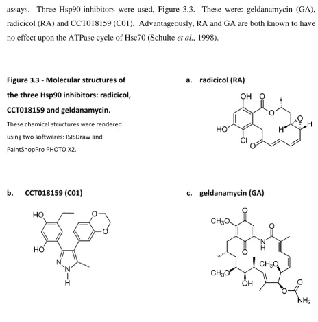
Figure 3.3 Molecular structures of the three Hsp90 inhibitors: radicicol, CCT018159 and
geldanamycin.
Figure 3.4 The Hsp90 inhibitor, geldanamycin (GA), sensitises cells to ricin.
Figure 3.5 The Hsp90 inhibitor, radicicol (RA), sensitises cells to ricin.
Figure 3.6 The Hsp90 inhibitor, CCT018159 (C01), sensitises cells to ricin.
Figure 3.7 N-ethylcarboxamidoadenosine (NECA) protects cells from ricin.
Figure 3.8 Cells incubated with either RA or GA display increased levels of Hsp72 and Grp94,
but constant expression of Hsp90
Figure 3.9 Chronic pre-treatment with both GA and RA protect cells from ricin
Figure 3.10 Chronic incubation with 4-PB leads to the reduced intensity of Hsc70 immunostaining & protects cells from intoxication.
Figure 3.11 A 10 minute heat shock at 45°C does not alter the susceptibility of cells to ricin Figure 3.12 The effect of DSG dominates the effect of radicicol
Figure 3.13 The effect of ricin incubation on Hsp90 / Hsp72 expression in HeLa cells.
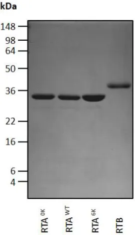
Figure 3.14 Purification of RTAvariants.
Figure 3.15 Purity of RTAWT, RTA0K and RTA6K preparations after centrifugal concentration.
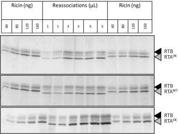
Figure 3.16 Quantification of reassociated holotoxin
Figure 3.17 Increasing the lysine complement of RTA reduces holotoxin cytotoxicity.
viii.
Chapter 4: An in vitro solubility assay for determining factors affecting the solubility of RTA
Figure 4.11 RTA can be heated and then split into soluble and aggregated fractions
Figure 4.2 Relative intensity of RTA from the pellet increases with temperature
Figure 4.3 Solubility of saporin after incubation for 15 minutes at 45°C.
Figure 4.4 Solubility of RTA correlates to enzymatic activity
Figure 4.5 The effect of macromolecular crowding upon RTA aggregation at 45°C and 37°C
Figure 4.6 The effect of [NaCl] upon RTA aggregation at 45°C
Figure 4.7 The effect of [CaCl2] upon RTA aggregation at 45°C
Figure 4.8 The effect of pH upon RTA aggregation at 37°C
Figure 4.9 The effect of glycerol on the solubility of RTA
Figure 4.10 The effect of glycerol on the cytotoxicity of ricin Figure 4.11 The effect of glycerol on the trafficking time of ricin Figure 4.12 The effect of POPS liposomes upon the solubility of RTA Figure 4.13 The effect of POPC liposomes upon the solubility of RTA Figure 4.14 Purification of Hsp40.
Figure 4.15 Purification of Hsc70
Figure 4.16 The effect of Hsc70 and Hsp40 upon the solubility of RTA after incubations at 45°C and 37°C
Figure 4.17 Effect of low BSA concentrations on the distribution of RTA between pellet and soluble fractions.
Figure 4.18 Higher concentrations of BSA inhibit the solubility of RTA
Figure 4.19 Silver-stained gels showing the effect of DSG upon the ability of Hsc70 and Hsp40 to prevent aggregation of RTA.
Figure 4.20 The effect of chaperones upon saporin Figure 4.21 Production of cytosol from HeLa cells
Figure 4.22 The effect of a cytosolic extract upon the aggregation of RTA Figure 4.23 The effect of CCT upon the rescue of soluble RTA by cytosol.
Chapter 5: Co-factors of Hsc70 and Hsp90 modulate their in vitro and in vivo activities
Figure 5.1 Hsc70, Hsp40 and CHIP can target native clients for ubiquitination.
Figure 5.2 Hsc70, Hsp40 and CHIP targets denatured clients for ubiquitination.
Figure 5.3 RTA as a substrate of the Hsc70 / CHIP complex
Figure 5.4 The effect of lysine content upon ubiquitination of RTA by the Hsc70 / CHIP
complex
Figure 5.5 Sequence of Ricin A Chain highlighting the relative positions of the native lysines
Figure 5.6 Hsc70/CHIP-mediated ubiquitination of RTAWT and RTA6K
Figure 5.7 The effect of negatively charged POPS liposomes upon the susceptibility of RTA to
ix.
CHIP-mediated ubiquitination.
Figure 5.8 Increased CHIP-mediated ubiquitination of RTAWT in the presence of Hsc70 and
Hsp90, as published by Spooner et al. (2008).
Figure 5.9 The effect of cytosol on RTA
Figure 5.10 A comparison of the domain architecture of human BAG domain proteins. Figure 5.11 BAG-5 inhibits the Hsc70/CHIP-mediated ubiquitination of clients Figure 5.12 The effect of BAG-5 upon ubiquitination by Hsc70/CHIP
Figure 5.13 The effect of BAG-5 upon ubiquitination of RTA by Hsc70/CHIP
Figure 5.14 The effect of BAG-5 upon the ability of Hsc70 / Hsp40 to keep RTA soluble at 45°C. Figure 5.15 The effect of BAG-5 alone upon the aggregation of RTA at 45°C.
Figure 5.16 Optimisation of over-expression conditions Figure 5.17 Over-expression of HOP protects cell from ricin
Figure 5.18 Over-expression of HOP, BAG-1, BAG-5, and CHIP sensitises cells to ricin treatment.
Figure 5.19 Over-expressing BAG-2 and HIP sensitise cells to ricin treatment.
Figure 5.20 Putative over-expression of an arbitrary protein, LacZ, does not affect ricin sensitivity.
Figure 5.21 The effect of SV40 virus infection upon the sensitivity of cells to ricin.
Chapter 6: Final Discussion
Figure 6.1 A model for the triage of RTA by cytosolic chaperones.
Chapter 7: References
Nil.
Chapter 8: Appendices
Figure 8.1 The activity of deoxyspergualin degrades on prolonged storage
Figure 8.2 The increasing turbidity of an RTA solution during a heat treatment
200
202 204 206 208 210 214 216 218 220 223
226 228
230
236
246
249
276
x.
List of Tables
Table heading Page
Chapter 1: Introduction
Table 1.1 Examples of ribosome inactivating proteins
Table 1.2 Protein import Pathways and their driving forces
Table 1.3 Select factors implicated in both Yeast and Mammalian ERAD
Chapter 2: Materials & Methods
Table 2.1 List of primary antibodies used
Table 2.2 List of secondary antibodies used
Table 2.3 DNA vectors used
Chapter 3: Pharmacological inhibition of cytosolic chaperones influences the cytotoxicity of ricin
Table 3.1 Chaperone inhibitors and their targets
Chapter 4: An in vitro solubility assay for determining factors affecting the solubility of RTA
Table 4.1 Conversion of chaperone molarity into absolute concentration
Chapter 5: Co-factors of Hsc70 and Hsp90 modulate their in vitro and in vivo activities
Table 5.1 Co-chaperones of Hsc70.
Table 5.2 Co-chaperones of Hsp90.
Table 5.3 Ubiquitination and its various cellular roles.
Chapters 7-8
Nil.
1
3 23 25
43
44 44 45
60
64
106
150
174
176 177 181
xi.
List of Abbreviations
The following is a complete list of uncommon abbreviations used in this thesis. It excludes those covered by the Système international d'unités
2YT 2× yeast extract, tryptone medium Derlin degradation in the ER associated protein
4-PB 4-phenyl butyrate DHFR dihydrofolate reductase
AAA ATPase associated with various cellular activities DMSO dimethyl sulfoxide
a.a. amino acid Doa dead on arrival
AP alkaline phosphatase DOI duration of infection
ADP adenosine diphosphate DSG deoxyspergualin
Aha Activator of Hsp90 ATPase Dsk dark skin
ALLN N-Acetyl-Leucinyl-Leucinyl-Norleucinyl-CHO DTT dithiothreitol
AMPK Adenosine monophosphate activated protein kinase DUB de-ubiquitinating factors
ApoB apolipoprotein B EE early endosomes
AT antitrypsin EDEM ER-resident degradation enhancing mannosidase
ATP adenosine triphosphate ER endoplasmic reticulum
ATX ataxin ERAD endoplasmic reticulum associated degradation
a.u. arbitrary units ERAD-L ~ (lumenal faults)
BAG Bcl-2 associated athanogene ERAD-M ~ (membrane faults)
BCIP 5-bromo-4-chloro-3-indolyl phosphate ERAD-C ~ (cytosolic faults)
BiP binding protein FBX F-box
β-me β-mercaptoethanol FGF fibroblast growth factor
BSA bovine serum albumin g gravity
°C degrees Celsius GA geldanamycin
C01 CCT018159 gp glycoportein
cAMP cyclic adenosine monophosphate GFP green fluorescence protein
cGMP cyclic guanosine monophosphate GTP guanosine triphosphate
CCT chaperone containing TCP-1 Grp glucose regulated protein
Cdc cell division cycle h hours
cf. confer Hdj human DnaJ homologue
CFTR cystic fibrosis transmembrane conductance regulator HERP homocysteine-induced ER stress regulated protein CHIP C-terminus of Hsc70 interacting protein HIP Hsc70 interacting protein
Ci Curies Hmg- 3-hydroxy-3-methyl-glutaryl-
Cnx calnexin HOP Hsp70/90 organising protein
CoA co-enzyme A Hrd Hmg-CoA reductase degradation
COP coat protein HRP horseradish peroxidise
CPI Complete protease inhibitors Hsc heat shock cognate
CPY carboxipeptidase Y HSF heat shock factor
Crt calreticulin Hsp heat shock protein
CTA1 Cholera toxin A chain HSPBP hsp binding protein
CTD c-terminal domain Htm homologous to mannosidase
Cue coupling of ubiquitination to ER degradation IC50 concentration required to effect 50% inhibition
xii.
IPTG isopropyl β-D-1-thiogalactopyranoside Rpt regulatory particle
Kar karyogamy RTA ricin toxin A chain
kDa kilodaltons RTB ricin toxin B chain
Lipofect. lipofectamine S Svedberg units (always prefixed by a number)
Log logarithm S soluble (never prefixed by a number)
Lys lysine SD standard deviation
mRNA messenger ribonucleic acid SDS sodium dodecyl sulfate
Min minutes Sec secretory
Mns mannosidase SEL suppressor of lin-12-like
MOPS 3-(N-morpholino)propanesulfonic acid SEM standard error of the mean
n number SERCA sarcoplasmic / ER Ca2+ ATPase
NAC nascent chain associated complex SF9 Spodoptera frugiperda cell line 9
N.B. nota bene (note well) Skp S-phase kinase-associated protein
NBT nitroblue tetrazolium SOB super optimal broth
NECA N-ethyl-5’-carboxamido adenosine Ssa stress seventy sub-family A
NEF nucleotide exchange factor Ste sterile
NLS nuclear localisation signal SV40 Simian vacuolating virus 40
Npl nuclear protein localisation t time
OS Oligosaccharide sensing TAg Large T antigen
P pellet (note capital) Tat twin arginine transport
p particle (note lower case) TCA trichloroacetic acid
p probability value (note italics) TEB thioesterase B gene
Ψ water potential TEMED tetramethylethylenediamine
PAGE polyacrylamide gel electrophoresis TGN trans-Golgi network
PAP pokeweed antiviral protein TPR tetratricopeptide repeat
PDI protein disulfide isomerise TPST tyrosyl protein sulfotransferasse
pH potenz hydrogen TRC tumour repressor in renal carcinoma
pI isoelectric point Tris Tris-(hydroxymethyl)-aminomethane
POPC 1-palmitoyl-2-oleoyl-sn-glycero-3-phosphocysteine Ub ubiquitin
POPS 1-palmitoyl-2-oleoyl-sn-glycero-3-phosphoserine Ubc ubiquitin conjugating
PPI peptide prolyl isomerise Ubx ubiquitin regulatory ‘x’
Pre pre-incubation UCSF University of California, San Francisco
Rab Ras-like proteins from rat brain UDP uridine disphosphate
RA radicicol UGGT UDP-glucose:glycoprotein transferase
Rad radiation sensitivity abnormal Ufd ubiquitin fusion degradation
RE recycling endosomes UPS ubiquitin-proteasome system
Rel. relative v/v volume/volume
RFP RING-finger protein v/w volume/weight
RING really interesting new gene WT wild-type
RIP ribosome inactivating protein Ydj yeast DnaJ homologue
RMA RING-finger motif with membrane anchor Yos yeast oligosaccharide sensing RNA ribonucleic acid
xiii.
Acknowledgements
Thanking everyone who has helped me form this thesis is an impossible task, so I have to thank the most important people. Professors Mike Lord and Lynne Roberts and Doctor Robert Spooner have all been monumentally kind to me during my four years of study. This kindness has been manifested in many forms: freely discussing speculation, evaluating results, the occasional beer and the accommodation of my work- and writing-styles. I’m told the latter can be unnecessarily abstruse, so thank you for your patience!
I must also thank Professor Jörg Höhfeld of the University of Bonn and his protégés, Doctors Christian Rogon, Michael Dreiseidler and Verena Arndt. They were especially helpful in providing constructs and stocks of purified protein. At Warwick, I would like to thank Kat Moore for inducting me into the lab and tutoring me for my first year, as well as Dr. J. Cook, Dr. Alice Rothnie and Dr. Paola Pietroni for technical assistance and advice. Professor John Ellis also deserves thanks for helping us to critique the initial stages of the project and for rigorously defending a lot of the core precepts that lie at the heart of chaperone biology (especially whenever a slip-of-the-tongue was made).
xiv.
Declaration
All the results presented in this thesis were obtained by the author at the University of Warwick, whilst under the supervision of Professors J.M. Lord and L.M. Roberts. This is the case unless it is explicitly stated otherwise. All sources of information have been specifically acknowledged by means of reference.
Results in this thesis which have been published before are highlighted by the publication attached at the rear. These published results fundamentally include: co-chaperone over-expression experiments; Hsc70 assays where the inhibitor, deoxyspergualin, has been used in vivo; and Hsp90 inhibitor work using acute geldanamycin and radicicol treatments in vivo (with wild-type toxin).
xv.
Summary
Ricin is a heterodimeric, toxic plant protein. It is able to deliver its catalytic A chain (RTA) into the cytosol of target cells. RTA crosses the endoplasmic reticulum (ER) membrane into the cytosol, masquerading as a substrate of ER-associated degradation (ERAD) to do so. Therein, RTA inactivates ribosomes. This thesis
shows that RTA is prone to lose solubility in vitro near the physiological temperature and pH of target cells.
This instability is hypothesised to cause RTA to misfold in the ER lumen, promoting chaperone and membrane interactions therein. Fittingly, this thesis shows that Grp94, a lumenal chaperone, promotes the toxicity of RTA, and that liposomes constructed of negatively-charged phospholipid interact with RTA in vitro. This instability resembles that of other toxic A chains that exploit ERAD. Cholera toxin A chain and pertussis toxin A chain, for instance, are also relatively unstable (Pande et al., 2007 & 2008).
The Hrd1 complex now seems the strongest candidate for retrotranslocating RTA from the ER lumen (Li et al., 2010). In the cytosol, the proteasomal cap has been shown to be involved in downstream processing of RTA – enabling toxicity (Li et al., 2010). This thesis reports that, in mammals, the balance of cytosolic chaperones and their co-factors helps to dictate the success of retrotranslocated RTA, putatively by determining its escape from terminal degradation in the proteasomal core. The effect of these chaperones occurs at a stage beyond access of the toxin subunit to the ER, and can result in both activation and inactivation of cytosolic RTA.
It has been shown that, on one hand, Hsc70 is responsible for activating RTA. Hsc70 may aid RTA in attaining an active conformation in the cytosol after retrotranslocation. Alternatively, it might supplant the effectors of its degradation. On the other hand, Hsc70 also enters RTA into a sequential triage with Hsp90. Unlike Hsc70, Hsp90 deactivates RTA. This effect is dependent upon the lysines of this toxin subunit, suggesting Hsp90 may participate in the lysine-ubiquitination of RTA. Supporting this conclusion, Hsc70 and Hsp90 can both ubiquitinate RTA in vitro. This ubiquitination can be promoted if RTA is first incubated with liposomes. This implies that RTA may be particularly vulnerable to ubiquitination during retrotranslocation, where it might also be partially solvated by phospholipid. Contrasting to RTA, Hsp90 actually aids the toxicity and dislocation of
ER-retrotranslocating cholera toxin A chain (Taylor et al., 2010). It seems that RTA may have fortuitously
evolved to exploit Hsc70 rather than Hsp90 to promote its cytosolic activation. Provocatively, the hydrophobic C-terminal tail of RTA demarcates it from a homologous toxin, saporin, which does not exploit ERAD to achieve toxicity. Indeed, this region may be an adaptation RTA has acquired to promote interaction of the toxin subunit with Hsc70, which even seems to occur in RTA’s native, folded state. This interaction may be another reason why the region is apparently significant to the cytotoxicity of the protein (Simpson et al., 1995)
Page 1 CHAPTER 1: Introduction
1.0 Ricin: occurrence, mode of action and the original applications of its study
Ricin is a potently toxic protein of the castor oil plant, Ricinus communis. The mature protein is found throughout the tissues of its native host at low levels. However, it is especially concentrated in the endosperm of the plant‟s seeds, from which commercially valuable castor oil can also be extracted (Alexander et al., 2008). Consumption of as few as four of these poisonous seeds can be a mortal dose for an adult (Wedin et al., 1986). This extreme toxicity first provoked Stillmark (1888) to discover the agent responsible: the protein, ricin.
The protein itself is a heterodimer, comprising a galactose-binding lectin moiety of 34kDa disulfide-bonded to an rRNA N-glycosidase of 32kDa (Olsnes & Pihl, 1973). These are denoted ricin toxin B chain (RTB) and ricin toxin A chain (RTA) respectively. Its bipartite structure and function makes ricin analogous to a broad class of two-component complexes with similar operational arrangement – known collectively as AB-toxins. The B chain of these complexes generally has a cell-binding role, helping to localise the holotoxin to the site where the A chain thereafter exerts a cytotoxic activity. This arrangement is also true of ricin (Lord & Robert, 1998). Ricin is also member to a large family of ribosome inactivating proteins (RIPs), which are subdivided into two main types (I & II) by the presence or absence of a B chain. Type I RIPs do not possess a lectin B chain, whereas type II RIPs, like ricin, do (Lord et al, 1994). It is thought that these proteins serve an integral function in their native hosts as they are found in well over 50 different species spread among 13 taxonomic families (Hartley et al., 1996).
RTB binds specifically to β1-4 linked galactosides. Therefore, it is able to bind glycoproteins
Page 2
1987). After this depurination, the ribosome irreversibly stalls during translation as it can no longer bind mechanistically-essential elongation factors (Stirpe & Battelli, 2006).
As the production of ribosomal RNA accounts for 70% of all transcription even under unstressed conditions (Granneman & Tollervey, 2007), the cell has only a limited capacity to compete against the catalytic activity of RTA. To emphasise the overwhelming extent of this burden, a single RTA is able to inactivate 2000 ribosomes per minute in vitro (Sandvig & Van Deurs, 2002). The target cell, unable to maintain its protein complement, nor to adapt its protein expression in response to dynamic environmental and internal cues, ultimately dies. Indeed, after intoxication of an animal host by any major route, both apoptosis and necrosis are observed in affected tissues (Sha et al., 2010). Rao et al. (2005) show that, in a simplified culture of HeLa cells, viability is lost through a primarily apoptotic route.
Surprisingly, despite their wide distribution among plants and the attention of significant research, the physiological role of RIPs in their native hosts remains a subject of debate (Hartley et al., 1996). Foremost, ricin has been suggested to have a role in warding off herbivores (Olsnes & Pihl, 1982). However, it seems inconsistent that as many as 36 hours may pass after ingestion of a lethal dose before the first signs of toxicity become manifest (Wedin et al., 1986). By this time a greedy herbivore may have already consumed a significant quantity of the plant, although it is possible that toxicity could have influenced forager behaviour over a greater timescale. Nevertheless, why synthesise a large, metabolically-costly protein to fulfil this job? Why not synthesise a more prudent secondary plant substance (examples of which are reviewed in Hruska, 1988), which could achieve a similar and more immediate reduction in edibility?
Page 3
[image:19.595.67.520.229.510.2]viruses and fungi in their native contexts (as reviewed in Hartley et al., 1996). Ultimately, it may be that a combination of these activities explains the pervasiveness of RIPs and the heavy accumulation of ricin in castor seeds, which can be as much as 5% of total protein content (Lord et al., 1994).
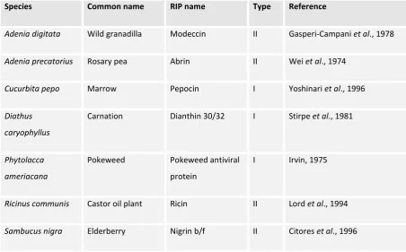
Table 1.1 – Examples of ribosome inactivating proteins
Species Common name RIP name Type Reference
Adenia digitata Wild granadilla Modeccin II Gasperi-Campani et al., 1978
Adenia precatorius Rosary pea Abrin II Wei et al., 1974
Cucurbita pepo Marrow Pepocin I Yoshinari et al., 1996
Diathus
caryophyllus
Carnation Dianthin 30/32 I Stirpe et al., 1981
Phytolacca
ameriacana
Pokeweed Pokeweed antiviral
protein
I Irvin, 1975
Ricinus communis Castor oil plant Ricin II Lord et al., 1994
Sambucus nigra Elderberry Nigrin b/f II Citores et al., 1996
Regardless of the physiological role of ricin in Ricinus communis, its potent toxicity has been exploited by humans in a number of therapeutic, investigative and criminal applications (reviewed in both Hartley et al., 1996; and Lord et al., 1994). Some examples of its use include, as:
(1) An active moiety in antibody-conjugated immunotoxins that are used to destroy cancerous cells, or else allogeneic T lymphocytes from donated bone marrow. (2) A tool in developmental biology used to ablate cell lineages under the control of
stage- or tissue-specific promoters.
Page 4 (5) An abortifacient.
(6) A way to kill other humans.
For these reasons, and because of the potential future uses of the toxin in related contexts, the mechanism of ricin is the subject of ongoing scientific interest.
1.1 Biosynthesis of ricin in its native Ricinus communis
The overall route by which ricin invades a target cell has been introduced, but the molecular mechanisms governing this feat have not been elaborated upon. To appreciate the fortuitously ingenious trafficking route the holotoxin undertakes, the biogenesis of the protein should be made clear. Unlike its heterodimeric, mature form, ricin is initially translated as one contiguous chain with a signal peptide – holistically known as „preproricin‟ (Hartley et al., 1996). During its synthesis, the emergence of an N-terminal signal peptide from the exit tunnel of the ribosome ensures the translational complex is targeted to the endoplasmic reticulum membrane (Martoglio & Dobberstein, 1998). There, the nascent polypeptide is co-translationally channelled into the lumen of the endoplasmic reticulum (ER) through the Sec61 translocon. As the translating polypeptide is incrementally driven through this membrane pore by the machinations of the ribosome, several modifications occur.
First, the signal peptide is cleaved by the lumen-facing activity of signal peptidase. Then, as four primary-sequence embedded N-glycosylation sites emerge from the translocon, they become core-glycosylated (Lord, 1985)1 by a complex adjacent to the translocon, the oligosaccharyl transferase, OST, complex. Finally, the signal peptide-cleaved, glycosylated polypeptide folds into its native secondary and tertiary structural elements, which are stabilised by the formation of five intra-chain disulfide bonds (Rutenber & Robertus, 1991).
This folded, signal-peptide cleaved protein, referred to as „proricin‟, exits the ER by vesicular flow and is targeted to vacuolar protein bodies by a twelve-residue region linking RTA and RTB (Frigerio et al., 2001). Upon entry to this vacuolar compartment, the targeting sequence is proteolytically excised, liberating the familiar, heterodimeric form of the toxin that we refer to as ricin (Butterworth & Lord, 1983). The five disulfide bonds that originally formed in the ER lumen persist in the final structure: four stabilise RTB, whilst the fifth maintains
1
Page 5
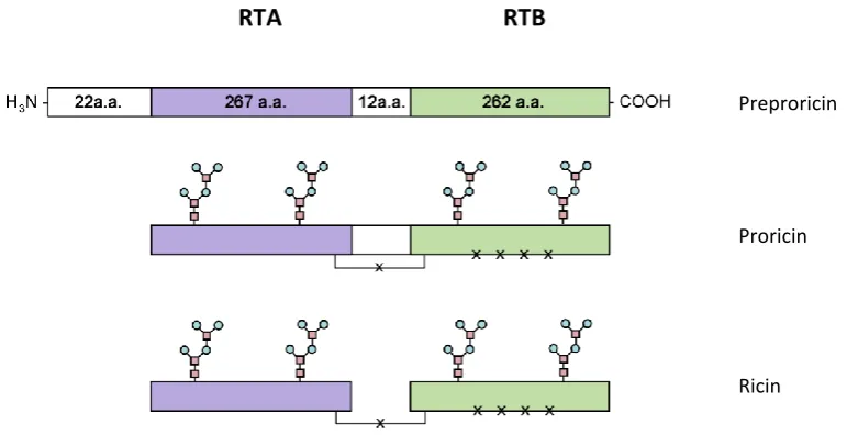
the only remaining covalent bridge between RTB to RTA. The differences between these three forms are summarised graphically in Figure 1.1.
[image:21.595.74.460.475.678.2]This series of post-translational processing events minimises the possibility that the toxic activity of RTA is ever accidentally exposed to the cytosol of the host cell. Not only is the protein compartmentally sealed off from host ribosomes at the moment of its synthesis, but even if it were inadvertently translated into the cytosol, uncleaved preproricin is unable to depurinate the ribosomes which are the substrate of the mature toxin (Richardson et al., 1989). This, along with the fact that many plant ribosomes are RIP-resistant (Frigerio et al., 1998; Hartley et al., 1996), prevents the accidental death of the host organism.
Figure 1.1 - The various forms of ricin during biogenesis of the mature holotoxin
Showing preproricin, proricin and ricin. Core N-glycosylation is indicated by the chain of attached circles and
squares. Disulfide bonds are indicated by “x”. One of these bridges RTA & RTB. The 22 amino acid signal
peptide and the 12 amino acid linker are shown by the white rectangles, which are progressively excised in the
subsequent maturation. RTA is shown in purple. RTB is shown in green. The graphics for the image below
were produced using Paintshop Pro X2, Corel.
RTA RTB
Preproricin
Proricin
Page 6 1.2 The three-dimensional structure of ricin holotoxin
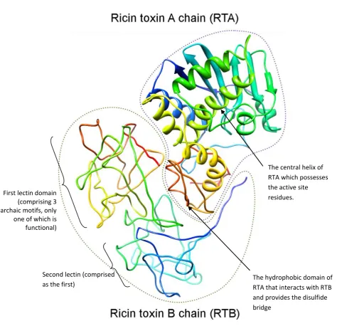
The fully-folded structure of mature ricin was determined to a resolution of 2.5Å by Rutenber & Robertus (1991). A rendering of this structure is printed in Figure 1.2. The detail in the following section provides a summary of this structural information, with interpretation derived from it paraphrased from Lord et al. (1994).
Ricin toxin A chain (RTA) – The enzymatically active, N-glycosidase moiety of the
holotoxin possesses eight α-helices and a single β-sheet. These elements account for roughly half of the 267 amino acid subunit. The N-terminal 40% of the polypeptide folds into a discrete conformational unit comprising two of these α-helices and the β-sheet. Adjunct to this domain, the polypeptide is arranged into a bundle of five α-helices, which nestles behind
the N-terminal β-sheet. The central helix of this α-helical cluster bears the active site residues of the N-glycosidase activity (Glu177 and Arg180), to which the appropriate stem-loop structure of rRNA must be accessible. The C-terminal portion of the polypeptide folds into another, independent conformational unit centred upon the eighth α-helix. This discrete
region, apposed to the active site helical bundle, helps produce the binding cleft of the enzyme. Importantly for future reference, the opposite side of this cleft-forming domain (the C-terminus) is hydrophobic and interacts with RTB. This region also houses the disulfide bridge which covalently links the two subunits.
Ricin toxin B chain (RTB) – The lectin moiety of the ricin holotoxin is 262 amino acids
Page 7
Figure 1.2 - The three-dimensional structure of the ricin holotoxin
Showing the three-dimensional structure of ricin, as published by Rutenber & Robertus (1991). The
polypeptide chain is coloured so that the C-terminus region of each chain is blue – progressing through the
spectrum to red at the N-terminal of both RTA and RTB. This image was produced using the UCSF Chimera
package from the Resource for Biocomputing, Visualization, and Informatics at the University of California, San
Francisco. Depth is implied by the fading in colour saturation.
First lectin domain (comprising 3 archaic motifs, only one of which is functional)
Second lectin (comprised
as the first) The hydrophobic domain of RTA that interacts with RTB
and provides the disulfide bridge
Page 8 1.3 Intoxication of a target cell
When mature ricin is exposed to target cells, intoxication proceeds via a series of events that can be divided into distinct stages. These stages are:
(1) Binding at the target cell‟s surface, followed by indiscriminate endocytosis.
(2) Retrograde trafficking to an endomembrane compartment where the phospholipid bilayer can be crossed.
(3) Translocation of active RTA across the membrane into the cytosol. (4) Inactivation of target ribosomes.
The novel content of this thesis focuses upon putative molecular interactions the toxin might make on either side of the translocated membrane and the ramifications this may have upon downstream processing of the protein by the target cell. To provide appropriate context to these unique findings, the earlier stages of toxin trafficking are summarised below.
1.3.1 Transit of ricin from the cell surface to the translocation-competent compartment Estimates suggest that the average HeLa cell possesses as many as ~3107 independent cell-surface galactosides that may be bound by RTB (Spooner et al., 2004). This abundance causes the holotoxin to be broadly adsorbed to the cell surface, resulting in its internalisation by a variety of endocytic routes – both clathrin-dependent and clathrin-independent (Simpson et al., 1998). Some of these routes deliver ricin to the lysosome, wherein it is degraded. Others deliver the holotoxin to early and recycling endosomes (EEs & REs, respectively) that stain positively for the trafficking molecule, Rab5 (Moisenovich et al., 2004).
Page 9
Ricin is subsequently transported from the TGN to the ER, wherein fluorescence can be observed if cells are intoxicated with a GFP-ricin construct (Liu et al., 2006). Only a minuscule quantity of toxin eventually achieves a cytosolic destination relative to that which is internalised at the cell surface. Data from Rapak et al. (1997) also corroborated the trafficking route described above in a series of elegant experiments. They produced a holotoxin where the constituent RTA was tagged with vacant glycosylation and sulfation sites. After intoxication of mammalian cells with this holotoxin, they observed that glycosylated, sulfated RTA could be retrieved from the cytosol. These modifications are performed by oligosaccharyl transferase of the ER (Yan et al., 1999) and tyrosyl-protein sulfotransferate (TPST) of the Golgi (Spooner et al., 2008a), respectively. Thus, it showed that toxin which had gained access to the cytosol had, at one stage during intoxication, co-localised with both of these activities. Supporting this hypothesis, increasing the transport of ricin to the Golgi and the ER under experimentally-controlled conditions increases the cytotoxicity of ricin. Indeed, Morre et al. (1987) observed that mammalian cells were sensitised to ricin when pre-treated with monensin (which increases the flow of protein traffic into the Golgi cisternae). Consistent with these findings, addition of a C-terminal Golgi retention motif to ricin, YQRL, had a predictably sensitising effect (Zhan et al., 1998), as did addition of the ER retrieval motif, KDEL (Wales et al., 1993). As native ricin lacks these sequences, it is unknown how it achieves an ER localisation. Contrastingly, cholera toxin, another example of an AB-toxin where the A chain dislocates from the ER into the cytosol, actually possesses a C-terminal KDEL (Majoul et al., 1998). Curiously, however, cholera toxin can also navigate to the ER if its KDEL sequence is removed (Lencer et al., 1995). In such a case it might be transported to the ER in a more serendipitous fashion, like ricin. For instance, these toxins might hitch-hike upon other proteins that, themselves, have appropriate retrieval sequences.
Day et al. (2001) hypothesised that ricin might disengage from its original, cell-surface binding partner after endocytosis. This would enable it to re-engage with escapee residents of the ER or Golgi which possess KDEL motifs. Indeed, Day et al. (2001) demonstrated that ricin could bind to an iconic ER chaperone, calreticulin, in vitro.2 Curiously, however, cells
2 N.B. This binding occurred only between holotoxin and calreticulin and not between RTA and calreticulin.
Page 10
remain sensitive to ricin when wider KDEL retrieval is prevented by inhibition of the COPI apparatus, preventing Golgi to ER transport (Chen et al., 2003). This suggests that there is a significant alternative route to the ER that remains to be investigated.
1.3.2 Rationalising the ER as the ultimate endomembrane destination
Whilst the exact nuances of ricin‟s retrograde trafficking remain obscure, the final destination
of the holotoxin in the secretory pathway seems very likely to be the ER lumen (Rapak et al, 1997; Liu et al., 2006; Sandvig et al., 1991). This finding is consistent with a multitude of other AB-toxins that are also transported to the ER (e.g. cholera toxin – Majoul et al., 1998; Pseudomonas exotoxin A and Shiga toxins – as reviewed in Watson & Spooner 2006). Fittingly, there are abundant machineries in the ER which facilitate the translocation of polypeptides across the membrane, which is what RTA must do to reach cytosolic ribosomes. Foremost among these machineries is the Sec61 translocon and its accessory factors, which are responsible for importing proteins into the ER. Complementing this apparatus are the components of endoplasmic reticulum associated degradation (ERAD), which collectively promote the export of undesirable polypeptides to the cytosol. There, they are hydrolysed by the 26S proteasome (Meusser et al., 2005; Werner et al., 1996).
From the perspective of the toxin, the directionality of polypeptide movement during ERAD seems the most appropriate to exploit. However, how could it evade the proteasomal destruction which is usually the tacit outcome of the process? Hazes & Read (1997) were first to report that RTA and the active moieties of many other ER-trafficking toxins (e.g. cholera toxin; shiga toxin; pseudomonas exotoxin, pertussis toxin) have low lysine complements compared to polypeptides of similar length. This implied the physiological role of these toxins exposed them to a situation where the replacement of these residues would be of benefit to the protein‟s function. As proteasomal degradation is greatly promoted by
lysine-linked polyubiquitination (Thrower et al., 2001), it seemed the toxins had evolved to escape this modification. Perhaps they had done so to evade polyubiquitination during a particularly vulnerable, ERAD-like translocation into the cytosol? Summarily, investigative interest grew out of this hypothesis into two areas of research:
(1) what features of ricin could make it appear like an ERAD substrate; and, moreover,
Page 11
Before experimental observations that probe these quandaries are introduced, this chapter continues by providing a background to the physiological role, nature and dynamics of this essential quality control pathway.
1.4 Endoplasmic reticulum associated degradation (ERAD)
The endoplasmic reticulum is the first endomembrane compartment which proteins of the secretory pathway encounter. Whether these proteins are co- or post-translationally translocated into the ER from their site of synthesis, they are all transiently accommodated by the narrow aperture of the Sec61 translocon en route to this organelle (Van den Berg et al., 2004; Tian & Andricioaei, 2006). This means that all polypeptides that are secreted into the lumen emerge into it in a substantially unfolded, translocation-competent state. In this state they transiently expose hydrophobic regions to the lumenal environment. These regions have a tendency to self-aggregate, forestalling the acquisition of native structures, so promoting the accumulation of dysfunctional ones. To mitigate these detrimental side-reactions, there are abundant chaperones in the ER lumen that temporarily bind these regions. This shield of chaperones persists until enough contiguous polypeptide has emerged from the translocon to enclose hydrophobic stretches within appropriately hydrophilic encasements. Indeed, the lumenal equivalent of the Hsc70 molecular chaperone, BiP (Kar2 in yeast), binds imported polypeptides as they emerge, ensuring just that (Hammon & Helenius, 1994).
Nevertheless, the folding process often goes awry if the capacity of the cell to successfully nurture the folding of nascent protein is exceeded. This occurs under conditions of, for instance, heat or oxidative stress, nutrient starvation; or after translational errors and mutation (Schröder & Kaufman, 2004; McClellan et al., 2005). Moreover, under unstressed conditions, the subunits of multimeric complexes can be synthesised in incorrect ratios, yielding orphan polypeptides that lack the partners that usually complement their native state (Vanhove et al., 2001). Even after having assumed a native state, proteins remain subject to varying degrees of regressive unfolding – whether these are stochastic or stress-induced. This results in the ongoing potential for terminally-misfolded conformers to be generated.
Page 12
(Kincaid & Cooper, 2007). Often, however, misfolded proteins are isolated in the early stages of the secretory pathway, preventing their transmission to downstream organelles and neighbouring cells. These proteins are degraded by cytosolic 26S proteasomes, the process which is designated „ERAD‟ (Werner et al., 1996).
However, removal of unwanted polypeptides from the ER lumen to the cytosol is no trivial task. Indeed, each ERAD client investigated to date is managed by a – sometimes vastly, sometimes subtly – distinct retinue of facilitators (as reviewed by Nakatsukasa & Brodsky, 2008). Still, there are common procedures that unite all ERAD pathways. In brief:
(1) Misfolded substrates have to be recognised, which is usually achieved by a combination of membrane factors, lingering associations with chaperones and lumenal lectins.
(2) The recognition factors bridge an interaction of the substrate with the retrotranslocation machinery. This machinery is contributed to by a miscellany of membrane-associated and membrane-integral complexes that may accommodate exit of the polypeptide from its ER locale (i.e. form a pore).
(3) Finally, upon the cytosolic side of the ER membrane, there are co- and post-translocational processing events which effectively drive extraction of the misfolded polypeptide from the membrane compartment and prepare it for degradation by the 26S proteasome.
The variety of ways in which these steps are achieved is outlined in the following sections.
1.4.1 How the cell identifies ERAD candidates
Quality control pathways in the ER must resolve terminally-misfolded polypeptides from those which are only transiently unfolded and those which are actually appropriately folded. However, the nature of a conformational faults can vary greatly, a corollary to the variety of stresses, mutations and chance events a cell may endure. Moreover, a fault can be localised to distinct phases of the ER environment: the lumen, the core of the lipid bilayer itself, or even to the cytosolic side of the ER membrane. Because of this diversity of „symptoms‟, the
Page 13
All the same, detection of ERAD clients relies upon two related, general attributes: chronic residence in the ER lumen and chronic exposure of hydrophobic regions (Määttänen et al., 2010). Retention in the ER can occur if a conformer fails to display appropriate export motifs on its surface (Ellgaard & Helenius, 2003). Retention would also occur if a client had a lingering interaction with ER chaperones, as would be the case when a misfolded protein recurrently displays hydrophobic regions to the solvent (Vanhove et al., 2001).
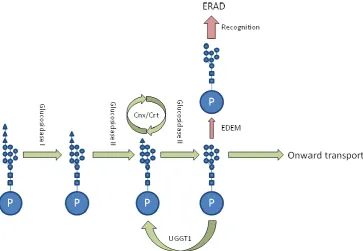
One of the best-elaborated mechanisms measuring prolonged ER-residency is the so-called „mannose timer‟ shown in Figure 1.3. The information that follows is discussed more
broadly in reviews by Wang & Ng (2008); Eriksson et al. (2004); and Määttänen et al. (2010). First, nascently secreted proteins with appropriate N-X-(S/T) glycosylation sites become core-glycosylated in the ER ([N-acetyl-glucosamine]2[mannose]9[glucose]3). This
Page 14 Figure 1.3 – Schematic of the mannose timer.
The pathway is described in text above. The large blue circle denoted ‘P’ represents the client protein in the
ER. The smaller blue squares attached represent N-acetyl glucosamine residues; blue circles mannose
residues; blue triangles represent glucose residues. “Onward transport” can be facilitated by vesicle sorting
receptors that recognise protein cargoes and interact with the cytosolic COPII machinery. Note EDEM is a
Page 15
Other routes by which clients may be co-opted onto ERAD pathways are less clear. However, they must exist to cater for proteins which are not glycosylated. What is more, not every glycoprotein is subjected to the mannose timer mechanism. Indeed, some glycoproteins are able to persist in the ER for long periods of time, in complexes with chaperones. For instance immunoglobulin heavy chains, which are retained in complex with BiP until their light chain binding partners are secreted into the lumen (Vanhove et al., 2001). Alternative mechanisms determining ERAD candidacy may be stochastically driven like the mannose timer. However, they might rely more simply upon prolonged chaperone interaction rather than a deterministic glycan switch. Indeed, OS-9 – which interacts with membrane integral ERAD machinery – can be found in complexes with two lumenal chaperones: Grp94 and BiP. The joint properties of these proteins may form an integral client-sensing gating complex to downstream ERAD effectors (Eletto et al., 2010; Kabani et al., 2003).
Unfortunately, it remains difficult to differentiate potential gating functions of lumenal chaperones from purely chaperoning activities, given that aggregation prevents ERAD (McCracken & Brodsky, 2006). It is also unclear whether Grp94 has a role in „herding‟ the
client to the chaperone machinery, or whether it is itself responsible for maintaining the complexes involved in handling the client (an argument made in a review by Nakatsukasa & Brodsky, 2008). The line therefore blurs between chaperone, gating arbiter and complex maintainer.
1.4.2 Membrane complexes involved in ERAD – multi-functional pores?
Page 16
1.4.2.1 ERAD-L & ERAD-M: The Hrd1 complex of Saccharomyces cereivisiae
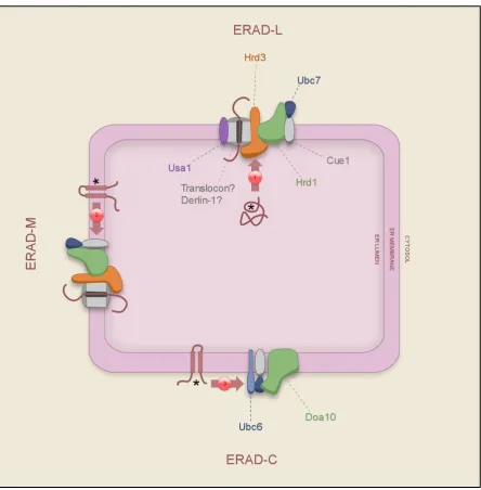
Substrates with conformational faults localised to the lumen are directed to the Hrd1 pathway (Figure 1.4). They are initially recognised by a presumptive „gating complex‟ that may comprise Yos9, Kar2, and Grp94 (Eletto et al., 2010; Kabani et al., 2003). This triumvirate of factors associates with the lumenal domain of an integral membrane protein, Hrd3, to which they are thought to pass clients like a mutant of the soluble protein α1-antitrypsin, i.e. α1-antitrypsin null Hong Kong (α1-AT NHK; Christianson et al., 2008), and glycoproteins
(Eletto et al., 2010). Hrd3 can also independently recognise and retain misfolded ER proteins, e.g. soluble, mutant carboxipeptidase Y (CPY*) without the direct aid of the gating complex (Gauss et al., 2006; and references in Hirsch et al., 2009). However, it seems reasonable to think that it works synergistically with the soluble chaperone complex to audit the contents of the ER. Hrd3, in turn, is bound stoichiometrically to the transmembrane E3 ligase, Hrd1 (Gardner et al., 2000). The E3 ligase activity of Hrd1 is required for the degradation of CPY* in the cytosol (Li et al., 2010; Bordallo & Wolf, 1999). This shows that degradation by other ERAD machinery cannot compensate in this context. Gardner et al. (2000) show that Hrd1 over-expression without the correlate up-regulation of Hrd3 leads to the promiscuous degradation of folded lumenal proteins. This demonstrates that the task of selecting misfolded clients is delegated to the other, upstream, factors.
The Hrd1 complex is also involved in the degradation of proteins with transmembrane aberrations, such as the Parkin-associated endothelin-like receptor, Pael-R (Omura et al., 2008). Problematic transmembrane domains might precipitate irregular structural phenotypes in connected lumenal domains. These would be recognised by soluble machinery. However, it has also been shown that a transmembrane helix of Hrd1 can detect tell-tale hydrophilic residues embedded in the otherwise hydrophobic core of the membrane, facilitating their transfer of the offending protein to a degradation pathway (Omura et al., 2008).
How Hrd1 substrates are channelled through the membrane after their recognition – a process known as „dislocation‟ or „retrotranslocation‟ – is unknown. It has been hypothesised that
Page 17
lumenal (rather than intra-membrane) faults, such as the transmembrane human V2 vasopressin receptor (Schwieger et al., 2008). It could be that Derlin-1 specifically tailors the dimensions of the membrane pore for such clients (Yihong et al., 2004).
After partially emerging onto the cytoplasmic face of the ER membrane, dislocated proteins are thought to be subjected to lysine-ubiquitination by the cytoplasm-facing RING finger ubiquitin ligase of Hrd1 (Hirsch et al., 2009). The ubiquitination cascade that leads to the priming of transferred ubiquitin residues is also enabled by membrane-associated proteins. An E2 enzyme, Ubc7, is tethered to the membrane by interaction with the transmembrane protein called Cue1, which interacts with Hrd1 (Bazirgan & Hampton, 2008). E2 ubiquitin-activating enzymes are involved in the sequence of reactions which prime ubiquitin for the ligation reaction that is catalysed by an E3 ubiquitin ligase‟s activity. After ubiquitination,
the substrates of the pathway converge with those of the Doa10 route, which is outlined next.
1.4.2.2 ERAD-C: The Doa10 complex of Saccharomyces cerevisiae
ERAD clients that are embedded in the ER membrane and which have cytosolic faults are degraded by a complex including Doa10 (Figure 1.4). As Doa10 contains fourteen transmembrane domains, it has been suggested that it may form a pore (Kreft et al., 2006). Membrane integral substrates of this complex include the mutant α-factor transporter,
designated Ste6*, and the plasma membrane H+-ATPase, abbreviated to PMA1 (Ravid et al., 2006). Much like Hrd1, Doa10 is accompanied by membrane-integral proteins which activate its E3 ligase activity. These include: Ubc7, Ubc6 & Cue1 (Huyer et al., 2004). Interestingly, Doa10 also contributes to the degradation of soluble cytoplasmic proteins, like the Mat alpha2 receptor (Swanson et al., 2001). However, such substrates do not appear to require downstream extraction machinery to facilitate this process (Ravid et al., 2006). Metzger et al. (2008) studied an example of this pathway, using a soluble, cytosolic orotidine 5-phosphate decarboxylase with a degradation-promoting C-terminal tag (Ura3-CL).
Page 18
Figure 1.4 – The ERAD-C, ERAD-L and ERAD-M complexes
The following figure is heavily influenced by the Cell Snapshot produced by Kawaguchi & Davis (2007). Unlike
their figure, machinery downstream of the E3 ligase is omitted for clarity (as these are discussed in the next
section of this introduction). ERAD substrates are the fleshy-pink wiggles and transmembrane domain
proteins marked with asterisks (the asterisk denotes the topology of the conformational fault the substrates
bears). 1) Substrates are recognised for ERAD-L by a combination of the mannose timer and the gating
complex formed by chaperones Kar2, Grp94 & Yos9 (Christianson et al., 2008). 2) Substrates are recognised
for ERAD-M by a transmembrane domain of Hrd1 (Omura et al., 2008), as well as by the mannose time
mechanism and Yos9. 3) Substrates are recognised for ERAD-C by a combination of Doa10 and putatively a
Page 19
1.4.2.3 A role for the Sec61 translocon in polypeptide export?
Early in the ERAD field it was proposed that the Sec61 translocon might simply be co-opted for the retro-translocation process. With the aid of supplementary, ERAD-specific factors it could become a „dislocon‟. However, as Sec61 is important for the initial translocation of
these polypeptides into the ER, it has been difficult to differentiate import failure from export success. Nakatsukasa & Brodsky (2008) make a comprehensive review of arguments for and against its involvement. Pilon et al. (1997) note that conditional Sec61 mutants are deficient in the retrotranslocation of ERAD clients like CPY* when shifted to Sec61-inactive conditions. Additionally, if the ERAD of a glycoprotein is reconstituted in vitro using microsomes, retrotranslocation can be prevented if translating ribosomes are first bound to Sec61 (Gillece et al., 2000).
Hebert et al. (2010) argue against Sec61 involvement because the minimal diameter of a glycoprotein is 20Å. They compare this to the aperture of the related, Archaeal SecY, which is too narrow, at 3Å (Van den Berg et al., 2004). Prior investigators, however, have shown that the mammalian translocon may be more flexible, its inner enclosure stretching to a diameter of 40-60Å during polypeptide import (Hamman et al., 1997). Nevertheless, Nakatsukasa & Brodsky (2008) review how experimenters have consistently failed to isolate Hrd1/Doa10 complexes containing Sec61. Moreover, they cite numerous studies where integral membrane substrates are efficiently degraded even when the translocon has been disrupted (using conditional mutants). One thought they express is that formation of Sec61-inclusive complexes may be transient, explaining why they may not have been observed to date.
Need there be a physical pore at all for some substrates? Ploegh (2007) speculates that
misfolded proteins might exit the ER environment attached to lipid droplets. His model suggests that insertion of neutral lipid into the bilayer can form discrete, lens-like domains. These could be ejected into the cytosol as lipid droplets. Microdomains of these droplets could possess bilayer-like properties, or „wrinkles‟. Such domains would permit the accommodation of integral membrane proteins, including chaperones like calnexin.3 This would expose their lumenal domains to the cytosol, where they could be recognised by quality control. Ploegh (2007) argues that it is plausible that soluble lumenal protein could be transported as well, piggy-backing upon integral-membrane proteins with chaperoning
Page 20
properties. Correct or not, this creative speculation highlights how a definitive pore has not yet been identified by the field. This particular issue, therefore, is in need of both clarification and – perhaps – novel thinking like Ploegh‟s.
1.4.3 Driving extraction from the ER membrane in Saccharomyces cerevisiae
The components mentioned so far recognise ERAD clients and accommodate them through the membrane. They also ubiquitinate them as they emerge into the cytosol. However, how client proteins are driven through the membrane remains to be explained. Other modes of protein translocation require the expenditure of energy to determine their unidirectionality. Indeed, transport is often coupled to the hydrolysis of ATP or GTP by the facilitating translocase and client-bound chaperones, or by the coupling of the process to the dissipation of an electrochemical gradient (Table 1.2).
The provision of energy during ERAD falls to different factors according to the substrate in question. Two complexes containing ATPases associated with assorted cellular activities (AAA proteins) are generally involved. These are Cdc48, or else subunits of the 19S proteasomal cap (Nakatsukasa & Brodsky, 2008). It is thought that these might act as motors, dragging substrates from the membrane (Figure 1.5). In some cases, the progressive polyubiquitination of client lysines might help ratchet the client into the cytosol. The same modification also improves the ability of Cdc48 and the proteasomal cap subunits to bind the substrate (Meusser et al., 2005).
Cdc48 is a soluble, hexameric protein that is both an „unfoldase‟ and a protein complex „segregase‟ (Schuberth & Buchberger, 2005; Ye et al., 2004). Given these generic properties,
its role extends beyond ERAD. It is involved in, for instance: membrane fusion, transcriptional activation, cell cycle control, apoptosis and is also functionally implicated as a molecular chaperone (Wang et al., 2004). Tailored to these various roles, Cdc48 has multiple co-factors which „plug-in‟ to its fundamental activity. During ERAD, Cdc48 is tethered closely to the Doa10/Hrd1 core membrane complexes by the transmembrane protein, Ubx2 (Schuberth & Buchberger, 2005). Beyond Ubx2, there are broadly three classes of Cdc48 ERAD co-factors:
Page 21
(2) A substrate-processing enzyme, PNGase (peptide-N4
-(acetyl-β-glucosaminyl)-asparagine amidase), removes N-linked glycans from glycoproteins. This is a prerequisite for their proteasomal degradation (Fagioli et al., 2001; Kim et al., 2006). PNGase and Ufd1/Npl4 can bind to Cdc48 at the same time (Raasi & Wolf, 2007), suggesting the tight coupling of binding and deglycosylation.
(3) The co-factors Rad23/Dsk2 bridge Cdc48 to the proteasome, with built-in ubiquitin-like domains (Raasi & Wolf, 2007).
Holistically, the Cdc48 complex therefore links three processes that facilitate proteasomal degradation: substrate binding, substrate processing and, ultimately, substrate transfer to the proteolytic complex. Substrates that use Cdc48 can be any of the three classes of ERAD substrate: soluble and lumenal e.g. CPY*; transmembrane with lumenal faults, e.g. Hmg-CoA reductase; or transmembrane with cytosolic faults, e.g. CFTR∆F508 (Rabinovich et al., 2002; Gnann et al., 2004).
A certain subset of clients can be extracted from the ER by the proteasome alone, e.g. pro-alpha-factor (Lee et al., 2004; reviewed in Meusser et al., 2005). Indeed, proteasomes associate very closely to Sec61 in vivo (Ng et al., 2007). Lee et al. (2004) pinpointed that the 19S cap of the proteasome alone is sufficient to facilitate the extraction of pro-alpha-factor from ER-derived microsomes. Subsequent addition of the 20S core complex caused the degradation of the retrotranslocated substrate, reconstituting the ERAD pathway entirely. This same finding showed that degradation was not intrinsically coupled to extraction. The cap complex comprises 19 individual proteins. Nine of these segregate into a polyubiquitin-binding lid (the first example of a ubiquitin polyubiquitin-binding protein was mammalian s5a / yeast Rpn10 of this lid complex – Van Nocker et al., 1996). A decameric base unit contacts the core particle; 6 of these are AAA proteins, like Cdc48. This ring helps to unfold substrates, an activity which may be useful for prising a protein from the ER membrane (Lee et al., 2004).