ILC2 orchestration of local immune function in adipose tissue 1
Cécile Bénézech1*and Lucy Jackson-Jones2* 2
1BHF Centre for Cardiovascular Science, University of Edinburgh, Scotland, EH16 4TJ, UK 3
2Division of Biomedical and Life Sciences, Lancaster University, England, LA1 4YG, UK. 4
5
*Correspondence: 6
Dr Lucy Jackson-Jones 7
Dr Cécile Bénézech 9
11
Running Title: ILC2 orchestration of immune adipose function 12
13
Abstract 14
ILC2s were originally identified as IL-5 and IL-13 secreting ‘natural helper cells’ present 15
within the fat associated lymphoid clusters of the mesenteries in both mouse and man. The 16
presence of ILCs in adipose tissue has more recently expanded to include all ILC groups. Since 17
their initial discovery, our knowledge of these cells and their role in adipose immune responses 18
has expanded significantly. In this review we summarise the current literature on the role that 19
ILC2s play in orchestrating adipose tissue function in both lean and obese states. We go on to 20
address new data detailing interactions of adipose ILCs with innate like B-cells (IBC) and 21
discuss how this interaction results in localised protection of mucosal sites during infection and 22
inflammation via the production of innate antibodies. 23
24
Introduction 25
Innate lymphoid cells are the newest kids on the block in terms of innate immune cell function, 26
however the previous 8 years have revealed a wealth of information on these previously 27
enigmatic lymphocyte like cells. In a non-activated state, ILCs possess lymphocyte 28
morphology but lack the expression of surface markers used to define other immune cell 29
populations. ILCs are thus described as ‘lineage negative’. Non-cytotoxic ILCs are currently 30
segregated into three transcriptionally defined groups that mirror the four major T-helper cell 31
subsets. Tbet dependent ILC1s which secrete IFNγ and TNFα, GATA3 dependent ILC2s which 32
secrete IL-5/IL-13 (and can secrete IL-10(Seehus, Kadavallore et al. 2017)) , RORγt dependent 33
ILC3s which secrete IL-17A/IL-22 and include a population of Lymphoid tissue inducer (LTi) 34
cells which are critical for secondary lymphoid organ development (Artis and Spits 2015) and 35
finally, Id3 dependent ILCregs which produce IL-10 and require autocrine TGF-β1(Wang, Xia 36
et al. 2017). In addition to these non-cytotoxic cell types, are the classical cytotoxic NK cells 37
that are important for protection against viruses and cancer. Although ILCs were first described 38
as natural helper cells (ILC2) in the fat associated lymphoid clusters of the mesenteries where 39
they support antibody responses, their presence and importance has since been extended to the 40
whole adipose organ with ILCs having been reported in most fat depots. ILCs are now 41
considered as key regulators of adipose tissue function. IBCs are B cells with ‘innate like’ 42
properties; they have a poly-specific B-cell receptor repertoire and rapidly produce polyclonal 43
IgM in response to both self and microbial antigens(Jackson-Jones and Benezech 2018). Here 44
we will discuss 1) the central regulatory role of ILC2 in the regulation of adipose tissue 45
homeostasis and 2) the key role of ILCs in activation of the IBC compartment during infection 46
at mucosal sites. 47
48
ILC2s are critical regulators of type 2 immune cells to maintain white adipose tissue 49
Recently, it has become apparent that type 2 immune cells play a critical role in the 1
maintenance of homeostasis in lean, healthy adipose tissue and that ILC2 are central regulators 2
of this function. Type 2 immune cells including ILC2, T regulatory cells (Treg), T-helper type 3
2 cells (Th2), Eosinophils, mast cells and M2 macrophages are prevalent in healthy adipose 4
tissue where they contribute to adipose tissue remodelling, counteracting the inflammatory 5
effect of obesity and inducing browning of white adipose tissue (Odegaard and Chawla 2015, 6
Villarroya, Cereijo et al. 2018). Here, we will concentrate on the role of ILC2 in orchestrating 7
the function of type 2 immune cells in adipose tissue. 8
9
ILC2s and immune homeostasis in white adipose tissue 10
ILC2s are present within visceral adipose tissue (VAT), where they are the predominant 11
producers of IL-5 and IL-13 at homeostasis and following prolonged exposure to IL-33 or 12
helminth infection (Hams, Locksley et al. 2013, Molofsky, Nussbaum et al. 2013). Th2 cells 13
remain a minor population of IL-5 and IL-13 producing cells within the VAT even during 14
helminth infection (Molofsky, Nussbaum et al. 2013). In lean adipose tissue, IL-33 drives the 15
recruitment and/or proliferation of ILC2 but the cellular origin of IL-33 and the mechanisms 16
leading to its secretion at homeostasis remains poorly understood. While we reported that 17
Gp38+ stromal cells of fat associated lymphoid clusters (FALCs) express high levels of IL-33, 18
others showed that IL-33 is also expressed by Gp38+ fibroblasts, Cadherin-11+ mesenchymal 19
cells, or endothelial cells of the stromal vascular fraction of adipose tissue (Kolodin, van 20
Panhuys et al. 2015, Molofsky, Van Gool et al. 2015, Jackson-Jones, Duncan et al. 2016, 21
Kohlgruber, Gal-Oz et al. 2018). It is likely that the relevant source of IL-33 in adipose tissue 22
is context dependent and further work is needed to elucidate the mechanism of IL-33 action in 23
adipose tissue. Tissue ILC2s are key producers of systemic IL-5 required for homeostatic 24
eosinophil maintenance (Nussbaum, Van Dyken et al. 2013). In adipose tissue, secretion of IL-25
5 by ILC2 is essential for the recruitment and maintenance of eosinophils (Molofsky, 26
Nussbaum et al. 2013) and is dependent on IL-33 (Molofsky, Nussbaum et al. 2013) (Figure 27
1). Secretion of IL-13 and IL-4 by ILC2 and eosinophils is critical for the maintenance of 28
alternatively activated or M2-like adipose tissue macrophages and glucose homeostasis (Wu, 29
Molofsky et al. 2011, Molofsky, Nussbaum et al. 2013). The precise phenotype and origin of 30
these macrophages is not known. Interestingly IL-33 has been shown to be competent to induce 31
macrophage proliferation independently of IL-4Rα expression in other non-adipose 32
macrophages populations{Jackson-Jones, 2016 #3) (Jackson-Jones, Ruckerl et al. 2016) and 33
whether IL-33 can directly activate adipose tissue macrophages remains to be investigated. 34
35
Pioneering work by the group of Diane Mathis demonstrated the existence of a unique subset 36
of GATA-3+ PPARγ+ regulatory T cells in adipose tissue important for preventing insulin 37
resistance(Feuerer, Herrero et al. 2009, Cipolletta, Feuerer et al. 2012). Regulatory T cells in 38
adipose tissue express the IL-33 receptor ST2 and require IL-33 for their maintenance 39
(Vasanthakumar, Moro et al. 2015). Additionally, expression of ICOSL by adipose tissue ILC2 40
provides additional signalling through ICOS in regulatory T cells for their accumulation within 41
VAT (Molofsky, Van Gool et al. 2015). Halim et al elegantly advance these finding by showing 42
that in the absence of ILC2s or specifically the absence of OX40L expression by ILC2s there 43
is a significant deficit in the number of GATA3+ T-regulatory cells within the perigonadal 44
adipose tissue following IL-33 delivery(Halim, Rana et al. 2018). 45
46
ILC2s and adipose tissue browning 47
Brown and beige adipose tissue are fat depots specialised in the dissipation of energy for the 48
production of heat. While brown adipose tissue is mostly found in infants and regresses with 49
thermogenic protein Ucp1 during exposure to cold (Poher, Altirriba et al. 2015). Two distinct 1
mechanisms involving ILC2s have been implicated in the browning of adipose tissue. 2
Mechanism one relies on the IL-33 dependent induction of methionine-enkephalin peptide 3
release from ILC2s that acts directly on adipocytes to upregulate UCP-1 and induce beiging 4
(Brestoff, Kim et al. 2015). The second published mechanism involves pharmacologic 5
expansion and activation of ILC2 with IL-33 in thermoneutral mice which induces the 6
proliferation of adipocytes and their differentiation into beige adipocytes (Lee, Odegaard et al. 7
2015). This is dependent on the release of IL-4 and IL-13 by ILC2 and the direct activation of 8
adipocyte precursor cells via the IL-4Rα (Lee, Odegaard et al. 2015). ILC2 may also be 9
important for the activation of eosinophils during acute cold exposure and the secretion of IL-10
4/13, which have been reported to induce browning through activation of alternatively 11
activated macrophage production of catecholamines (Qiu, Nguyen et al. 2014). However, the 12
mechanisms leading to secretion of IL-33 upon cold exposure were not elucidated. The 13
production of catecholamines by alternatively activated macrophages is controversial with a 14
recent report stating that alternatively activated macrophages do not produce catecholamines 15
and are thus unlikely to have a direct role in adipocyte metabolism or adaptive thermogenesis 16
(Fischer, Ruiz et al. 2017). 17
18
Is there a link between the gut mucosa and the metabolic regulatory function of ILC2 in adipose 19
tissue? 20
In the small intestine, the release of IL-5 and IL-13 by ILC2 is increased by food intake, leading 21
to fluctuation in the levels of circulating eosinophils during the day (Nussbaum, Van Dyken et 22
al. 2013). It would be interesting to know if the secretion of IL-5 and IL-13 or other important 23
mediators such as methionine-enkephalin peptides by adipose tissue ILC2s fluctuates with food 24
intake, thus allowing the synchronisation of adipose tissue function with food intake via 25
immune regulation. 26
27
A link between adipose tissue ILC2s and metabolic dysfunction 28
During obesity the number of ILC2s decreases in adipose tissue both in mouse and human, 29
leading to decrease in overall Type-2 immunity and increased inflammation in adipose tissue. 30
Importantly, the loss of ILC2 in obesity can be reversed by IL-33 injection in obese mice 31
restoring glucose tolerance and insulin sensitivity. However, the mechanisms leading to the 32
loss of ILC2 during obesity are not well understood. Interestingly, a population of ILC1s 33
expand in the adipose tissue during diet-induced obesity and produce IFN-γ in response to IL-34
12, contributing to inflammation and insulin resistance (O'Sullivan, Rapp et al. 2016). IFN-γ 35
has an antagonistic effect on ILC2 (Molofsky, Van Gool et al. 2015) which may be responsible 36
for the loss of ILC2 during obesity. It is also possible that IFNγ and or IL-12 drives the 37
conversion of ILC2 towards ILC1 during diet-induced obesity, as described in response to IL-38
12(Lim, Menegatti et al. 2016). In addition, upregulation of PD-1 expression on ILC2 and its 39
engagement via PD-L1hi M1 macrophages has recently been described to inhibit the protective 40
function of ILC2s during obesity. Within obese adipose, increased PD-1 expression on ILC2s 41
was dependent on TNFα and IL-33 (Oldenhove, Boucquey et al. 2018). 42
43
In the second half of this mini-review, the original role of ILCs in the initiation of local immune 44
function in FALCs is discussed and extended to include the newly described pleural FALCs 45
(Elewa, Ichii et al. 2014, Benezech, Luu et al. 2015, Jackson-Jones, Duncan et al. 2016); finally 46
we discuss the interaction between ILC2s, IBCs and IgM during atherosclerosis. 47
48
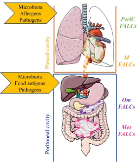
The peritoneal and pleural cavities, primarily considered as sites of macrophage (Bain and 1
Jenkins 2018) and B1 cell residence represent compartments that demarcate, contain and 2
protect the boundaries between three major mucosal sites directly exposed to environmental 3
antigens; namely the lungs, the intestines and the reproductive tract (of females)(Figure 2). 4
Immune protection within the body cavities is co-ordinated by small, inducible lymphoid 5
clusters found within specialised small adipose tissues (the mediastinum, pericardium, 6
mesenteries and omentum). Initially described as ‘milky-spots’ within the omentum(Dickinson 7
1906) these inducible structures were rebranded in 2010 as Fat Associated Lymphoid Clusters 8
or FALCs (Moro, Yamada et al. 2010). FALCs are local hubs that are important for providing 9
a second line of defence between the mucosal surfaces and a systemic immune response, 10
working to compartmentalise antibody mediated immune responses within body cavities. 11
Evidence supporting FALC orchestration of antibody responses within the body cavities is 12
mounting, with multiple reports linking FALCs to the initiation of independent and T-13
dependent immune responses (Rangel-Moreno, Moyron-Quiroz et al. 2009, Moro, Yamada et 14
al. 2010, Benezech, Luu et al. 2015, Jones, Racine et al. 2015, Jackson-Jones, Duncan et al. 15
2016). 16
17
FALCs, ILCs and the initiation of innate like B cell responses 18
Intestinal barrier functions 19
FALCs were identified as immune cell aggregates within the mesenteries, that were enriched 20
in lineage negative, c-Kit+,Sca-1+ cells; these cells are now known as ILC2s(Moro, Yamada et 21
al. 2010, Neill, Wong et al. 2010, Price, Liang et al. 2010). ILC2s are potent producers of IL-22
5 and IL-13; detectable levels of both cytokines are induced in the peritoneal lavage of Rag2 -/-23
mice which do not have mature T or B cells, but are absent from γc-/-Rag2-/- following infection 24
with the tissue migrating parasite Nippostrongylus brasiliensis (Moro, Yamada et al. 2010). 25
This result highlighted the potency of common-gamma chain receptor dependent innate 26
immune cells for the initiation of immune responses within the peritoneal cavity in the context 27
of intestinal worm infection. IL-5 is a critical growth factor for B1 B cells (Erickson, Foy et al. 28
2001); Moroand colleagues showed, using elegant in vivo transfers and in vitro co-cultures of 29
ILC2 with peritoneal B-cells in the presence or absence of a blocking antibody against IL-5, 30
that ILC2s provide support for B1 cell self-renewal (Moro, Yamada et al. 2010). ILC2s isolated 31
from mesenteric FALCs were also shown to be competent for the induction of IgA secretion 32
by peritoneal B cells in vitro (Moro, Yamada et al. 2010). Peritoneal B1 cells have been shown 33
to migrate to the intestinal lamina propria in order to secrete IgA (Fagarasan, Kawamoto et al. 34
2010, Baumgarth 2011). In addition to the conventional ‘Type-2’ cytokines described above, 35
ILC2 have also been shown to secrete IL-6 (Mjosberg, Bernink et al. 2012)(Salimi, Barlow et 36
al. 2013). As IL-6 has been described to induce antibody production by B-cells, as well as act 37
as a growth factor for plasmablasts (Jego, Bataille et al. 2001) and contribute to the regulation 38
of T follicular helper cells (Eto, Lao et al. 2011), it is plausible that ILC2 secretion of this 39
cytokine locally modifies FALC B-cell function; a hypothesis that warrants further 40
experimental investigation to confirm. Contrary to secondary organs, the development of 41
FALCs is not dependent on ILC3 as shown by the normal development and composition of 42
FALCs in Rorc-/- mice (Benezech, Luu et al. 2015). However, studies in germ free mice 43
revealed that the number of FALCs forming in the mesenteries is decreased indicating that 44
factors derived from the commensal flora are important to drive the formation of FALCs. 45
ILC3s are an important innate source of GM-CSF, a cytokine required for the induction of IgM 46
by innate response activator (IRA) B cells (Rauch, Chudnovskiy et al. 2012). Competency to 47
support IgA secretion by B1 was also reported for peritoneal macrophages, which had been 48
exposed to omentum culture supernatant (Okabe and Medzhitov 2014). Given the almost 49
know what component of the IgA secretion mediated by peritoneal macrophages is in part 1
dependent upon ILCs. A thorough characterisation of the ILC occupation of the murine 2
omentum has not been carried out; however a recent report characterised the presence of ILCs 3
in multiple human tissues including detailing the presence of ILC1 like cells within the 4
omentum (Simoni, Fehlings et al. 2017). 5
6
Pulmonary barrier functions 7
IgM is a large antibody and as such secretion of IgM into the circulation does not guarantee its 8
presence at tissue sites where it is required. In the global absence of the IL-33R ST2, the 9
secretion of IgM from FALCs within the pleural cavity is ablated (Jackson-Jones, Duncan et 10
al. 2016). This is not a direct effect on the B-cells as co-transfer of IL-33R sufficient and 11
deficient B-cells resulted in comparable induction of B-cell activation following Alternaria 12
alternata delivery. Utilising blocking antibodies against IL-5 delivered directly into the pleural 13
space, we concluded that the IL-33 was acting via an IL-5 producing intermediate population 14
of cells. ILC2s were the only cells found to be expressing IL-5 within FALCs of the pleural 15
cavity during type-2 inflammation (Jackson-Jones, Duncan et al. 2016). Thus, the presence of 16
IgM secreting B-cells within FALCs in the context of type-2 inflammation is assumed to 17
depend upon IL-5 secretion from IL-33 activated ILC2s. The link between ILC2 and antibody 18
production within the thoracic cavity was also made by Drake et al 2016 who showed that in 19
vitro culture of lung derived ILCs with splenic B cells resulted in antibody production (Drake, 20
Iijima et al. 2016). However, as there are fewer B-cells within the lungs and because fluid phase 21
B cells isolated from the pleural space do no secrete antibodies, it is likely that pleural FALCs 22
are the sites where the ILC/B cell interactions take place in the thoracic cavity. In support of a 23
tight immune crosstalk between lung and pleural space is a report showing that delivery of 24
GM-CSF secreting IRA B cells into the pleural space mediates protection from pneumonia 25
(Weber, Chousterman et al. 2014). Neither the role of FALCs in the activation of the transferred 26
IRA B cells nor the requirement for lung or FALC resident ILCs in this process was 27
investigated. This study serves to further highlights the crosstalk which occurs between 28
mucosal tissues and their associated serous cavities. 29
30
Is FALC derived IgM Atheroprotective? 31
Innate like B-cells (IBCs) can be both protective and pathogenic in atherosclerosis. 32
Recognition of oxidation specific epitopes on low density lipoproteins (LDL) (Binder, 33
Hartvigsen et al. 2004) by natural IgM plays a protective role in atherosclerosis and clinical 34
studies show that lower levels of IgM correlates with increased risk of cardiovascular diseases. 35
The production of atheroprotective IgM by IBCs is dependent on IL-33 (Miller, Xu et al. 2008), 36
IL-5 and IL-5 producing ILC2 (Perry, Oldham et al. 2013, Newland, Mohanta et al. 2017), a 37
signaling loop that is active in FALCs (Jackson-Jones, Duncan et al. 2016). Importantly, it has 38
been shown that the number of FALCs in the para-aortic adipose of ApoE-/- mice increases in 39
the vicinity of atherosclerotic lesions (Newland, Mohanta et al. 2017) and that they contain 40
IBC producing atheroprotective IgM (Srikakulapu, Upadhye et al. 2017). This suggests that 41
ILC2 regulation of local IgM secretion by FALC IBCs could be key to IBC mediated 42
atheroprotection and that loss of ILC2 during the development of obesity could contribute to 43
accelerated atherosclerosis. 44
45
Summary 46
Since their initial discovery 8 years ago, ILC2s have emerged as major regulators of type-2 47
immunity in adipose tissue where they co-ordinate eosinophil, macrophage, adipocyte and IBC 48
function. FALCs are specialised hubs that act as a second line of immune defence sitting behind 49
cytokines by FALC resident ILCs, which kick-start the ensuing immune response following 1
detection of a danger signal (eg IL-33). 2
3
Acknowledgements 4
Research in CBs laboratory is funded Medical Research Council (MRC) UK Grant 5
(MR/M011542/1); Research in LJJs laboratory is funded by Lancaster University. Figure 1 & 6
2 were designed using Servier Medical Art (www.servier.com) and used under a creative 7
commons license. 8
9
Author Contributions 10
CB & LJJ shared authorship of this review. 11
12
Figure Legends 13
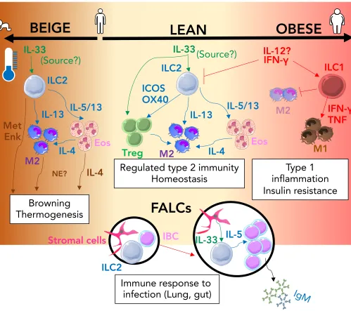
Figure 1. The ILC2 driven interactions that regulate immune adipose function. In the lean state 14
(centre; cream) IL-33 action (green arrows) signals to both T-regulatory cells (Treg) and ILC2 15
resulting in regulated Type-2 immunity via the activity of secreted and membrane bound type-16
2 signals (blue arrows); this response is amplified in the presence of lower ambient living 17
temperature and during infancy and can result in browning thermogenesis within adipose tissue 18
(Left; brown). Type-2 signals that can control browning are shown (brown arrows). In the 19
obese state (right; pink) Inflammation mediated by type-1 signals (red arrows) promotes the 20
activation of ILC1 and the inhibition of ILC2 which results in inhibition of M2 and expansion 21
of the M1 macrophage population which contribute to the development of insulin resistance. 22
During Type-2 inflammation within the lung or gut, ILC2 containing FALCs (Black circles) 23
expand; IL-33 produced by stromal cells (green arrow) increases IL-5 secretion (blue arrow) 24
from ILC2 which induces innate like B cell (IBC) proliferation and secretion of IgM. (MetEnk= 25
methionine-enkephalin peptides, NE= norepinephrine, Eos = Eosinophils, IBC = Innate Like 26
B cell, M1/M2 = M1 or M2 macrophage) 27
28
Figure 2. Compartmentalized protection of mucosal sites by fat associated lymphoid clusters 29
within body cavities. Within the pleural cavity, protection from/regulation of, microbiota, 30
infection, inflammation and damage is mediated by inducible FALCs within the pericardium 31
(green) and mediastinum (orange). Within the peritoneal cavity, protection from/regulation of 32
microbiota, infection, inflammation and damage is mediated by FALCs within the omentum 33
(purple) and mesenteries (pink) m= mediastinal, PeriC= pericardial, om=omental, 34
mes=mesenteric. 35
36 37 38
REFERENCES 39
40
Artis, D. and H. Spits (2015). "The biology of innate lymphoid cells." Nature 517(7534): 293-41
301. 42
Bain, C. C. and S. J. Jenkins (2018). "The biology of serous cavity macrophages." Cell 43
Immunol 330: 126-135. 44
Baumgarth, N. (2011). "The double life of a B-1 cell: self-reactivity selects for protective 45
effector functions." Nat Rev Immunol 11(1): 34-46. 46
Benezech, C., N. T. Luu, J. A. Walker, A. A. Kruglov, Y. Loo, K. Nakamura, Y. Zhang, S. 47
Nayar, L. H. Jones, A. Flores-Langarica, A. McIntosh, J. Marshall, F. Barone, G. Besra, K. 48
Miles, J. E. Allen, M. Gray, G. Kollias, A. F. Cunningham, D. R. Withers, K. M. Toellner, N. 49
"Inflammation-induced formation of fat-associated lymphoid clusters." Nat Immunol 16(8): 1
819-828. 2
Binder, C. J., K. Hartvigsen, M. K. Chang, M. Miller, D. Broide, W. Palinski, L. K. Curtiss, 3
M. Corr and J. L. Witztum (2004). "IL-5 links adaptive and natural immunity specific for 4
epitopes of oxidized LDL and protects from atherosclerosis." J Clin Invest 114(3): 427-437. 5
Brestoff, J. R., B. S. Kim, S. A. Saenz, R. R. Stine, L. A. Monticelli, G. F. Sonnenberg, J. J. 6
Thome, D. L. Farber, K. Lutfy, P. Seale and D. Artis (2015). "Group 2 innate lymphoid cells 7
promote beiging of white adipose tissue and limit obesity." Nature 519(7542): 242-246. 8
Cipolletta, D., M. Feuerer, A. Li, N. Kamei, J. Lee, S. E. Shoelson, C. Benoist and D. Mathis 9
(2012). "PPAR-gamma is a major driver of the accumulation and phenotype of adipose tissue 10
Treg cells." Nature 486(7404): 549-553. 11
Dickinson, G. K. (1906). "II. The Omentum and its Functions." Ann Surg 44(5): 652-665. 12
Drake, L. Y., K. Iijima, K. Bartemes and H. Kita (2016). "Group 2 Innate Lymphoid Cells 13
Promote an Early Antibody Response to a Respiratory Antigen in Mice." J Immunol 197(4): 14
1335-1342. 15
Elewa, Y. H., O. Ichii, S. Otsuka, Y. Hashimoto and Y. Kon (2014). "Characterization of mouse 16
mediastinal fat-associated lymphoid clusters." Cell Tissue Res 357(3): 731-741. 17
Erickson, L. D., T. M. Foy and T. J. Waldschmidt (2001). "Murine B1 B cells require IL-5 for 18
optimal T cell-dependent activation." J Immunol 166(3): 1531-1539. 19
Eto, D., C. Lao, D. DiToro, B. Barnett, T. C. Escobar, R. Kageyama, I. Yusuf and S. Crotty 20
(2011). "IL-21 and IL-6 are critical for different aspects of B cell immunity and redundantly 21
induce optimal follicular helper CD4 T cell (Tfh) differentiation." PLoS One 6(3): e17739. 22
Fagarasan, S., S. Kawamoto, O. Kanagawa and K. Suzuki (2010). "Adaptive immune 23
regulation in the gut: T cell-dependent and T cell-independent IgA synthesis." Annu Rev 24
Immunol 28: 243-273. 25
Feuerer, M., L. Herrero, D. Cipolletta, A. Naaz, J. Wong, A. Nayer, J. Lee, A. B. Goldfine, C. 26
Benoist, S. Shoelson and D. Mathis (2009). "Lean, but not obese, fat is enriched for a unique 27
population of regulatory T cells that affect metabolic parameters." Nat Med 15(8): 930-939. 28
Fischer, K., H. H. Ruiz, K. Jhun, B. Finan, D. J. Oberlin, V. van der Heide, A. V. Kalinovich, 29
N. Petrovic, Y. Wolf, C. Clemmensen, A. C. Shin, S. Divanovic, F. Brombacher, E. 30
Glasmacher, S. Keipert, M. Jastroch, J. Nagler, K. W. Schramm, D. Medrikova, G. Collden, S. 31
C. Woods, S. Herzig, D. Homann, S. Jung, J. Nedergaard, B. Cannon, M. H. Tschop, T. D. 32
Muller and C. Buettner (2017). "Alternatively activated macrophages do not synthesize 33
catecholamines or contribute to adipose tissue adaptive thermogenesis." Nat Med 23(5): 623-34
630. 35
Halim, T. Y. F., B. M. J. Rana, J. A. Walker, B. Kerscher, M. D. Knolle, H. E. Jolin, E. M. 36
Serrao, L. Haim-Vilmovsky, S. A. Teichmann, H. R. Rodewald, M. Botto, T. J. Vyse, P. G. 37
Fallon, Z. Li, D. R. Withers and A. N. J. McKenzie (2018). "Tissue-Restricted Adaptive Type 38
2 Immunity Is Orchestrated by Expression of the Costimulatory Molecule OX40L on Group 2 39
Innate Lymphoid Cells." Immunity 48(6): 1195-1207 e1196. 40
Hams, E., R. M. Locksley, A. N. McKenzie and P. G. Fallon (2013). "Cutting edge: IL-25 41
elicits innate lymphoid type 2 and type II NKT cells that regulate obesity in mice." J Immunol 42
191(11): 5349-5353. 43
Jackson-Jones, L. H. and C. Benezech (2018). "Control of innate-like B cell location for 44
compartmentalised IgM production." Curr Opin Immunol 50: 9-13. 45
Jackson-Jones, L. H., S. M. Duncan, M. S. Magalhaes, S. M. Campbell, R. M. Maizels, H. J. 46
McSorley, J. E. Allen and C. Benezech (2016). "Fat-associated lymphoid clusters control local 47
IgM secretion during pleural infection and lung inflammation." Nat Commun 7: 12651. 48
Jackson-Jones, L. H., D. Ruckerl, F. Svedberg, S. Duncan, R. M. Maizels, T. E. Sutherland, S. 49
delivery induces serous cavity macrophage proliferation independent of interleukin-4 receptor 1
alpha." Eur J Immunol 46(10): 2311-2321. 2
Jego, G., R. Bataille and C. Pellat-Deceunynck (2001). "Interleukin-6 is a growth factor for 3
nonmalignant human plasmablasts." Blood 97(6): 1817-1822. 4
Jones, D. D., R. Racine, S. T. Wittmer, L. Harston, A. M. Papillion, L. M. Dishaw, T. D. 5
Randall, D. L. Woodland and G. M. Winslow (2015). "The omentum is a site of protective IgM 6
production during intracellular bacterial infection." Infect Immun 83(5): 2139-2147. 7
Kohlgruber, A. C., S. T. Gal-Oz, N. M. LaMarche, M. Shimazaki, D. Duquette, H. N. Nguyen, 8
A. I. Mina, T. Paras, A. Tavakkoli, U. von Andrian, A. S. Banks, T. Shay, M. B. Brenner and 9
L. Lynch (2018). "gammadelta T cells producing interleukin-17A regulate adipose regulatory 10
T cell homeostasis and thermogenesis." Nat Immunol 19(5): 464-474. 11
Kolodin, D., N. van Panhuys, C. Li, A. M. Magnuson, D. Cipolletta, C. M. Miller, A. Wagers, 12
R. N. Germain, C. Benoist and D. Mathis (2015). "Antigen- and cytokine-driven accumulation 13
of regulatory T cells in visceral adipose tissue of lean mice." Cell Metab 21(4): 543-557. 14
Lee, M. W., J. I. Odegaard, L. Mukundan, Y. Qiu, A. B. Molofsky, J. C. Nussbaum, K. Yun, 15
R. M. Locksley and A. Chawla (2015). "Activated type 2 innate lymphoid cells regulate beige 16
fat biogenesis." Cell 160(1-2): 74-87. 17
Lim, A. I., S. Menegatti, J. Bustamante, L. Le Bourhis, M. Allez, L. Rogge, J. L. Casanova, H. 18
Yssel and J. P. Di Santo (2016). "IL-12 drives functional plasticity of human group 2 innate 19
lymphoid cells." J Exp Med 213(4): 569-583. 20
Miller, A. M., D. Xu, D. L. Asquith, L. Denby, Y. Li, N. Sattar, A. H. Baker, I. B. McInnes 21
and F. Y. Liew (2008). "IL-33 reduces the development of atherosclerosis." J Exp Med 205(2): 22
339-346. 23
Mjosberg, J., J. Bernink, K. Golebski, J. J. Karrich, C. P. Peters, B. Blom, A. A. te Velde, W. 24
J. Fokkens, C. M. van Drunen and H. Spits (2012). "The transcription factor GATA3 is 25
essential for the function of human type 2 innate lymphoid cells." Immunity 37(4): 649-659. 26
Molofsky, A. B., J. C. Nussbaum, H. E. Liang, S. J. Van Dyken, L. E. Cheng, A. Mohapatra, 27
A. Chawla and R. M. Locksley (2013). "Innate lymphoid type 2 cells sustain visceral adipose 28
tissue eosinophils and alternatively activated macrophages." J Exp Med 210(3): 535-549. 29
Molofsky, A. B., F. Van Gool, H. E. Liang, S. J. Van Dyken, J. C. Nussbaum, J. Lee, J. A. 30
Bluestone and R. M. Locksley (2015). "Interleukin-33 and Interferon-gamma Counter-31
Regulate Group 2 Innate Lymphoid Cell Activation during Immune Perturbation." Immunity 32
43(1): 161-174. 33
Moro, K., T. Yamada, M. Tanabe, T. Takeuchi, T. Ikawa, H. Kawamoto, J. Furusawa, M. 34
Ohtani, H. Fujii and S. Koyasu (2010). "Innate production of T(H)2 cytokines by adipose 35
tissue-associated c-Kit(+)Sca-1(+) lymphoid cells." Nature 463(7280): 540-544. 36
Neill, D. R., S. H. Wong, A. Bellosi, R. J. Flynn, M. Daly, T. K. Langford, C. Bucks, C. M. 37
Kane, P. G. Fallon, R. Pannell, H. E. Jolin and A. N. McKenzie (2010). "Nuocytes represent a 38
new innate effector leukocyte that mediates type-2 immunity." Nature 464(7293): 1367-1370. 39
Newland, S. A., S. Mohanta, M. Clement, S. Taleb, J. A. Walker, M. Nus, A. P. Sage, C. Yin, 40
D. Hu, L. L. Kitt, A. J. Finigan, H. R. Rodewald, C. J. Binder, A. N. J. McKenzie, A. J. 41
Habenicht and Z. Mallat (2017). "Type-2 innate lymphoid cells control the development of 42
atherosclerosis in mice." Nat Commun 8: 15781. 43
Nussbaum, J. C., S. J. Van Dyken, J. von Moltke, L. E. Cheng, A. Mohapatra, A. B. Molofsky, 44
E. E. Thornton, M. F. Krummel, A. Chawla, H. E. Liang and R. M. Locksley (2013). "Type 2 45
innate lymphoid cells control eosinophil homeostasis." Nature 502(7470): 245-248. 46
O'Sullivan, T. E., M. Rapp, X. Fan, O. E. Weizman, P. Bhardwaj, N. M. Adams, T. Walzer, A. 47
J. Dannenberg and J. C. Sun (2016). "Adipose-Resident Group 1 Innate Lymphoid Cells 48
Odegaard, J. I. and A. Chawla (2015). "Type 2 responses at the interface between immunity 1
and fat metabolism." Curr Opin Immunol 36: 67-72. 2
Okabe, Y. and R. Medzhitov (2014). "Tissue-specific signals control reversible program of 3
localization and functional polarization of macrophages." Cell 157(4): 832-844. 4
Oldenhove, G., E. Boucquey, A. Taquin, V. Acolty, L. Bonetti, B. Ryffel, M. Le Bert, K. 5
Englebert, L. Boon and M. Moser (2018). "PD-1 Is Involved in the Dysregulation of Type 2 6
Innate Lymphoid Cells in a Murine Model of Obesity." Cell Rep 25(8): 2053-2060 e2054. 7
Perry, H. M., S. N. Oldham, S. P. Fahl, X. Que, A. Gonen, D. B. Harmon, S. Tsimikas, J. L. 8
Witztum, T. P. Bender and C. A. McNamara (2013). "Helix-loop-helix factor inhibitor of 9
differentiation 3 regulates interleukin-5 expression and B-1a B cell proliferation." Arterioscler 10
Thromb Vasc Biol 33(12): 2771-2779. 11
Poher, A. L., J. Altirriba, C. Veyrat-Durebex and F. Rohner-Jeanrenaud (2015). "Brown 12
adipose tissue activity as a target for the treatment of obesity/insulin resistance." Front Physiol 13
6: 4. 14
Price, A. E., H. E. Liang, B. M. Sullivan, R. L. Reinhardt, C. J. Eisley, D. J. Erle and R. M. 15
Locksley (2010). "Systemically dispersed innate IL-13-expressing cells in type 2 immunity." 16
Proc Natl Acad Sci U S A 107(25): 11489-11494. 17
Qiu, Y., K. D. Nguyen, J. I. Odegaard, X. Cui, X. Tian, R. M. Locksley, R. D. Palmiter and A. 18
Chawla (2014). "Eosinophils and type 2 cytokine signaling in macrophages orchestrate 19
development of functional beige fat." Cell 157(6): 1292-1308. 20
Rangel-Moreno, J., J. E. Moyron-Quiroz, D. M. Carragher, K. Kusser, L. Hartson, A. Moquin 21
and T. D. Randall (2009). "Omental milky spots develop in the absence of lymphoid tissue-22
inducer cells and support B and T cell responses to peritoneal antigens." Immunity 30(5): 731-23
743. 24
Rauch, P. J., A. Chudnovskiy, C. S. Robbins, G. F. Weber, M. Etzrodt, I. Hilgendorf, E. Tiglao, 25
J. L. Figueiredo, Y. Iwamoto, I. Theurl, R. Gorbatov, M. T. Waring, A. T. Chicoine, M. 26
Mouded, M. J. Pittet, M. Nahrendorf, R. Weissleder and F. K. Swirski (2012). "Innate response 27
activator B cells protect against microbial sepsis." Science 335(6068): 597-601. 28
Salimi, M., J. L. Barlow, S. P. Saunders, L. Xue, D. Gutowska-Owsiak, X. Wang, L. C. Huang, 29
D. Johnson, S. T. Scanlon, A. N. McKenzie, P. G. Fallon and G. S. Ogg (2013). "A role for IL-30
25 and IL-33-driven type-2 innate lymphoid cells in atopic dermatitis." J Exp Med 210(13): 31
2939-2950. 32
Seehus, C. R., A. Kadavallore, B. Torre, A. R. Yeckes, Y. Wang, J. Tang and J. Kaye (2017). 33
"Alternative activation generates IL-10 producing type 2 innate lymphoid cells." Nat Commun 34
8(1): 1900. 35
Simoni, Y., M. Fehlings, H. N. Kloverpris, N. McGovern, S. L. Koo, C. Y. Loh, S. Lim, A. 36
Kurioka, J. R. Fergusson, C. L. Tang, M. H. Kam, K. Dennis, T. K. H. Lim, A. C. Y. Fui, C. 37
W. Hoong, J. K. Y. Chan, M. Curotto de Lafaille, S. Narayanan, S. Baig, M. Shabeer, S. E. S. 38
Toh, H. K. K. Tan, R. Anicete, E. H. Tan, A. Takano, P. Klenerman, A. Leslie, D. S. W. Tan, 39
I. B. Tan, F. Ginhoux and E. W. Newell (2017). "Human Innate Lymphoid Cell Subsets Possess 40
Tissue-Type Based Heterogeneity in Phenotype and Frequency." Immunity 46(1): 148-161. 41
Srikakulapu, P., A. Upadhye, S. M. Rosenfeld, M. A. Marshall, C. McSkimming, A. W. 42
Hickman, I. S. Mauldin, G. Ailawadi, M. B. S. Lopes, A. M. Taylor and C. A. McNamara 43
(2017). "Perivascular Adipose Tissue Harbors Atheroprotective IgM-Producing B Cells." 44
Front Physiol 8: 719. 45
Vasanthakumar, A., K. Moro, A. Xin, Y. Liao, R. Gloury, S. Kawamoto, S. Fagarasan, L. A. 46
Mielke, S. Afshar-Sterle, S. L. Masters, S. Nakae, H. Saito, J. M. Wentworth, P. Li, W. Liao, 47
W. J. Leonard, G. K. Smyth, W. Shi, S. L. Nutt, S. Koyasu and A. Kallies (2015). "The 48
transcriptional regulators IRF4, BATF and IL-33 orchestrate development and maintenance of 49
Villarroya, F., R. Cereijo, J. Villarroya, A. Gavalda-Navarro and M. Giralt (2018). "Toward an 1
Understanding of How Immune Cells Control Brown and Beige Adipobiology." Cell Metab 2
27(5): 954-961. 3
Wang, S., P. Xia, Y. Chen, Y. Qu, Z. Xiong, B. Ye, Y. Du, Y. Tian, Z. Yin, Z. Xu and Z. Fan 4
(2017). "Regulatory Innate Lymphoid Cells Control Innate Intestinal Inflammation." Cell 5
171(1): 201-216 e218. 6
Weber, G. F., B. G. Chousterman, I. Hilgendorf, C. S. Robbins, I. Theurl, L. M. Gerhardt, Y. 7
Iwamoto, T. D. Quach, M. Ali, J. W. Chen, T. L. Rothstein, M. Nahrendorf, R. Weissleder and 8
F. K. Swirski (2014). "Pleural innate response activator B cells protect against pneumonia via 9
a GM-CSF-IgM axis." J Exp Med 211(6): 1243-1256. 10
Wu, D., A. B. Molofsky, H. E. Liang, R. R. Ricardo-Gonzalez, H. A. Jouihan, J. K. Bando, A. 11
Chawla and R. M. Locksley (2011). "Eosinophils sustain adipose alternatively activated 12
macrophages associated with glucose homeostasis." Science 332(6026): 243-247. 13
BEIGE
IL-33
IL-5
IgM
IFN-
γ
Τ
reg
Regulated type 2 immunity
Homeostasis
inflammation
Type 1
Insulin resistance
LEAN
IL-5/13
IL-13
ICOS
OX40
IL-4
IL-33
(Source?)
M2
Eos
IL-5/13
IL-13
IL-4
IL-33
(Source?)
Eos
NE?
IL-4
Met
Enk
Browning
Thermogenesis
OBESE
FALCs
ILC1
ILC2
ILC2
M2
M1
IBC
ILC2
Stromal cells
IFN-
γ
TNF
IL-12?
[image:11.540.22.521.74.517.2]Immune response to
infection (Lung, gut)
Figure 2. Compartmentalized protection of mucosal sites by fat associated lymphoid clusters within body cavities. Within the pleural cavity, protection from/regulation of, microbiota, infection, inflammation and damage is mediated by inducible FALCs within the pericardium (green) and mediastinum (orange). Within the peritoneal cavity, protection from/regulation of microbiota, infection, inflammation and damage is mediated by FALCs within the omentum (purple) and mesenteries (pink) m= mediastinal, PeriC= pericardial, om=omental, mes=mesenteric
Om FALCs
M FALCs
PeriC FALCs
Pl
eur
al
c
avi
ty
Pe
ri
tone
al
c
avi
ty
Mes FALCs
Microbiota Food antigens
Pathogens Microbiota