ISSN Print: 2164-3024
DOI: 10.4236/ojrad.2018.82011 Jun. 26, 2018 91 Open Journal of Radiology
Development of the Press-Through Package
Recognizable with Abdominal X-Ray Image of
Abdominal Soft Tissue Phantom
Kyoichi Ito
1*, Morio Shimada
2, Kiyomi Sadamoto
3, Mikio Murata
3, Kiyoshi Kubota
4,
Motoyoshi Itoh
4, Kohki Yoshikawa
21Department of Radiation, International Goodwill Hospital, Yokohama, Japan
2Department of Radiological Sciences, Faculty of Health Sciences, Komazawa University, Tokyo, Japan 3Department of Clinical Pharmacy, Yokohama University of Pharmacy, Yokohama, Japan
4Sanko Alumi Inc., Konosu, Saitama, Japan
Abstract
Background: Patients with dementia who accidentally swallowed a press- through package (PTP) have been increased in recent years. Swallowed PTP is usually detected with CT having a risk of radiation exposure since PTP is very difficult to detect with abdominal X-ray image. Purpose: This study is aimed at developing the new PTP sheet recognizable with abdominal X-ray by im-proving the conventional PTP sheet. Material and Methods: The PTP sheet samples used in this study are: No. 1 Control: Commercially available tablet 100 mg, No. 2 Triple-stacked aluminum foil, 6-overcoated with BaSO4 10.3
g/m2, No. 3 Aluminum foil, 6-overcoated with BaSO
4 10.3 g/m2, No. 4
Five-stacked aluminum foil, 6-overcoated with BaSO4 No. 5 Aluminum foil,
single-coated with WO3 2.55 g/m2, No. 6 Aluminum foil, double-coated with
WO3 5.1 g/m2, No. 7 Aluminum foil, triple-coated with WO3 7.66 g/m2, No. 8
Three-stacked aluminum foil, triple-coated with WO3. PTP sheets (No. 1 -
No. 8) were placed inside the abdominal soft tissue phantom, and the images were obtained using FUJIFILM DR CALNEO PT. Region of interest (ROI) was placed on PTP sheets (No. 1 - No. 8) and abdominal soft tissue liquid phantom, and each contrast was measured from the average pixel of the two ROIs. Contrast was calculated by the calculation formula. Each ROI was measured three times, and their average value and standard deviation were es-timated. Results: Statistical significance in contrast was not observed in com-mercial PTP sheet (No. 1), PTP sheet (No. 3), and PTP sheet (No. 5), while there was a significant difference between PTP sheet (No. 1) and PTP sheet (No. 2), PTP sheet (No. 4) PTP sheet (No. 6), PTP sheet (No. 7) (p < 0.05). Significant difference in contrast was observed also between commercial PTP How to cite this paper: Ito, K., Shimada,
M., Sadamoto, K., Murata, M., Kubota, K., Itoh, M. and Yoshikawa, K. (2018) Devel-opment of the Press-Through Package Recognizable with Abdominal X-Ray Image of Abdominal Soft Tissue Phantom. Open Journal of Radiology, 8, 91-98.
https://doi.org/10.4236/ojrad.2018.82011
Received: May 27, 2018 Accepted: June 23, 2018 Published: June 26, 2018
Copyright © 2018 by authors and Scientific Research Publishing Inc. This work is licensed under the Creative Commons Attribution International License (CC BY 4.0).
http://creativecommons.org/licenses/by/4.0/
DOI: 10.4236/ojrad.2018.82011 92 Open Journal of Radiology sheet (No. 1) and PTP sheet (No. 8) (P < 0.01). PTP sheet (No. 1), PTP sheet (No. 3), and PTP sheet (No. 5) were not recognized at all by plain abdominal X-ray photography. Conclusion: The other all prototype PTP sheets (No. 2, No. 4, No. 6, No. 7 and No. 8) were well recognized. PTP sheets (No. 2, No. 4) of BaSO4 is considered to be more suitable than PTP sheets of WO3 (No. 6,
No. 7, No. 8) because it is easier to produce. We will develop oral PTP sheets for patients with dementia for abdominal X-ray in the future.
Keywords
Press-Through Package (PTP), Soft Tissue Phantom, Abdominal X-Ray, Development, BaSO4, WO3
1. Introduction
Patients with dementia who accidentally swallowed a press-through package (PTP) have been increased in recent years. Swallowed PTP is usually detected with CT having a risk of radiation since PTP is very difficult to detect with ab-dominal X-ray image. PTP consists of lids coated with a heat-sealed material on an aluminum leaf and a dome of vinylchloride. These materials are widely used in Japan to enclose drugs due to their features of cleanliness, hermetic sealing, easy handling, toughness, and low cost. A risk of accidental swallow of PTP in the elderly individuals and the subjects with mental or visual disturbances is a serious issue for emergency physicians. Swallowed PTP with sharp corners may cause gastrointestinal bleeding, mediastinitis, intestinal perforation, and ob-structive ileus, although such severe complications are extremely rare [1] [2] [3]. Endoscopic removal should be carried out if PTP exists in the esophagus or sto-mach. Early laparotomy should be considered if PTP passes through the pyloric ring and symptoms are observed [4]. Therefore, it is very important to make an early and correct diagnosis of the location of the PTP. PTP material is difficult to detect directly by plain radiography due to its high radiolucency, while there are increasing reports that the location of a PTP can be detected by multidetector computed tomography (MDCT) [4] [5] [6] [7] [8]. Therefore, we considered to make PTP sheet which can be recognized by plain X-ray examination with less exposure because CT examination receives much exposure.
2. Purpose
When we diagnose accidentally swallowing of patient’s PTPs, we always use CT to diagnose that PTPs are in the stomach or intestinal tract. In these cases, CT scans give patients about 10 times more exposure than plain abdominal X-ray radiography.
DOI: 10.4236/ojrad.2018.82011 93 Open Journal of Radiology of this new PTP sheets were launched by a conference of these pharmaceutical experts and radiology experts together. As far as we search, there is no paper on the development of PTP sheets that can be recognized with Plain X-ray radio-graphy.
The degree of X-ray detection is considered to be proportional to the fourth power of atomic number. From this, it turned out that barium and tungsten are the ones that can be painted as ink on the aluminum sheet of PTP.
This study is aimed at developing the new PTP sheet recognizable with plai-nabdominal X-ray by improving the conventional PTP sheet.
3. Materials and Methods
3.1. Investigation of X-Ray Blocking Substances
Compounds containing lead, barium, barium sulfate, titanium, iodine com-pounds, tin, lead, tungsten, bismuth, bismuth nitrite are considered as X-ray blocking substances. Barium sulfate, tungsten carbide, tin oxide, and bismuth oxide of a simple substance or compound having an atomic number of 47 or more are considered as candidates. X-ray detection sensitivity is proportional to the fourth power of atomic number. Candidates for X-ray blocking substances include iodine, barium, and tungsten. Bismuth is toxic and excluded. Iodine was excluded this time because atom number is smaller than barium. Barium and tungsten were finally selected as X-ray blocking substances. The ink containing blocking material (barium sulfate or tungsten trioxide) was coated on the cover material (aluminum foil) of PTP sheet in the form of gravure print.
3.2. Method of Overcoating with a Coater
Possibility of industrialization (commercialization) by gravure ink Atomic ratio of tungsten carbide [WC] W = 183.8/195.851
Compared with this, the atomic ratio of barium sulfate Ba = 137.3/233.43 X-ray shielding effect W:Ba = 29.99:9.83
50 g/m2 BaSO
4 (Solid) → WC 50 × 0.328 × (1.066/1.700) = 10.283 g/m2
The present prototype PTP aluminum was tungsten trioxide WO3 = 231.84 atomic ratio of tungsten W = 183.8/231.84 = 0.7928
Therefore, neodarum sol (BaSO4: Kaigen Pharma CO., LTD. Tokyo Japan)
solution 2-fold dilution → “WO3 = 12.093 g/m2
We prepared prototype PTP sheets using the inks of barium sulfate and tungsten trioxide ink. Commercially available medicinal zyrolic tablets (hyper-uricemia drug: Daiichi Sankyo Company, Limited. Tokyo Japan).
Japan) were used as a control.
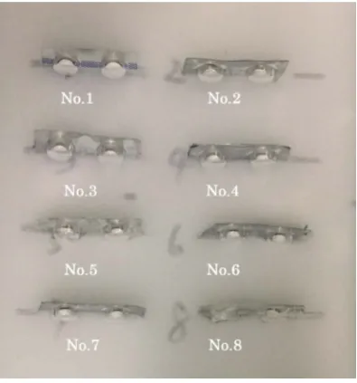
PTP sheet samples are No. 1 to No. 8 below (Figure 1).
No. 1: Control: commercially available zyrolic tablet 100 mg PTP
No. 2: Triple-stacked aluminum foil, 6-overcoated with BaSO4 10.3 g/m2 (solid
15.0 g/m2)
DOI: 10.4236/ojrad.2018.82011 94 Open Journal of Radiology
Figure 1. Samples of PTP sheets (No. 1 - No. 8).
No. 4: Five-stacked aluminum foil, 6-overcoated with BaSO4
No. 5: Aluminum foil, single-coated with WO3 2.55 g/m2 (solid 3.5 g/m2)
No. 6: Aluminum foil, double-coated with WO3 5.1 g/m2
No. 7: Aluminum foil, triple-coated with WO3 7.66 g/m2
No. 8: Three-stacked aluminum foil, triple-coated with WO3
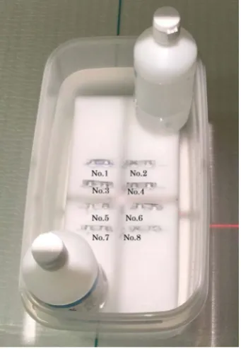
3.3. Method of Preparation of Abdominal Soft Tissue Phantom
A polyethylene container was used for preparation of a soft tissue equivalent phantom. The size of the container is 320 mm (sideway) × 170 mm (height) × 160 mm (length), and the volume is 8704 ml. In this volume 6144 ml, 1081 g of granulated sugar and 79.87 g of salt were added at a ratio of granulated sugar 176 mg/mL water vs salt 13 mg/mL water to prepare an abdominal soft tissue phan-tom. Two melamine foams in a size of 30 mm (height) × 300 mm (width) × 135 mm (length) were overpositioned, slits were made, and PTP sheets (No. 1 - No. 8) were inserted through these slits avoiding their floating (Figure 2).3.4. Measurement Method of Contrast of PTP Sheets
PTP sheets (No. 1 - No. 8) were placed inside the abdominal soft tissue phan-tom, and the images were obtained at the specified condition of voltage 76 Kv, electric current 200 mA, time 160 msec, using FUJIFILM DR CALNEO PT (Fu-jifilm Corporation Tokyo Japan). Region of interest (ROI) was placed on PTP sheets (No. 1 - No. 8) and abdominal soft tissue liquid phantom, and each con-trast was measured from the average pixel of the two ROIs. Concon-trast was calcu-lated by the calculation formula below.
Contrast = (ROIptp − ROIphantom)/ROIphantom
DOI: 10.4236/ojrad.2018.82011 95 Open Journal of Radiology
Figure 2. Abdominal soft tissue phantom
with PTP sheets (No. 1 - No. 8). Above and below bottles containing water are the weights not to float melamine foam.
3.5. Statistical Analysis
Differences in Contrast between the commercial PTP and our prototype PTPs were compared using an unpaired t-test. The P-value was assessed. All statistical analyses were performed with Excel Statistics 2010 for Windows version 1.13.
4. Results
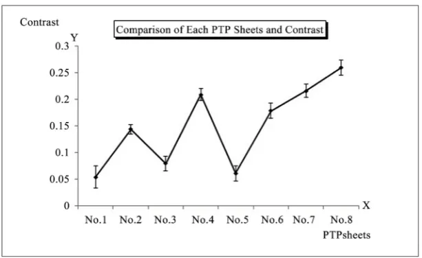
PTP of No. 1 (Control: commercially available zyrolic tablet) was hardly de-tected.
Contrast of PTP (No. 1) was as low as 0.01501.
PTP of No. 2 (Triple-stacked aluminum foil, 6-overcoated with BaSO4) was
moderately recognized. Contrast of PTP (No. 2) was a moderate value of 0.1146. PTP of No. 3 (Aluminum foil, 6-overcoated with BaSO4) was hardly detected as
same as PTP (No. 1). Contrast of PTP (No. 3) was as low as 0.03729. PTP of No. 4 (Five-stacked aluminum foil, 6-overcoated with BaSO4) was clearly detected.
Contrast of PTP (No. 4) was relatively higher as 0.1694. PTP of No. 5 (Alumi-num foil, single-coated with WO3) was hardly detected as same as PTP (No. 1).
Contrast of PTP (No. 5) was as low as 0.03223. PTP of No. 6 (Aluminum foil, double-coated with WO3) was able to be recognized moderately. Contrast of
PTP (No. 6) was a moderate value of 0.1331. PTP of No. 7 (Aluminum foil, triple-coated with WO3) was clearly detected. Contrast of PTP (No. 7) was
DOI: 10.4236/ojrad.2018.82011 96 Open Journal of Radiology among all. There were no significant differences in contrast among commercial-ly available PTP (No. 1), PTP (No. 3) and PTP (No. 5). However there was a sig-nificant difference between PTP sheet (No. 1) and PTP sheet (No. 2), PTP sheet (No. 4) PTP sheet (No. 6), PTP sheet (No. 7) (p < 0.05). Significant differences in contrast were observed also between commercially available PTP (No. 1) and PTP (No. 8) (P < 0.01) (Figure 3) (Figure 4).
5. Discussion
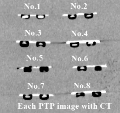
[image:6.595.272.474.298.477.2]MDCT was performed using a 64-detector-row system (CT LightSpeed VCT; GEHealthcare, Milwaukee, USA). Imaging and reconstructionparameters for the 40-section device were a beamcollimation of 8 × 2.5 mm, a beam pitch of 1.35, a gantryrotation time of 0.6 s, 120 kVp, 199 mAs, a section thicknessof 1.25 mm and a reconstruction interval of 1.25 mm using same PTP sheets (No. 1 - No. 8) (Figure 5).
Figure 3. Abdominal X-ray image of each PTP sheets
in the phantom.
[image:6.595.220.527.519.707.2]DOI: 10.4236/ojrad.2018.82011 97 Open Journal of Radiology
Figure 5. CT coronal reconstraction image of PTP
sheets (No. 1 - No. 8).
All PTP sheets (No. 1 - No. 8) depict very clearly by CT scan. We always use CT to diagnose that PTPs are in the stomach or intestinal tract. In these cases, CT scans give patients about 10 times more exposure than plain abdominal X-ray radiography.
Inadvertent PTP ingestion is being diagnosed more frequently in cases of for-eign bodies in the digestive tract [7] [8].
Radiological tests play a very important role in revealing the location of PTPs that have been inadvertently swallowed. However, contrary to popular opinion, preoperative confirmation of PTPs in the UGI tract is difficult due to their radi-olucency.
DOI: 10.4236/ojrad.2018.82011 98 Open Journal of Radiology practically difficult.
6. Conclusions
PTP (No. 1), PTP (No. 3), and PTP (No. 5) were not recognized at all by simple X-ray photography. At the present time, BaSO4PTP sheet above the
concentra-tion of PTP sheet (No. 2) (Triple-stacked aluminum foil, 6-overcoated with Ba-SO4 10.3 g/m2) is considered most suitable.
The other all prototype press-through packages (No. 2, No. 4, No. 6, No. 7, No. 8) were well recognized. From now on, we will further improve these PTP sheets recognizable by plain X-ray and develop PTP sheet that can be orally ad-ministered to patients.
There were no significant differences in contrast among commercially availa-ble PTP (No. 1), PTP (No. 3) and PTP (No. 5). There was a significant difference between PTP (No. 1) and PTP (No. 2), PTP (No. 4) PTP (No. 6), PTP (No. 7) (p < 0.05). Significant difference in contrast was observed also between commercial PTP (No. 1) and PTP (No. 8) (P < 0.01).
All PTP sheets (No. 1 - No. 8) depict very clearly by CT scan.
References
[1] Norstein, J., Krajci, P., Bergan, A. and Geiran, O. (1995) Intestinal Perforation after Igestion of a Blister-Wrapped Tablet. Lancet, 346, 1308.
https://doi.org/10.1016/S0140-6736(95)91918-X
[2] Imaizumi, H., Yamauchi, M., Namiki, A., Takahashi, H. and Hatakeyama, K. (1997) Obstructive Ileus Caused by a Swallowed Foreign Body (a “Press-Through” Pack-age) and Preexisting Adhesions. American Journal of Emergency Medicine, 15, 52-53. https://doi.org/10.1016/S0735-6757(97)90048-6
[3] Sudo, T., Sueyoshi, S., Fujita, H., Yamana, H. and Shirouzu, K. (2003) Esophageal Perforation Caused by a Press through Pack. Diseases of the Esophagus, 16, 169-172. https://doi.org/10.1046/j.1442-2050.2003.00320.x
[4] Domen, H., Ohara, M., Noguchi, M., et al. (2011) Inadvertent Ingestion of a Press-Through Package Causing Perforation of the Small Intestine within an Inci-sional Hernia and Panperitonitis. Case Reports in Gastroenterology, 5, 391-395.
https://doi.org/10.1159/000330290
[5] Hou, S.K., Chern, C.H., How, C.K., Wang, L.M., Huang, C.I. and Lee, C.H. (2006) Press through Pack-Age Miss-Swallowing. International Journal of Clinical Practice, 60, 234-237. https://doi.org/10.1111/j.1742-1241.2006.00766.x
[6] Bosmans, J.M., Spinhoven, M.J., Deckers, F.P., Pouillon, M.M., Borre, F.J. and Pari-zel, P.M. (2006) Ac Cidental Ingestion of a Press-Through Package: An underesti-mated Cause of Serious Iatrogenic Disease in the Elderly? Journal of the American Geriatrics Society, 54, 1467-1468. https://doi.org/10.1111/j.1532-5415.2006.00858.x
[7] Takada, M., Kashiwagi, R., Sakane, M., Tabata, F. and Kuroda, Y. (2000) 3D-CT Diagnosis for Ingested Foreign Bodies. American Journal of Emergency Medicine, 18, 192-193. https://doi.org/10.1016/S0735-6757(00)90018-4
[8] Yamaguchi, H., Yamashita, H., Yamauchi, H., Suzuki, T., Ishimaru, M. and Naga-wa, H. (2005) Intestinal Perforation Caused by Stagnated Press-Though Packages.