ECG is not a reliable predictor of sudden cardiac death in
the general population
*
Juliane Theilade1,2#, Redi Pecini2#†, Jacob L. Marott3, Gorm B. Jensen3,4
1The Danish National Research Foundation Centre for Cardiac Arrhythmia (DARC), Copenhagen, Denmark
2Department of Cardiology, The Heart Centre, Copenhagen University Hospital—Rigshospitalet, Copenhagen, Denmark 3Copenhagen City Heart Study, Bispebjerg Hospital, Bispebjerg, Denmark
4Department of Cardiology, Copenhagen University Hospital, Hvidovre, Denmark Email: †redpecini@me.com
Received 15 January 2013; revised 12 March 2013; accepted 14 April 2013
Copyright © 2013 Juliane Theilade et al. This is an open access article distributed under the Creative Commons Attribution License, which permits unrestricted use, distribution, and reproduction in any medium, provided the original work is properly cited.
ABSTRACT
Objectives: To determine the predictive value of the ECG for sudden death in the general population. De- sign: In the Copenhagen City Heart Study, a ran- domly selected population sample in Copenhagen, Denmark has been followed prospectively since 1976. From this population sample, we analyzed ECGs of individuals who had suffered sudden cardiac death (SCD) before the age of 50 years and compared them with ECGs of a randomly selected control individuals from the same population sample. Specific ECG signs that could point toward a condition associated with a risk of SCD were noted. Results: From a total of 18,974 individuals in the cohort, 207 had died at an age younger than 50 years. Among these, 24 persons with SCD were identified. The most prevalent ECG abnormality was QRS fragmentation. We found no ECGs with long or short QTc, Brugada sign or WPW. The prevalence of signs of left ventricular hyper- trophy, early repolarization, or fragmentation was not different from the prevalence of these signs in the control group. Conclusion: In the Copenhagen City Heart Study, the ECG failed to predict SCD in per- sons who died before the age of 50 years.
Keywords: Sudden Cardiac Death; ECG; Predictive
Value of the ECG; General Population; Prediction of SCD; Arrhythmia; Cardiomyopathy; Ischemic Heart Disease; Copenhagen City Heart Study; Prospective
1. INTRODUCTION
Coronary heart disease is by far the leading cause of the sudden cardiac death (SCD) and becomes more common as age advances [1]. In the young, hypertrophic cardio- myopathy (HCM), genetic arrhythmias and, to a lesser extent, Wolf-Parkinson-White syndrome (WPW), con- stitute a considerable proportion of the SCD causes [2-6]. Individuals with these diseases can be asymptomatic for a long time and SCD can be their first presentation of disease. For this reason, they are difficult to identify among the general population. Nevertheless, even in the asymptomatic persons, the ECG may show characteristic signs of HCM [7], long QT-syndrome [8], short QT-syndrome, arrhythmogenic right ventricular cardio-myopathy (ARVC) [9] Brugada syndrome [10], early repolarization, [11] or WPW [12]. As such, the ECG can identify individuals at risk for SCD. However, ECG screening in the general population is unproven and is only performed in specific groups and for specific pur-poses, such as employment, health insurance or partici-pation in competitive sports. Therefore, the prevalence of characteristic ECG changes of diseases that are associ-ated with SCD in young persons is not well studied. In the present study, we analyzed ECGs of individuals from the general population who had died at a young age, with particular emphasis on specific ECG changes that can point toward a disease associated with a risk of SCD.
2. METHODS
2.1. Study Population
*This study was supported by a grant from the fondsbørsvekselerer
Henry Hansen og hustrus legat. The Copenhagen City Heart Study is supported by the Danish Heart Association.
#The first two authors have contributed equally in the manuscript.
†Corresponding author.
risk factors and diseases, which was started in 1976 with the invitation of a random population sample of healthy participants of either sex, aged 20 or above, from a well defined area of the city of Copenhagen. The first survey was carried out in 1976-1978. Of the 19,698 persons, who were invited to participate, 14,223 accepted the in-vitation. To date, three follow-ups of the Copenhagen City Heart Study have taken place. At each follow-up, the participants from the previous examinations, who were alive and accepted the invitation, underwent the examinations together with a supplement of new partici-pants of young age. At the second examination in 1981-1983, 12,698 persons participated, at the third ex-amination in 1991-1994, 10,135 participated, and at the fourth examination, 2001-2003, 6238 persons partici-pated. At each examination, a 12-lead ECG was recorded in each participant. Follow-up was close to 100% by means of public registers.
Each person in Denmark is provided with a unique identification number upon birth or, if immigrant, upon the date of entry in Denmark. This identification number is used in the majority of public registers in order to identify and store data about a single person. Two of the public registers are the National Death Register, which, among other data, includes the cause of death and the National Patient Register. Each time a person in Den-mark is hospitalized or is seen in an outpatient clinic, the diagnose(s) for the particular disease(s) that were the cause of the hospitalization or the visit are added to the National Patient Register. In the present study, we se-lected participants in the Copenhagen City Heart Study, who had died before the age of 50. Using the above- mentioned public registers and the identification number we were able to track data about their cause of death and about their health status while alive. For each index case, we selected an age- and sex-matched control was se-lected from the same population sample. Controls were alive at least until the age of 50.
2.2. Definition of Sudden Cardiac Death
We based the definition of sudden cardiac death on a combination of data from the National Patient Register and from the National Death Register. First, we selected all the individuals in the database of Copenhagen City Heart Study that had died before the age of 50 years. From these individuals we selected those, who had no records in the National Patient Register or had only re-cords of non-serious diseases. Finally, we looked at the cause of death in the National Death Register for the remaining individuals. Those individuals with a diagno-sis of sudden death not attributed to trauma, a cere-brovascular incident, suicide, drowning or drug overdose were selected for the ECG analyses. As such the
defini-tion of SCD in our study was: individual died before the age of 50, with no records of serious disease and with no clear established non-cardiac death diagnosis. The cause of death in the death certificate was established by the patient’s caring physician, the patient’s primary physi-cian, or by the physiphysi-cian, who performed the autopsy.
2.3. ECG Analyses
Two cardiology fellows (JT and RP) examined the ECGs together. In the individuals who had participated in more than one examination, the most recent ECG was chosen. We looked for specific ECG signs that could indicate increased risk for life threatening arrhythmias and sud-den cardiac death. Regarding HCM, we used the Soko-low-Lyon criteria for left ventricular hypertrophy (sum of S wave in V1 and R wave in V5 or V6 ≥ 3.5 mV). We calculated the corrected QT interval (QTc) using the Bazett’s formula (QT interval divided by the square root of the RR interval, both measured in seconds) [15]. As a sign of ARVC we only looked for an epsilon wave [9]. For signs of Brugada syndrome we looked for coved or saddleback configuration of the ST-segment in the leads V1, V2 and V3 [10]. For signs of WPW configuration we looked for a short PQ interval together with a delta wave. Finally, we looked for signs of fragmentation in the QRS complex defined as a slurring and slight prolongation of the final portion of the QRS complex without necessarily producing a broadening of the QRS complex [16]. Early repolarization was defined as an elevation of the J point at least 1 mm above the baseline level, either as QRS slurring (a smooth transition from the QRS segment to the ST segment) or notching (a positive J deflection in-scribed on the S wave) in the inferior lead (II, III, and aVF), lateral leads (I, aVL), and V3 to V6 [11]. Abnor- mal T-waves were defined as negative, symmetric T waves, at least 3 mm in depth in all leads, except in leads III, aVR, V1 and V2.
In addition, we measured all the following intervals in milliseconds: RR, P wave width, PQ and QRS width. All the mentioned measurements from the case persons were compared with the measurements from the control per-sons. The two examiners had to agree whether a specific sign was present on the ECG. The intervals were meas-ured separately, with each examiner measuring all of the ECGs.
The study was performed in a triple-blind fashion. During the ECG selection, ECG analyzes and the data processing the examiners were not aware of to which group the ECGs belonged.
2.4. Statistical Analysis
was 360 ms. Examples of the ECGs are presented in
Figure 2.
comparison between continuous variables we used a two-sided t test. For the comparison between discrete variables we used the Fisher exact test. A two-sided p value of less than 0.05 was considered to indicate statis-tical significance.
The time of death from the first ECG registration var-ied from one to seven years.
4. DISCUSSION
3. RESULTS
We analyzed ECGs of persons, who had died suddenlybefore the age of 50. These persons belonged to a large group chosen randomly from the general population, which has been followed prospectively since 1976. In the ECGs we looked specifically for signs that are related to conditions with increased risk for SCD. None of the ECGs signs were found more frequently in the case group compared to the control group. Different explana-tions may be possible for our findings.
Data about the selection mode of the deceased partici-pants of the Copenhagen City Heart study, their death diagnose and past medical history are presented in Table 1 and Figure 1. As of June 1, 2010 we identified 207
persons that had died before the age of 50. 143 persons with records of chronic conditions in the National Patient Register were excluded. From the remaining 64 persons, 32 had no medical records about past diseases from hos- pitals or clinics in Denmark. The other 32 had medical records of non-chronic conditions. Examples of the latter were eye infection, appendicitis, fractures of the extremi- ties, acute back pain. From this group of 64 persons, 24 individuals had suffered sudden death. The diagnoses in the death certificates or autopsy reports in these indi- viduals were as follows: 8 ischemic heart disease, 13 SCD, 2 unspecified heart disease (ICD 8 code 429.9), and one unspecified acute heart failure (ICD 8 code 782.4).
[image:3.595.312.538.335.482.2]First, we made the diagnosis of sudden death retro-spectively, based on data from registries. Therefore, we
Table 1. Overview of general characteristics of individuals
studied.
Persons died before the age of 50 Number
Total 207
Excluded persons 143
Known chronic conditions Malignancy Alcohol or drug abuse Cardiovascular diseases Other
47 38 36 18
Trauma, suicide, homicide 4
No chronic conditions 32
Never hospitalized/seen in a clinic 32 Data about the latest ECG findings and measurements
are presented in Table 2. No parameters were different
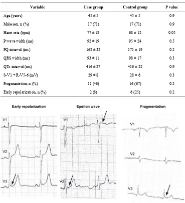
[image:3.595.102.504.493.720.2]between the case and the control groups. QRS fragmen- tation was the most frequent finding in the two groups. We found no WPW configuration and no Brugada sign in the ECGs. We found an epsilon wave configuration in only one of the ECGs, which belonged to a control per- son. The longest QTc interval was 471 ms, the shortest
Table 2. Comparisons between individuals with SCD and controls.
Variable Case group Control group P value
Age (years) 45 ± 5 45 ± 5 0.9
Male sex, n (%) 17 (71) 17 (71) 0.9
Heart rate (bpm) 77 ± 18 68 ± 12 0.05
P wave width (ms) 92 ± 19 95 ± 24 0.5
PQ interval (ms) 162 ± 32 171 ± 19 0.2
QRS width (ms) 93 ± 11 98 ± 17 0.3
QTc interval (ms) 416 ± 27 416 ± 22 0.9
S-V1 + R-V5-6 (mV) 29 ± 8 28 ± 6 0.3
Fragmentation, n (%) 11 (46) 16 (67) 0.2
Early repolarization, n (%) 2 (8) 6 (25) 0.2
Figure 2. Examples of abnormal ECGs.
could not follow the definition of SCD strictly [1]. As such, it is possible that the individuals in our cohort might not have suffered cardiac death, but died from cerebrovascular causes. However, we believe that this problem was minimal, as none of the diagnoses in the death certificate described death from cerebrovascular cause. Furthermore, the prevalence of death from cardiac causes in individuals from 20 to 50 years old is about five times higher than the prevalence of death from cere-brovascular causes. [17]
Second, the age limit for inclusion in our cohort was 10 - 15 years higher than the age limit of most other SCD studies. This may have resulted in a greater number of SCD due to coronary heart disease, as this disease be-comes more prevalent with increasing age. This is not surprising as even in studies, in which the age limit has been 35 - 40 years, coronary heart disease has been one
of the most prevalent etiologies of SCD. [2-5,18] The higher age limit in our study was dictated by the low numbers of SCD in the younger participants. In our co-hort, in the age groups younger than 40 we only found two persons with SCD.
[image:4.595.113.483.101.292.2]these studies are biased towards patients with a worse prognosis. ECG failed to predict SCD in our cohort. Therefore, although ECG is invaluable in symptomatic patients, it may be unsuitable as a screening tool in large asymptomatic populations.
Most importantly, it is probable that the prevalence of the genetic arrhythmias was low in our cohort. Sudden death occurs most often in persons under the age of 40 in patients with genetic arrhythmias [19,21-23]. Neverthe- less, we only found two persons with SCD before the age of 40 and, as such, the result may reflect the relative rar- ity of genetic arrhythmias in Denmark. This may par- ticularly be true for the Brugada syndrome. In an analy-sis of the ECGs from the same cohort we found a lower prevalence of the Brugada sign than that reported from the majority of other studies [24]. Another recent study from Denmark also reported a lower number of HCM and ARVC than that reported in other studies in persons who had suffered SCD before the age of 35 [18].
5. LIMITATIONS
Autopsy was not performed systematically in all patients with sudden death. However, it is unlikely that a system-atic autopsy would have influenced our results. A sys-tematic autopsy might have resulted in the exclusion of some cases and the number of SCD in our cohort was already low from the start.
Not all the ECGs in our cohort were examined. De-spite this limitation, the results of the study are still valid. The study was designed to look for ECG abnormalities in persons that had suffered SCD before the age of 50. Even if we had found abnormalities in the other ECGs, this would have been in persons who were alive after the age of 50. As such, the ECG would not have gained any fur-ther predictive value, as far as SCD is concerned.
6. CONCLUSION
In the Copenhagen City Heart Study, the ECG did not have a predictive value for SCD.
REFERENCES
[1] Priori, S.G., Aliot, E., Blomstrom-Lundqvist, C., Bossaert, L., Breithardt, G., Brugada, P., et al. (2002) Task force on sudden cardiac death, European society of cardiology. Europace, 4, 3-18. doi:10.1053/eupc.2001.0214
[2] Fragkouli, K. and Vougiouklakis, T. (2010) Sudden car- diac death: An 11-year postmortem analysis in the region of Epirus, Greece. Pathology—Research and Practice,
206, 690-694. doi:10.1016/j.prp.2010.05.005
[3] Doolan, A., Langlois, N. and Semsarian, C. (2004) Causes of sudden cardiac death in young Australians. Medical Journal of Australia, 180, 110-112.
[4] Puranik, R., Chow, C.K., Duflou, J.A., Kilborn, M.J. and
McGuire, M.A. (2005) Sudden death in the young. Heart Rhythm, 2, 1277-1282. doi:10.1016/j.hrthm.2005.09.008
[5] Drory, Y., Turetz, Y., Hiss, Y., Lev, B., Fisman, E.Z., Pines, A., et al. (1991) Sudden unexpected death in per- sons less than 40 years of age. American Journal of Car- diology, 68, 1388-1392.
doi:10.1016/0002-9149(91)90251-F
[6] Basso, C., Corrado, D., Rossi, L. and Thiene, G. (2001) Ventricular preexcitation in children and young adults: Atrial myocarditis as a possible trigger of sudden death. Circulation, 103, 269-275. doi:10.1161/01.CIR.103.2.269
[7] Savage, D.D., Seides, S.F., Clark, C.E., Henry, W.L., Maron, B.J., Robinson, F.C., et al. (1978) Electrocardio- graphic findings in patients with obstructive and nonob- structive hypertrophic cardiomyopathy. Circulation, 58,
402-408. doi:10.1161/01.CIR.58.3.402
[8] Monnig, G., Eckardt, L., Wedekind, H., Haverkamp, W., Gerss, J., Milberg, P., et al. (2006) Electrocardiographic risk stratification in families with congenital long QT syndrome. European Heart Journal, 27, 2074-2080. doi:10.1093/eurheartj/ehl159
[9] Marcus, F.I., McKenna, W.J., Sherrill, D., Basso, C., Bauce, B., Bluemke, D.A., et al. (2010) Diagnosis of ar- rhythmogenic right ventricular cardiomyopathy/dysplasia: proposed modification of the task force criteria. Euro- pean Heart Journal, 31, 806-814.
doi:10.1093/eurheartj/ehq025
[10] Antzelevitch, C., Brugada, P., Borggrefe, M., Brugada, J., Brugada, R., Corrado, D., et al. (2005) Brugada syn- drome: Report of the second consensus conference: En- dorsed by the Heart Rhythm Society and the European Heart Rhythm Association. Circulation, 111, 659-670.
doi:10.1161/01.CIR.0000152479.54298.51
[11] Haissaguerre, M., Derval, N., Sacher, F., Jesel, L., Deis- enhofer, I., de Roy, L., et al. (2008) Sudden cardiac arrest associated with early repolarization. The New England Journal of Medicine, 358, 2016-2023.
doi:10.1056/NEJMoa071968
[12] Fitzsimmons, P.J., McWhirter, P.D., Peterson, D.W. and Kruyer, W.B. (2001) The natural history of Wolff-Parkin- son-White syndrome in 228 military aviators: A long- term follow-up of 22 years. American Heart Journal, 142,
530-536. doi:10.1067/mhj.2001.117779
[13] Appleyard, M. (1989) The Copenhagen city heart study: Østerbroundersøgelsen: A book of tables with the data from the first examination (1976-1978) and a five-year follow-up (1981-1983). Scandinavian Journal of Social Medicine, 41, 1-160.
[14] Schnohr, P., Jensen, G., Lange, P., Scharling, H. and Appleyard, M. (2001) The Copenhagen city heart study. Tables with data from the third examination 1991-1994. European Heart Journal, 3, 1-83.
[15] Funck-Brentano, C. and Jaillon, P. (1993) Rate-corrected QT interval: Techniques and limitations. The American Journal of Cardiology, 72, 17B-22B.
doi:10.1016/0002-9149(93)90035-B
[17] Heron, M. (2011) Deaths: leading causes for 2007. Na- tional Vital Statistics Reports, 59, 1-95.
[18] Winkel, B.G., Holst, A.G., Theilade, J., Kristensen, I.B., Thomsen, J.L., Ottesen, G.L., et al. (2010) Nationwide study of sudden cardiac death in persons aged 1-35 years. European Heart Journal, 32, 983-990.
[19] Priori, S.G., Schwartz, P.J., Napolitano, C., Bloise, R., Ronchetti, E., Grillo, M., et al. (2003) Risk stratification in the long-QT syndrome. The New England Journal of Medicine, 348, 1866-1874. doi:10.1056/NEJMoa022147
[20] Sauer, A.J., Moss, A.J., McNitt, S., Peterson, D.R., Za- reba, W., Robinson, J.L., et al. (2007) Long QT syndro- me in adults. Journal of the American College of Cardi- ology, 49, 329-337. doi:10.1016/j.jacc.2006.08.057
[21] Priori, S.G., Napolitano, C., Gasparini, M., Pappone, C., Della Bella, P., Giordano, U., et al. (2002) Natural his- tory of Brugada syndrome: Insights for risk stratification and management. Circulation, 105, 1342-1347.
doi:10.1161/hc1102.105288
[22] Zareba, W., Moss, A.J., Schwartz, P.J., Vincent, G.M., Robinson, J.L., Priori, S.G., et al. (1998) Influence of genotype on the clinical course of the long-QT syndrome. Interna- tional long-QT syndrome registry research group. New England Journal of Medicine, 339, 960-965.
doi:10.1056/NEJM199810013391404
[23] Tabib, A., Loire, R., Chalabreysse, L., Meyronnet, D., Miras, A., Malicier, D., et al. (2003) Circumstances of death and gross and microscopic observations in a series of 200 cases of sudden death associated with arrhyth- mogenic right ventricular cardiomyopathy and/or dyspla- sia. Circulation, 108, 3000-3005.
doi:10.1161/01.CIR.0000108396.65446.21
[24] Pecini, R., Cedergreen, P., Theilade, S., Haunso, S., Thei- lade, J. and Jensen, G.B. (2010) The prevalence and rele- vance of the Brugada-type electrocardiogram in the Dan- ish general population: data from the Copenhagen city heart study. Europace, 12, 982-986.