ORIGINAL RESEARCH
HEAD & NECK
MR Imaging Evolution of Endoscopic Cranial Defect
Reconstructions Using Nasoseptal Flaps and
Their Distinction from Neoplasm
K.O. Learned, N.D. Adappa, J.Y.K. Lee, J.G. Newman, J.N. Palmer, and L.A. Loevner
ABSTRACT
BACKGROUND AND PURPOSE: Endoscopic endonasal approach is the procedure of choice for the resection of ventral skull base neoplasms, with defect closure requiring multilayer reconstruction. This study evaluates the temporal MR imaging evolution of nasoseptal flaps and free grafts used in endoscopic skull base reconstruction.
MATERIALS AND METHODS: Sixty-nine follow-up brain MRIs of 22 patients who had endoscopic skull base reconstruction using 26 nasoseptal flaps combined with 8 collagen-matrix dural grafts, 10 fascia lata grafts, and 10 intracranial fat grafts were retrospectively reviewed. Temporal changes in signal intensity, enhancement, thickness, and the configuration of reconstructive layers were evaluated. Tissue with signal intensity or enhancement different from that of normal evolving reconstructive layers at the surgical bed was evaluated, and its association with clinically confirmed tumor was assessed with the Fisher exact test.
RESULTS: All normal reconstructive layers were retracted to cranial defects and showed maturation of imaging features within 2– 6 months. The immediate postoperative T2-isointensity to brain and enhancement of nasoseptal flaps persisted, but the flap thickness was reduced by 20%–30% (average thickness, 4.5⫾1.3 mm); additionally, the C shape and vascular pedicle of the nasoseptal flaps became indistinct, but the flap location remained unchanged. The initial appearance of the nonenhancing fascia lata with variable T2 signal intensity became enhancing with increasing T2-hypointensity and a graft-thickness reduction ofⱖ50% (average thickness, 3.5⫾1.6 mm). All fat grafts showed progressive resorption. In 6 patients, abnormal tissue represented residual or recurrent tumor (P⫽.0001).
CONCLUSIONS: Maturation and stability of multilayer endoscopic skull base reconstructions on MR imaging occurs within 2– 6 months postoperatively. Understanding of the normal imaging evolution of endoscopic skull base reconstructions is essential to distinguish them from neoplasms.
ABBREVIATIONS:ASB⫽anterior skull base; CSB⫽central skull base; EEA⫽endoscopic endonasal approach; ESBR⫽endoscopic skull base reconstruction; NSF⫽ nasoseptal flap
T
he traditional open craniofacial surgical approach to the ven-tral skull base allows a wide exposure, but at the cost of crani-otomy, brain and cranial nerve retraction, and facial incision. The endoscopic transsphenoidal adenohypophysectomy is the stan-dard in pituitary surgery. More recently, the use of the endoscopic endonasal approach (EEA) has expanded beyond the sella, and itis now routinely used for the resection of a spectrum of sinonasal, ventral skull base, and intracranial pathologies.1-6Endoscopic
skull base reconstruction (ESBR) with multilayer closure of the cranial base defect using vascularized tissues has become the re-pair method of choice in EEA, resulting in low CSF leak rates comparable with those in the open surgical approach.7-10The
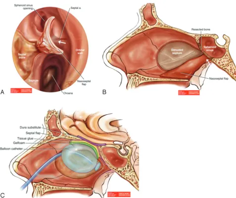
workhorse of ESBR in EEA is the vascularized pedicled nasoseptal flap (NSF), which is based on the nasoseptal branch of the sphe-nopalatine artery (Fig 1A, -B).11,12A combination of intradural
fat graft, inlay subdural collagen matrix, and/or the fascia lata with onlay NSF is effective in repairing cranial defects (Fig 1C).7-10
Most cranial defects are sufficiently closed by a single NSF ele-vated from one side of the nasal septum. Larger, extended cranial defects may require harvesting of bilateral nasoseptal flaps from both sides of the nasal septum and additional free mucosal grafts to cover the entire defect.
Kang et al13described the MR imaging appearance of ESBR
Received August 20, 2013; accepted after revision November 6.
From the Departments of Radiology (K.O.L., L.A.L.), Neurosurgery (J.Y.K.L.), and Otorhinolaryngology-Head and Neck Surgery (N.D.A., J.G.N., J.N.P., L.A.L.), Univer-sity of Pennsylvania School of Medicine, Philadelphia, Pennsylvania.
Paper previously presented as a scientific oral presentation at: Annual Meeting of the American Society of Neuroradiology and the Foundation of the American So-ciety of Neuroradiology Symposium, May 18 –23, 2013; San Diego, California. Please address correspondence to Kim O. Learned, MD, Department of Radiology, Neuroradiology Division, Hospital of the University of Pennsylvania, 219 Dulles Building, 3400 Spruce St, Philadelphia, PA 19104; e-mail: [email protected]
Indicates article with supplemental on-line tables.
using NSFs to repair the surgically created skull base defects in a small cohort of patients following transsphenoidal resection of pituitary adenomas.13Recently, Learned et al14reported the
im-mediate postoperative MR imaging evaluation of ESBR utilizing NSFs in a spectrum of sinonasal and skull base tumors resected using EEAs with defects along the entire ventral skull base. In the immediate postoperative period, NSFs have a C-shaped configu-ration on coronal and/or sagittal planes and avid enhancement, with the associated free grafts closely abutting the NSFs and show-ing no enhancement.13,14Follow-up MR imaging is routinely
per-formed for neoplastic surveillance in patients following EEA skull base resections and reconstructions. The normal evolution of the multilayer reconstructions following EEA is not well understood and can be mistaken for recurrent or persistent tumor. This study evaluates the temporal MR imaging appearances of grafts and NSFs used in ESBR.
MATERIALS AND METHODS
Subjects
Our institutional review board approved the retrospective review of our skull base surgery data base from January 2008 to May 2013 to identify 22 patients (13 men, 9 women; mean age, 54 years) who had undergone EEA skull base surgery, multilayer ESBR using NSFs, and brain MR imaging surveillance within 48 hours follow-ing surgery and thereafter for follow-up. The spectrum of pathol-ogies included sinonasal tumors (36%), clival chordomas and chondrosarcomas (18%), and intracranial neoplasms (46%). Sur-gically created skull base defects included anterior skull base (ASB) (41%), central skull base (CSB) (36%), and clival defects (23%). In 22 ESBRs, 26 NSFs (18 single and 8 bilateral flaps) were used with a combination of 8 collagen matrix dural grafts (Dura-Gen; Integra Life Sciences, Plainsboro, New Jersey; Surgisis Biodesign Dural graft; Cook Medical, Bloomington, Indiana), 10
[image:2.594.54.532.43.443.2]autologous fascia lata grafts, 10 intracranial fat grafts, and 4 addi-tional free mucosal grafts. Table 1 summarizes patient clinical data.
Imaging Methods
Sixty-nine follow-up MRIs were obtained at 2- to 6-month inter-vals, depending on tumor histology, adjuvant therapy, clinical status, and surveillance paradigm. The length of follow-up ranged from 3 to 41 months, with an average of 16.4 months. We categorized the follow-up MRIs of the entire cohort according to the time at which they were obtained after surgery: first follow-up at 2–3 months; subsequently, every 3-month-interval follow-up during the first 1–2 years after surgery; and every 6-month-interval follow-up thereafter. The graph in Fig 2 summarizes the percentage of serial follow-up MRIs after surgery and adjuvant treatment.
Brain MRIs were performed at 1.5T (n⫽55) and 3T (n⫽14) (Signa and Optima; GE Healthcare, Milwaukee, Wisconsin; and Espree, Avanto, Verio and Skyra; Siemens, Erlangen, Germany), including thin-section images of the sella turcica/ventral skull
base with parameters detailed in On-line Tables 1 and 2. All pa-tients received intravenous administration of 0.1-mmol/kg gado-linium (MultiHance; Bracco Diagnostics, Princeton, New Jersey).
Imaging and Statistical Analysis
All images were reviewed together in consensus by 2 neuroradi-ologists subspecialized in head and neck imaging. The NSFs and free grafts were evaluated for temporal changes in T2 signal inten-sity (in comparison with the gray matter and white matter of the brain parenchyma), the presence of enhancement, and the aver-age thickness. The C-shaped configuration of enhancing NSFs in our cranial base reconstructions was best demonstrated on an enhanced sagittal and/or coronal plane. The free grafts were lo-cated above the enhancing NSFs in multilayer ESBRs. Therefore, the average of 2 maximal flap and graft thickness measurements at 2 different locations on enhanced sagittal and/or coronal planes was recorded. All studies of each patient were evaluated at the same time in our retrospective study to assess the temporal change of ESBRs. Imaging assessment was correlated with surgical tech-niques and clinical follow-up. The presence of tissue with signal-intensity or enhancement different from that of normal evolving reconstructive layers was also evaluated, and its MR imaging char-acteristics were compared with those of the neoplasm on preop-erative MR imaging. The association of the abnormal tissue that was similar to the preoperative neoplasm and clinically confirmed tumor was assessed with the Fisher exact test, using the Statistical Package for the Social Sciences, Version 20.0 (IBM, Armonk, New York).
RESULTS
All normal reconstructions demonstrated retraction of the recon-structive layers to the cranial defects with reduction in thickness and showed maximal alterations of the imaging features within 2– 6 months, with little or no further change on subsequent im-aging. Fifty-five percent of follow-up MR imaging studies were performed before or without adjuvant treatment and 45%,
af-Table 1: Patient clinical data
Pathologies (No. of Patients and Average Tumor Size)
Skull Base Defects
ESBR Tissues (No. of Patients)
Length of MRI Follow-Up (mo)
Adjuvant Therapy (No. of Patients) Esthesioneuroblastoma (n⫽6; 2.5 cm) ASB F, FL, NSF, FM (n⫽1) 9–32 Radiation (n⫽4),
D, NSF (n⫽3) Chemoradiation (n⫽2)
D, NSF, FM (n⫽2) Sinonasal endocrine carcinoma
(n⫽1; 4.0 cm)
ASB F, FL, NSF, FM (n⫽1) 16 Chemoradiation (n⫽1)
Sinonasal undifferentiated carcinoma (n⫽1; 2.2 cm)
ASB D, NSF (n⫽1) 3 Chemoradiation (n⫽1)
Chondrosarcoma (n⫽1; 2.5 cm) Clivus D, NSF (n⫽1) 21 Radiation (n⫽1)
Chordoma (n⫽3; 2.0 cm) Clivus NSF (n⫽2) 9–21 Radiation (n⫽2)
F, FL, NSF (n⫽1)
Meningioma (n⫽4; 2.2 cm) ASB D, NSF (n⫽1) 3–41 Radiation (n⫽1)
CSB F, FL, NSF (n⫽2) Clivus F, FL, NSF (n⫽1)
Craniopharyngioma (n⫽1; 3.5 cm) CSB F, FL, NSF (n⫽1) 6
Pituicytoma (n⫽1; 3.1 cm) CSB F, FL, NSF (n⫽1) 13 Radiation (n⫽1) Pituitary macroadenoma
(n⫽4; 3.4 cm)
CSB NSF (n⫽1) 6–27 Radiation (n⫽1)
F, NSF (n⫽1) FL, NSF (n⫽1) F, FL, NSF (n⫽1)
Note:—ASB indicates cribriform plates, crista galli, and/or ethmoid fovealis; CSB, planum sphenoidale, tuberculum sella, sella, and/or dorsum sella; D, collagen matrix dural graft; F, intracranial fat; FL, fascia lata; FM, free mucosa.
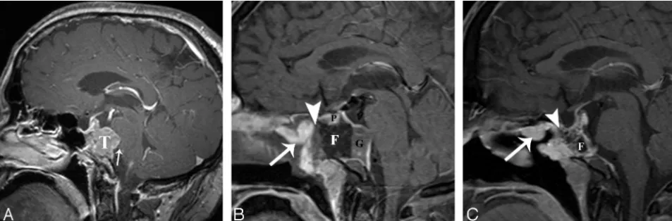
[image:3.594.54.530.54.271.2] [image:3.594.54.285.300.453.2]ter adjuvant treatment (Fig 2). The patients’ sinonasal inflam-matory mucosal changes and adjuvant radiation or chemora-diation had no appreciable effect on the normal imaging evolution of ESBRs (Figs 3 and 4). The appearance of the dif-ferent components of the flaps and their changes with time to become stable within 2– 6 months are summarized in Table 2 and presented in detail below.
The immediate postoperative T2-isointensity to the brain (mostly similar to gray matter) and solid enhancement of 21 NSFs persisted, and the flap thickness was reduced by 20%–30% (aver-age thickness, 4.5⫾1.3 mm) (Figs 3 and 4). Their immediate postoperative distinctive C shape and vascular pedicle configura-tions became less well-defined on follow-up imaging as the NSFs incorporated into the newly enhancing free grafts and granulation tissues, but the flap location remained unchanged (Fig 3). The markedly nodular appearance of the NSF was noted in 4 of our patients (18%) who did not have tumor recurrence (Fig 3C).
Five NSFs without enhancement on immediate postoperative MR imaging demonstrated 1- to 5-mm-thick solid enhancing tis-sue on follow-up. Similarly, 4 free mucosal grafts without en-hancement on immediate postoperative MR imaging showed 1-to 5-mm-thick solid enhancement on follow-up. Their enhance-ment correlated to clinically evident healing granulation tissue and/or mucosalization at the graft sites.
The immediate postoperative nonenhancing fascia lata with variable T2 signal intensity became mildly enhancing with in-creasing T2-hypointensity, and the graft thickness decreased by
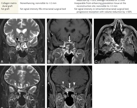
ⱖ50% (average thickness, 3.5⫾ 1.6 mm) (Figs 3 and 4). The collagen matrix dural grafts remained nonvisible toⱕ2 mm, and the immediate postoperative nonenhancing thin grafts became inseparable from enhancing granulation tissue at the recon-structed skull base on follow-up imaging (Fig 5). All fat grafts showed progressive resorption withⱖ50% reduction in volume within 2– 6 months and near disappearance by 14 months (Figs 3 and 4).
The nodular tissue with variable T2 signal intensity and
en-hancement different from the normal evolution of reconstructive layers was seen at the interface of the reconstruction and tumor bed in 6 patients (1 sinonasal carcinoma, 1 chordoma, 1 menin-gioma, and 3 pituitary tumors) (Fig 5). The MR imaging charac-teristics of the nodular tissue were similar to those of the primary neoplasm seen on the preoperative MR imaging. In the patients with sinonasal carcinoma, recurrence occurred at the margin of the radiation field. The presence of the abnormal tissue was asso-ciated with neoplastic residua in 5 patients and recurrence in 1 patient (P⫽.0001).
DISCUSSION
Endoscopic Skull Base Surgery and Cranial Defect Reconstruction
The endonasal approach provides a direct and less invasive surgi-cal corridor to primarily midline sinonasal, skull base, and intra-cranial neoplasms along the ventral intra-cranial base, with equivalent or better gross total resection compared with open surgery.1-7
Open skull base surgery, classically in the form of combined cra-niotomy and a transfacial or endonasal endoscope-assisted ap-proach, is repaired with the vascularized pericranial flap routinely sutured to the edge of the dural defect. Cranial defect closure in EEA has unique challenges, including the inability to suture the reconstructive layers and the difficulty in using a pericranial flap. A variety of biologic glues and temporary supportive nasal bol-sters has been used to secure the ESBR layers to the cranial defect, while awaiting granulation and incorporation of the reconstruc-tive tissues into the surgical bed.8-10The small dural defects have
been successfully closed with nonvascularized free grafts such as seen in pituitary surgery. With the increasing size of cranial de-fects in EEA for resection of larger tumors and with deeper intra-arachnoid dissections and increased exposure to the cis-terns, repair techniques using nonvascularized free grafts have shown unacceptable postoperative CSF leak rates.7,8To allow
maximal resection with CSF leak rates similar to those in open surgical approaches, ESBR in the EEA routinely necessitates
[image:4.594.54.531.48.205.2]multilayer reconstruction using a vascularized NSF. The NSF is a pedicled vascular flap based on the posterior septal artery, a branch of the sphenopalatine artery.11This robust
vascular-ized flap has become the mainstay in ESBR, with reported post-operative CSF leak rates in 5%–10% of cases, a range similar to that in open procedures.7,8
Our ESBRs were performed in a multilayer stepwise fash-ion, beginning intracranially and advancing out to the
sinona-sal mucosa, using a combination of autologous fat filling the intracranial surgical bed, subdural inlay collagen matrix un-derneath the edges of the dural defect and/or inlay-onlay fascia lata, and the final mucosal onlay closure with single or bilateral NSFs (Fig 1). Occasionally, additional free mucosal grafts taken from the nasal septum or turbinates were required to cover the far anterior edge of the cribriform defects, which were not reachable by the pedicled NSF (Table 1).
[image:5.594.53.533.49.529.2]MR Imaging Follow-Up of Endoscopic Skull Base Reconstruction
The imaging features of ESBR with the characteristic enhancing C shape and vascular pedicle configuration of the NSF and nonen-hancing free grafts on immediate postoperative MR imaging have been described previously.13,14Kang et al13reported NSFs used to
reconstruct defects following resection of pituitary adenomas and found stable signal intensity but variable changes in thickness and enhancement of NSFs on follow-up MRI at 3–7 months.13They
described 2 flaps without enhancement in the immediate
[image:6.594.54.534.136.515.2]postop-erative period that showed delayed enhancement on follow-up imaging and 8 flaps that enhanced in the immediate perioperative period with persistent enhancement in 7/8 flaps on follow-up imaging. Five of the initially enhancing NSFs became thinner; 2, thicker; and the average flap thickness varied from nonvisualiza-tion to 7 mm. They also noted reducnonvisualiza-tion in the thickness of the multilayer reconstruction between the graft and defect. Their cohort of pituitary adenomas did not require adjuvant chemoradiation.
Table 2: MR imaging evolution of ESBR from the immediate postoperative period to stability on follow-up
Immediate Postoperative MRI Follow-Up MRI at 2–6 Months
NSF Solid enhancement, T2-isointensity to brain (mostly similar to gray matter), distinctive C shape and vascular pedicle configurations
Persistent solid enhancement, persistent T2-isointensity to brain (mostly similar to gray matter), thickness reduction
by 20%–30%, average thickness of 4.5 mm
Less well-defined C shape and vascular pedicle configurations, unchanged flap location
Fascia lata Nonenhancing, variable T2 signal intensity Mildly enhancing, increasing T2-hypointensity, thickness reduction byⱖ50%, average thickness of 3.5 mm Collagen matrix
dural graft
Nonenhancing, nonvisible toⱕ2 mm Inseparable from enhancing granulation tissue at the reconstruction site, nonvisible toⱕ2 mm
Fat graft Fat signal intensity fills intracranial surgical bed Fat signal intensity in retracted intracranial surgical bed, progressive resorption with volume reduction byⱖ50%
Our long-term follow-up (range, 3– 41 months; average, 16.4 months) showed some differences from the published results of Kang et al.13None of our enhancing NSFs lost their immediate
postoperative solid enhancement, and all demonstrated mild re-duction in thickness. Our average flap thickness of 4.5 mm was comparable. None of our cases, including a similar cohort with a transellar approach without adjuvant treatment, had increases in flap thickness as seen in 2 patients in the study of Kang et al (an increase of 1 and 2 mm in flap thickness). This discrepancy may be explained by sampling error. During the immediate postoperative period, the NSF stood out as the only enhancing tissue among the nonenhancing free grafts and postoperative sinus contents. Therefore, its unique enhancing C-shaped configuration was eas-ily delineated. Delayed enhancement of the free grafts, retraction and reduction in the thickness of the reconstructive layers, and integration of the enhancing NSF into the reconstruction site re-sulted in its less distinct configuration on follow-up MR imaging (Fig 3). The nonenhancing NSFs on immediate postoperative MR imaging probably compromised the vascular pedicles. Theoreti-cally, they served as free mucosal grafts at the reconstructive site. Therefore, these nonenhancing NSFs behaved in a manner similar to those in the free mucosal grafts on follow-up; both appeared as thin enhancing granulation-mucosalization, radiographically and clinically.
The reduction in thickness of free grafts in our cohort varied. The fascia lata remained thicker than DuraGen, and its noticeable reduction in thickness probably reflects resolution of immediate postoperative swelling with increasing T2-hypointensity (Fig 4). Enhancement on follow-up reflects the incorporation of the fascia lata into the granulation tissue at the cranial defect. In reconstruc-tions using only inlay subdural collagen matrix as the dural graft, the nonvisible toⱕ2-mm nonenhancing collagen matrix became indistinguishable from the enhancing granulation at the defect (Fig 5).
In addition, the stabilization of the mature imaging features occurred within 2– 6 months following surgery, with little or no further change on subsequent imaging. Seventy-four percent of our follow-up MRIs were obtained within the first year after sur-gery, because the initial follow-up intervals were generally closer, with a resulting higher number of studies, especially those for oncologic surveillance. Close to equal proportions of the imaging studies were performed in patients after they received adjuvant chemo- and/or radiation treatment (45%) and in patients who did not or have not received adjuvant treatment (55%). Notably, the adjuvant radiation or chemoradiation and the sinonasal in-flammatory changes had no appreciable effect on the normal im-aging evolution of ESBRs in our oncologic patients (Fig 4).
Imaging studies evaluating postoperative endoscopic pituitary surgery demonstrated the involution of surgical materials and identification of residual-recurrent adenoma by location, charac-teristic signal intensity, and nodular enhancing patterns that were identical to those of the corresponding preoperative ade-noma.15,16 Our evaluation of parasellar neoplasms following
EEAs is concordant with those of MR imaging studies of transellar hypophysectomy. Our study also showed that the full-thickness enhancement of T2-isointense NSFs, which is different from the thin peripheral enhancement of the adjacent T2-hyperintense
si-nonasal mucosa, may mimic residual or recurrent tumor in pa-tients with sinonasal neoplasms if the radiologist is not familiar with the normal appearance and evolution of these reconstruc-tions (Fig 4). To avoid this pitfall, proper identification of the normal NSF on imaging and evaluation of the reconstructive margins for the presence of nodular tissue with signal character-istics and enhancement similar to that of preoperative neoplasm and different from that of the reconstructive layers are paramount (Fig 5).
Limitations and Implications for Patient Care
Limitations of our study include its retrospective nature and small sample size limited to an adult population with predominantly midline neoplasms. Even though the scanner variability makes our findings applicable to most practices, its effects on our results are uncertain, and that uncertainty may explain some of the mi-nor differences in our results compared with those of Kang et al.13
Our reported imaging features of free grafts were limited to col-lagen matrix dural grafts and the fascia lata, and different materi-als used at other institutions may appear different. However, these free tissue grafts are most commonly used at many centers.8-10
The vascularized NSF, the workhorse of ESBR in EEA, is always present irrespective of the type of free grafts and reconstruction techniques.
With the ongoing evolution of surgical navigation, endoscopic instrumentation, and the growing success of EEA and ESBR in achieving gross total resection and reliable closure similar to that in the open approach, EEA is becoming a surgical standard for carefully selected cases in the minimally invasive surgical arena.1
As these procedures are increasing across the country, neuroradi-ologists will increasingly face the challenge of postoperative sur-veillance following EEA and ESBR, which potentially have unique patterns of neoplastic residua and recurrence compared with those of traditional open surgery.
CONCLUSIONS
Understanding the normal imaging evolution of multilayer ESBR, specifically the persistent T2-isointensity with solid enhancement of the NSF and the maturation of the reconstructive layers within 2– 6 month follow-up, is critical in the imaging assessment for tumor surveillance following an EEA to skull base neoplasms. Recognition of typical patterns of evolution should help the in-terpreting radiologist distinguish expected posttreatment change from tumor recurrence.
Disclosures: John Y.K. Lee—UNRELATED:Payment for Lectures (including service on Speakers Bureaus): Baxter. Jason G. Newman—UNRELATED:Stock/Stock Options: Visionsense;Other: Intuitive Surgical,Comments: proctor for transoral robotic sur-gery. Laurie A. Loevner—UNRELATED:Grants/Grants Pending: American College of Radiology Imaging Network,*Payment for Lectures (including service on Speakers Bureaus): Continuing Medical Education, university visiting professorships, Interna-tional Diagnostics Course in Davos, Greece, and Hong Kong,Royalties: books au-thored—Temporal Bone Imaging(Thieme),Brain Case Review(Elsevier).*Money paid to the institution.
REFERENCES
1. Castelnuovo P, Dallan I, Battaglia P, et al.Endoscopic endonasal skull base surgery: past, present and future.Eur Arch Otorhinolar-yngol2010;267:649 – 63
for sinonasal malignancy. Otolaryngol Clin North Am 2011;44: 1081–140
3. Komotar RJ, Starke RM, Raper DM, et al.Endoscopic skull base surgery: a comprehensive comparison with open transcranial ap-proaches.Br J Neurosurg2012;26:637– 48
4. Koutourousiou M, Gardner PA, Tormenti MJ, et al.Endoscopic en-donasal approach for resection of cranial base chordomas: out-comes and learning curve.Neurosurgery2012;71:614 –24
5. Leng LZ, Greenfield JP, Souweidane MM, et al.Endoscopic, endo-nasal resection of craniopharyngiomas: analysis of outcome in-cluding extent of resection, cerebrospinal fluid leak, return to
preoperative productivity, and body mass index.Neurosurgery
2012;70:110 –23
6. Dehdashti AR, Ganna A, Witterick I, et al.Expanded endoscopic endonasal approach for anterior cranial base and suprasellar lesions: indications and limitations.Neurosurgery2009;64:677– 87 7. Kassam AB, Prevedello DM, Carrau RL, et al.Endoscopic endonasal
skull base surgery: analysis of complications in the authors’ initial 800 patients.J Neurosurg2011;114:1544 – 68
8. Harvey RJ, Parmar P, Sacks R, et al.Endoscopic skull base recon-struction of large dural defects: a systematic review of published evidence.Laryngoscope2012;122:452–59
9. Liu JK, Schmidt RF, Choudhry OJ, et al.Surgical nuances for naso-septal flap reconstruction of cranial base defects with high-flow
ce-rebrospinal fluid leaks after endoscopic skull base surgery. Neuro-surg Focus2012;32:E7
10. Kassam AB, Carrau RL, Snyderman CH, et al.Endoscopic recon-struction of the cranial base using a pedicled nasoseptal flap. Neu-rosurgery2008;63:ONS44 –52, discussion ONS52–53
11. Hadad G, Bassagasteguy L, Carrau RL, et al. A novel recon-structive technique after endoscopic expanded endonasal
approaches: vascular pedicle nasoseptal flap. Laryngoscope
2006;116:1882– 86
12. Palmer JN, Chiu AG.Atlas of Endoscopic Sinus and Skull Base Surgery.
Philadelphia: Elsevier; 2013
13. Kang MD, Escott E, Thomas AJ, et al.The MR imaging appearance of
the vascular pedicle nasoseptal flap. AJNR Am J Neuroradiol
2009;30:781– 86
14. Learned KO, Adappa ND, Loevner LA, et al.MR imaging evaluation of endoscopic cranial base reconstruction with pedicled nasoseptal flap following endoscopic endonasal skull base surgery.Eur J Radiol
2013;82:544 –51
15. Steiner E, Knosp E, Herold CJ, et al.Pituitary adenomas: findings of postoperative MR imaging.Radiology1992;185:521–27