ORIGINAL RESEARCH
Cerebellar Lesions in Multiple System Atrophy:
Postmortem MR Imaging
ⴚ
Pathologic Correlations
E. Matsusue S. Fujii Y. Kanasaki T. Kaminou E. Ohama T. Ogawa
BACKGROUND AND PURPOSE: Cerebellar atrophy and white matter T2-hyperintensities have been characterized as cerebellar lesions of multiple system atrophy (MSA). The aim of the study was to correlate MR images with histologic findings in cerebellar lesions of MSA.
MATERIALS AND METHODS: Postmortem T2-weighted images using 1.5T were compared with histo-logic findings in 7 postmortem-proved cases with MSA. The MR imaging findings in the cerebellar cortices and deep white matter dentate nucleus regions were compared with their histologic findings in each case.
RESULTS: We detected 3 types of cerebellar changes: type 1, no apparent atrophy or signal-intensity changes; type 2, cerebellar atrophy and inhomogeneous (patchy and/or confluent) cerebellar white matter hyperintensities; and type 3, cerebellar atrophy and diffuse white matter hyperintensities. Hypointensities were seen in the dentate nucleus regions. Atrophy of the cerebellar white matter was more severe than that of cerebellar cortices, and this anatomy was well depicted on coronal images. Histologically, degeneration was more severe in the cerebellar white matter than in the cerebellar cortices. Hyperintensities in the cerebellar white matter showed loss of myelinated fibers and gliosis. Hypointensities in the dentate nucleus regions revealed diffuse ferritin deposition in preserved dentate nuclei and white matter both around and within the nuclei.
CONCLUSIONS:Hyperintensities in the cerebellar white matter reflect degenerated white matter associated with loss of myelinated fibers and gliosis, whereas hypointensities in the dentate nucleus regions reflect diffuse ferritin deposition in preserved dentate nuclei and white matter around and within the nuclei. Degeneration is more severe in the cerebellar white matter than in the cerebellar cortices.
M
ultiple system atrophy (MSA) is a sporadic adult-onset progressive neurodegenerative disorder, characterized pathologically by degeneration of the basal ganglia and the olivopontocerebellar system and clinically known as parkin-sonism, cerebellar ataxia, and autonomic dysfunction. MSA has 3 subtypes: olivopontocerebellar atrophy (OPCA), stria-tonigral degeneration (SND), and Shy-Drager syndrome. Also, sporadic OPCA, SND, and Shy-Drager syndrome can all mutually exist both pathologically and clinically. The com-mon and specific histologic findings in MSA are glial cytoplas-mic inclusions (GCIs).1,2The major constituent of GCIs is ␣-synuclein, which is also a major component of the Lewy body, which is the pathologic hallmark lesion in Parkinson disease and dementia with Lewy bodies. Recently the term “synucleinopathy” has been proposed to encompass a pre-sumed common pathogenic process shared by these neurode-generative disorders.3,4On the basis of symptoms at onset, MSA has been divided into 2 forms: MSA-C, characterized by a predominance of cerebellar symptoms; and MSA-P, in which parkinsonism is prevalent.5,6Hyperintensities in the pons, middle cerebellar peduncles, and cerebellum, corresponding to pontocerebellar tract atrophy and hyper- and/or hypointensities associated with posterior putaminal atrophy on T2-weighted images,
have been described as characteristic findings on MR images in patients with MSA.7-12
Diffusion-weighted MR imaging (DWI) has emerged as a useful method for detecting early pathologic changes of vari-ous diseases based on water diffusivity.13Apparent diffusion coefficient (ADC) and fractional anisotropy (FA) values have been used to evaluate the degree of tissue degeneration in var-ious disorders. Recent evidence has demonstrated that ADC values in the pons, cerebellum, and putamen are significantly higher,14-16whereas FA values are lower in MSA than in Par-kinson disease or controls.14,17This difference suggests that DWI may be a useful tool in the diagnosis of MSA.
There have been several reports studying the correlation of antemortem MR images with histologic findings7-9in cerebel-lar lesions of MSA. However, there have been no reports di-rectly correlating postmortem MR images with histologic findings in cerebellar lesions. Our aim was to elucidate MR imaging⫺pathologic correlations on postmortem T2-weighted images and their histologic findings in 7 autopsy-proved cases of cerebellar lesions of MSA.
Materials and Methods
We retrospectively analyzed brain MR images in 7 postmortem-proved MSA cases (5 women, 2 men; mean age, 71 years; age range, 55– 83 years) and, with the consent of relatives, were able to conduct postmortem examination on the brains of the deceased. As for the histologic diagnosis of MSA, established MSA neuropathologic crite-ria were applied, checking the presence of GCIs with neuronal loss and gliosis in a number of structures including the putamen, substan-tia nigra, locus coeruleus, basis pontis, inferior olivary nucleus, and dorsal motor nucleus of the vagus.18The GCIs were examined by using␣-synuclein immunohistochemistry. Histologic findings also Received February 19, 2009; accepted after revision April 6.
From the Division of Radiology (E.M., S.F., Y.K., T.K., T.O.), Department of Pathophysio-logical and Therapeutic Science, Faculty of Medicine, Tottori University, Yonago, Tottori, Japan; and Department of Neuropathology (E.O.), Institute of Neurological Sciences, Faculty of Medicine, Tottori University, Yonago, Tottori, Japan.
Please address correspondence to Eiji Matsusue, MD, Division of Radiology, Department of Pathophysiological and Therapeutic Science, Faculty of Medicine, Tottori University. 36-1 Nishi-cho, Yonago, Tottori 683-8504, Japan; e-mail: [email protected] DOI 10.3174/ajnr.A1662
BRAIN
ORIGINAL
indicated 5 cases of OPCA, 1 case of SND, and 1 case of combined OPCA and SND.
T2-weighted images of 10% formalin-fixed brains at postmortem examination (postmortem MR images) were obtained in all cases by using a 1.5T imaging system (Symphony; Siemens, Erlangen, Ger-many). First, we washed the fixed brains by using running tap water for 24 consecutive hours. Each brain was then put into its original box, which was void of water. Therefore, the fixed brains were com-pletely surrounded by air with the exception of some residual water in the ventricles and sulci. The fixed brains in the box were positioned in a standard way in the 8-channel head coil, and axial and coronal fast spin-echo T2-weighted images (TR/TE, 4000/81; 320⫻512 matrix; 150-mm FOV; section 4/1 mm) were obtained. For gross examina-tion, each cerebellum was cut into 3-mm-thick coronal sections, each cerebrum was cut into 5-mm-thick coronal sections, and each brain stem had 3-mm-thick axial sections. Therefore, sagittal MR images were not obtained because each brain was not cut into sagittal sections for gross examination.
Evaluation of antemortem MR images was not performed because all cases belonged to other hospitals and the MR images were available for only 3 cases. However, the findings at postmortem T2-weighted imaging were qualitatively well correlated with the in vivo imaging findings, despite the shortening of T1 and T2 relaxation times second-ary to formalin fixation.19
For MR-pathologic direct correlation, neuropathologic examina-tions were performed in all cases by using 5 staining methods: hema-toxylin-eosin, Bielschowsky, Klu¨ver-Barrera, glial fibrillary acidic protein (GFAP), and ferritin immunohistochemistry. The Biels-chowsky method was used to evaluate axons, and the Klu¨ver-Barrera method was used to evaluate myelin. GFAP immunohistochemistry was used to evaluate gliosis, and ferritin immunohistochemistry was used to reveal ferritin deposits.
Evaluation of Postmortem MR Imaging Findings
The presence of atrophy and signal-intensity change of cerebellar cor-tices, deep white matter and dentate nucleus regions was evaluated on postmortem coronal T2-weighted images. Cerebellar cortices were considered to be atrophied if there were dilations of folial fissures of the cerebellum. Cerebellar deep white matter was considered to be atrophied if there were loss of volume and a concaved contour of the white matter. To appreciate the increased signal intensity of the cer-ebellar deep white matter, signal-intensity changes of cercer-ebellar white matter were compared with those of cerebral white matter on coronal images. The extent of T2 hyperintensities of the cerebellar white mat-ter was graded as follows: none, inhomogeneous (presence of patchy and/or confluent hyperintense areas), and diffuse. Dentate nuclei were considered to be atrophied if they had a flattened appearance.
We also evaluated whether axial images were useful for the detec-tion of cerebellar cortices, deep white matter, and dentate nuclei. The presence of atrophy and signal-intensity change of the pons and mid-dle cerebellar peduncles was evaluated on postmortem axial T2-weighted images. In our final analysis, we evaluated cerebellar symp-toms and measured the duration of the disease for each case.
Postmortem MR Imaging and Pathologic Correlations
The MR imaging findings in the cerebellum were compared with their histologic findings in each case. The following cerebellar regions were histologically assessed for the correlations: 1) cerebellar cortices, 2) deep cerebellar white matter, and 3) dentate nucleus regions.
Results
Postmortem MR Imaging Findings
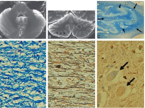
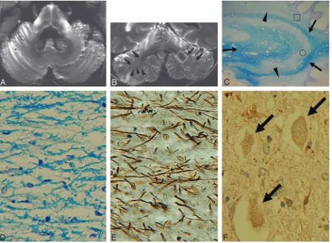
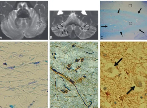
There were 3 types of cerebellar changes on postmortem coro-nal T2-weighted MR images: type 1, no apparent cerebellar atrophy or signal-intensity changes (case 1) (Fig 1A, -B); type 2, atrophy of cerebellar cortices and deep white matter and inhomogeneous (patchy and/or confluent) cerebellar deep white matter hyperintensities (cases 2– 4) (Fig 2A, -B); and type 3, atrophy of cerebellar cortices and white matter and diffuse cerebellar deep white matter hyperintensities (cases 5–7) (Fig 3A, -B). The atrophy of cerebellar deep white matter and cortices was equally apparent in both types 2 and 3. How-ever, the atrophy was more severe in cerebellar white matter than in cerebellar cortices in types 2 and 3. Diffuse hypointen-sities were seen in the dentate nucleus region in all types. At-rophy was not seen in types 1 and 2 but was present in type 3. Also, hypointensities of the dentate nucleus region were con-spicuous due to diffuse hyperintensities of cerebellar white matter in type 3. In all cases, cerebellar white matter and cor-tices and dentate nucleus regions were not precisely depicted on axial images, especially in types 2 and 3 (Figs 1A, 2A, and 3A). As to changes of the middle cerebellar peduncle and pon-tine base, no signal-intensity change or atrophy was seen in type 1. In types 2 and 3, there were diffuse hyperintensities with atrophy in middle cerebellar peduncles and crossed hy-perintensities in the atrophied pontine base. More severe at-rophy and increased signal intensities were seen in type 3 than in type 2.
Cerebellar symptoms such as ataxic gait, dysarthria, or nys-tagmus were observed in types 2 and 3, but not in type 1. The total duration of illness in all our cases was as follows: type 1, 5 years (case 1); type 2, 6 years (cases 3 and 4) and 7 years (case 2); type 3, 8 years (cases 5 and 6) and 13 years (case 7). Among the examined, case 1 (type 1) had the shortest duration of illness (5 years) and case 7 had the longest duration of illness (13 years).
Postmortem MR Imaging and Pathologic Correlations In the case of type 1, cerebellar cortices without atrophy showed histologically well preserved neurons without appar-ent gliosis. Cerebellar white matter without signal-intensity change or atrophy (Fig 1B) showed histologically mild loss of myelin and axons with mild gliosis (Fig 2C–E). Hypointensi-ties in the dentate nucleus regions (Fig 1B) reflected diffuse ferritin deposition and well-preserved dentate nuclei and white matter around and within the nuclei (Figs 2C, -F). His-tologically, the case was diagnosed as SND type (case 1).
In all cases of type 2, inhomogeneous (patchy and/or con-fluent) deep cerebellar white matter hyperintensities with at-rophy (Fig 2B) showed loss of myelin and axons with gliosis but no tissue rarefaction (Fig 3C–E). The atrophy was more severe in the cerebellar white matter compared with cerebellar cortices (Fig 2B). Hypointensities in the dentate nucleus re-gions (Fig 2B) showed well-preserved dentate nuclei and white matter within and surrounding the dentate nuclei and diffuse ferritin deposition (Figs 2C, -F). Histologically, they were di-agnosed as SND plus OPCA type (case 2) and OPCA type (cases 3 and 4), respectively.
hyper-intensities with atrophy (Fig 3B) showed tissue rarefaction with loss of myelin and axons as well as gliosis (Fig 3C–E). Atrophy was seen in both cerebellar white matter and cortices but was more severe in the cerebellar white matter compared with the cerebellar cortices (Figs 3B). Atrophied and hypoin-tense dentate nucleus regions (Fig 3B) showed atrophied but well-preserved neurons with gliosis and loss of myelin and axons of white matter within and surrounding the nuclei and diffuse ferritin deposition (Figs 3C, -F). Histologically, all cases were diagnosed as OPCA type (cases 5–7).
Discussion
Pathologic Distribution of the Olivopontocerebellar Region in MSA
In MSA, the olivopontocerebellar system is usually involved.20 Input cerebellar fibers, including pontocerebellar fibers (transverse pontine and middle cerebellar peduncle) and olivocerebellar fibers (inferior cerebellar peduncle), are mainly affected. In our cases with hyperintensities and atrophy in the deep cerebellar white matter (types 2 and 3), there were diffuse hyperintensities with atrophy in middle cerebellar
pe-duncles and crossed hyperintensities in the atrophied pontine base. Also, more severe atrophy and increased signal intensi-ties were seen in cases with diffuse hyperintensity of the cere-bellar white matter (type 3) than in cases with inhomogeneous hyperintensity of cerebellar white matter (type 2). These find-ings suggest that the degree of pontocerebellar fiber degener-ation is correlated with that of cerebellar white matter degeneration.
As to output cerebellar fibers, the fibers projecting from Purkinje cells to the dentate nuclei are affected in variable degrees, yet dentatorubral fibers (superior cerebellar pedun-cle) are well preserved.
Signal-Intensity Change and Atrophy of the Cerebellum Mizutani21 reported that in patients with MSA, cerebellar white matter is usually more affected than cerebellar cortices. Our MR images indicated that in cases with cerebellar white matter hyperintensities with atrophy (types 2 and 3), the atro-phy was more severe in the cerebellar white matter compared with the cerebellar cortices. In addition, the degeneration was histologically more severe in the white matter compared with
[image:3.594.57.536.40.396.2]the cortices. Even in the case without cerebellar atrophy or signal-intensity changes (type 1), cerebellar white matter showed histologically mild degeneration, including loss of myelin and axons with mild gliosis. All of these findings are consistent with predominant cerebellar white matter involve-ment in MSA.
Histologic findings also indicated that hyperintensities of cerebellar white matter showed loss of myelin and axons with gliosis, which reflected histologic changes in MSA. Also, dif-fuse cerebellar white matter hyperintensities seen in type 3 showed tissue rarefaction with severe loss of myelin and axons as well as gliosis. Severe tissue rarefaction in cerebellar white matter is sometimes seen in cases with MSA.21
As for duration of illness, more severe atrophy and in-creased signal intensities, suggesting more severe degenera-tion, were seen in cerebellar white matter as well as cerebellar cortices in our cases with a longer duration of illness.
Change of the Dentate Nucleus
In MSA, dentate nuclei are preserved, as well as their projec-tions to the red nuclei and the thalami through the superior
cerebellar peduncles. As to signal intensity of the dentate nu-clei on T2-weighted imaging, it decreases in proportion to ferritin concentration of the nuclei and becomes more prom-inent with age.22
In types 1 and 2, T2-weighted images showed diffuse hypointensities of dentate nucleus regions with their pre-served volume. Ferritin deposition of the dentate nucleus regions was diffuse in all cases. Even in type 3 with diffuse cerebellar white matter hyperintensities, the degeneration of the dentate nuclei and white matter both within and surrounding the nuclei was mild. Furthermore, neurons of the nuclei were well preserved.
Consequently, signal-intensity change of the dentate nu-cleus region in our cases was considered as a reflection of dif-fuse ferritin deposition in the preserved dentate nucleus and white matter both within and surrounding the nucleus, seem-ing to reflect age-related change. Also, in type 3, hypointensi-ties of the dentate nucleus region were conspicuous due to diffuse hyperintensities of cerebellar white matter, showing the severe degeneration of MSA.
[image:4.594.55.537.42.396.2]Usefulness of the Coronal Image
In all of our cases, cerebellar cortices, deep white matter, and dentate nucleus regions were well depicted on coronal images compared with axial ones. Coronal images were also useful to evaluate signal-intensity changes of cerebellar white matter precisely. Depicting cerebellar white matter and cortices and dentate nucleus regions, especially in cases with cerebellar at-rophy, is susceptible to the partial volume effects with cerebel-lar folia on axial images. Coronal images can precisely demon-strate those cerebellar anatomies without the partial volume effect with cerebellar folia, especially in the cases with severe cerebellar atrophy.
In the past, axial images were commonly used to evaluate cerebellar white matter in MSA; however, signal-intensity change and/or atrophy of cerebellar white matter on axial im-ages may not have been depicted in the early stage of MSA due to partial volume effects of cerebellar folia. Therefore, coronal images, including conventional MR imaging and physiologic MR imaging such as DWI, may be useful to detect signal-intensity change and/or atrophy of cerebellar white matter in
the early stages of MSA. To further clarify the diagnostic value of using coronal images in MSA, one would need to perform a prospective MR imaging study comparing the cerebellar le-sions that have MSA with age-matched controls on a larger pool of subjects, by using axial and coronal MR imaging.
Conclusions
Postmortem coronal T2-weighted images revealed that atro-phy was more severe in cerebellar white matter compared with cerebellar cortices in cases with MSA. Hyperintensities in the cerebellar white matter reflected degenerated white matter as-sociated with loss of myelinated fibers and gliosis, whereas hypointensities in the dentate nucleus regions reflected pre-served dentate nuclei and white matter both within and sur-rounding the nuclei. These findings reflected characteristic histologic findings of cerebellar lesions in MSA. The coronal images were useful in exactly depicting the location of cerebel-lar cortices and white matter as well as precisely detecting the location of dentate nucleus regions.
[image:5.594.57.536.42.393.2]References
1. Papp MI, Kahn JE, Lantos PL, et al.Glial cytoplasmic inclusions in the CNS of patients with multiple system atrophy (striatonigral degeneration, olivopon-tocerebellar atrophy and Shy-Drager syndrome).J Neurol Sci1989;94:79 –100 2. Dickson DW, Lin W, Liu WK, et al.Multiple system atrophy: a sporadic
synucleinopathy.Brain Pathol1999;9:721–32
3. Spillantini MG, Crowther RA, Jakes R, et al.Filamentous alpha-synuclein in-clusions link multiple system atrophy with Parkinson’s disease and dementia with Lewy bodies.Neurosci Lett1998;251:205– 08
4. Galvin JE, Lee VMY, Trojanowski JQ.Synucleinopathies: clinical and patho-logical implications.Arch Neurol2001;58:186 –90
5. Wenning GK, Ben Shlomo Y, Magalhaes M, et al.Clinical features and natural history of multiple system atrophy: an analysis of 100 cases. Brain
1994;117:835– 45
6. Gilman S, Low P, Quinn N, et al.Consensus statement on the diagnosis of multiple system atrophy.Clin Auton Res1998;8:359 – 62
7. Savoiardo M, Strada L, Girotti F, et al.Olivopontocerebellar atrophy: MR di-agnosis and relationship to multisystem atrophy.Radiology1990;174:693–96 8. Yagishita A.MR-pathologic correlations of pontocerebellar lesion in multiple
system atrophy.Pathol and Clin Med (Japanese) 1994;12:225–30
9. Schrag A, Kingsley D, Phatouros C, et al.Clinical usefulness of magnetic reso-nance imaging in multiple system atrophy.J Neurol Neurosurg Psychiatry
1998;65:65–71
10. Yekhlef F, Ballan G, Macia F, et al.Differentiation of atypical parkinsonian syndromes with routine MRI.Neurology2000;55:1239 – 40
11. Bhattacharya K, Saadia D, Eisenkraft B, et al.Brain magnetic resonance imag-ing in multiple-system atrophy and Parkinson disease: a diagnostic algo-rithm.Arch Neurol2002;59:835– 42
12. Yekhlef F, Ballan G, Macia F, et al.Routine MRI for the differential diagnosis of Parkinson’s disease, MSA, PSP, and CBD.J Neural Transm2003;110:151– 69 13. Le Bihan D, Turner R, Douek P, et al.Diffusion MR imaging: clinical
applica-tions.AJR Am J Roentgenol1992;159:591–99
14. Ito M, Watanabe H, Kawai Y, et al.Usefulness of combined fractional anisot-ropy and apparent diffusion coefficient values for detection of involvement in multiple system atrophy.J Neurol Neurosurg Psychiatry2007;78:722–28 15. Schocke MF, Seppi K, Esterhammer R, et al.Trace of diffusion tensor
differen-tiates the Parkinson variant of multiple system atrophy and Parkinson’s dis-ease.Neuroimage2004;21:1443–51
16. Kanazawa M, Shimohata T, Terajima K, et al.Quantitative evaluation of brain-stem involvement in multiple sybrain-stem atrophy by diffusion-weighted MR im-aging.J Neurol2004;251:1121–24
17. Shiga K, Yamada K, Yoshikawa K, et al.Local tissue anisotropy decreases in cerebellopetal fibers and pyramidal tract in multiple system atrophy.J Neurol
2005;252:589 –96. Epub 2005 Apr 18
18. Lowe J, Lennox G, Leigh PN.Disorders of movement and system degenera-tion.In: Graham DI, Lantos PL, eds.Greenfield’s Neuropathology. 6th ed. Lon-don, UK; Arnold; 1997:297–300
19. Blamire AM, Rowe JG, Styles P, et al.Optimising imaging parameters for post mortem MR imaging of the human brain.Acta Radiol1999;40:593–97 20. Wenning GK, Tison F, Elliott L, et al.Olivopontocerebellar pathology in
mul-tiple system atrophy.Mov Disord1996;11:157– 62
21. Mizutani T.Pathological issues of spinocerebellar degeneration.Clin Neurosci (Japanese) 1993;11:618 –25