Effect of spirometry on intra-thoracic pressures
TILLER, Nicholas <http://orcid.org/0000-0001-8429-658X> and SIMPSON,
Andrew J.
Available from Sheffield Hallam University Research Archive (SHURA) at:
http://shura.shu.ac.uk/18730/
This document is the author deposited version. You are advised to consult the
publisher's version if you wish to cite from it.
Published version
TILLER, Nicholas and SIMPSON, Andrew J. (2018). Effect of spirometry on
intra-thoracic pressures. BMC research notes, 11 (110).
Copyright and re-use policy
See
http://shura.shu.ac.uk/information.html
RESEARCH NOTE
Efect of spirometry on intra-thoracic
pressures
Nicholas B. Tiller
1,2*and Andrew J. Simpson
2,3Abstract
Objective: Due to the high intra-thoracic pressures associated with forced vital capacity manoeuvres, spirometry is contraindicated for vulnerable patients. However, the typical pressure response to spirometry has not been reported. Eight healthy, recreationally-active men performed spirometry while oesophageal pressure was recorded using a latex balloon-tipped catheter.
Results: Peak oesophageal pressure during inspiration was − 47 ± 9 cmH2O (37 ± 10% of maximal inspiratory pres-sure), while peak oesophageal pressure during forced expiration was 102 ± 34 cmH2O (75 ± 17% of maximal expira-tory pressure). The deleterious consequences of spirometry might be associated with intra-thoracic pressures that approach maximal values during forced expiration.
Keywords: Spirometry, Pressure, intra-thoracic pressure, Pulmonary function, Lung function, Balloon catheter
© The Author(s) 2018. This article is distributed under the terms of the Creative Commons Attribution 4.0 International License (http://creat iveco mmons .org/licen ses/by/4.0/), which permits unrestricted use, distribution, and reproduction in any medium, provided you give appropriate credit to the original author(s) and the source, provide a link to the Creative Commons license, and indicate if changes were made. The Creative Commons Public Domain Dedication waiver (http://creat iveco mmons .org/ publi cdoma in/zero/1.0/) applies to the data made available in this article, unless otherwise stated.
Introduction
Spirometry is the most common pulmonary function test for the diagnosis and monitoring of respiratory dis-orders. A forced vital capacity (FVC) manoeuvre is initi-ated via the co-contraction of several inspiratory muscles including the diaphragm, external intercostals, and the accessory inspiratory muscles (scalenes and sterno-cleidomastoids), causing a sharp fall in intra-thoracic pressure, and subsequent inspiratory airlow. Following the attainment of total lung capacity (TLC), the patient rapidly contracts the major expiratory muscles (e.g., rec-tus abdominis, internal intercostals, external obliques), which generates large positive pressures in the thorax, and a subsequent maximal forced expiration to residual volume (RV). In healthy participants, spirometry is con-sidered both safe and reproducible [1].
Spirometry is, however, contraindicated for vulner-able populations including patients with recent car-diac complications or those having recently undergone major surgery [1]. Moreover, spirometry is associated with bronchoconstriction [2], cardiac arrhythmia [3],
and gastro-oesophageal relux [4]. he mechanisms that underpin these negative consequences are unclear, although they may relate to the large intra-thoracic pressures associated with maximal, dynamic respira-tory manoeuvres. Intra-thoracic pressures during the FVC manoeuvre have not been characterised, but such data would inform our understanding of the respiratory-mechanical response to spirometry. Accordingly, we aim to report oesophageal pressure (Pes)—a common sur-rogate for intra-thoracic pressure—during spirometry in healthy men.
Main text
Methods
Study subjects
Eight healthy, non-smoking, recreationally-active men volunteered to participate (mean ± SD: age 24 ± 5 years; stature 1.79 ± 0.07 m; mass 74 ± 11 kg). Subjects com-pleted a pre-participation health questionnaire, and were free from any known cardiorespiratory disorders. At the time of testing, subjects were physically-active, but were not engaged in any specialist athletic training. Experi-mental procedures were approved by the institution Research Ethics Committee, performed according to the Declaration of Helsinki, and written informed consent was provided.
Open Access
*Correspondence: n.tiller@shu.ac.uk 1
Academy of Sport and Physical Activity, Sheffield Hallam University, Sheffield S10 2BP, UK
Page 2 of 4 Tiller and Simpson BMC Res Notes (2018) 11:110
Study design
Participants performed an FVC manoeuvre into a phlanged mouthpiece connected to a low-resistance, bidirectional turbine, with measurements recorded using an online gas analyser (Oxycon Pro #791965, Jaeger GmbH, Hoechberg, Germany). Intra-thoracic pressure was estimated via oesophageal pressure [4] measured using a balloon-tipped catheter (#47-9005-5Fr, Ackrad Labs, Cooper Surgical, Berlin, Germany) connected to a diferential pressure transducer (#DP45 LPV Reluctance Sensor; Validyne range ± 229 cmH2O), which was cali-brated across the physiological range. he catheter was inserted pernasally into the stomach, illed with 1 mL of air, and withdrawn until the diaphragm produced a negative pressure delection on inspiration. he balloon was then withdrawn a further 10 cm so that the distal end was situated in the lower one-third of the oesopha-gus. Oesophageal pressures during both the inspira-tory (Pes,insp) and expiratory (Pes,exp) portions of the FVC
manoeuvre were expressed in absolute terms and as a percentage of the maximal static inspiratory pressure (PImax) and expiratory pressure (PEmax) recorded from
residual volume and total lung capacity, respectively. All respiratory manoeuvres were performed in accordance with recommended standards [5].
Results
Pulmonary function was within normal limits (see Table 1) [6]. Oesophageal pressure during the inspira-tory portion of the FVC reached a peak value of − 47 ± 9 cmH2O, which was equivalent to 37 ± 10%
PImax. Oesophageal pressure during the expiratory
por-tion of the FVC reached a peak value of 102 ± 34 cmH2O,
which was equivalent to 75 ± 17% PEmax. Consequently,
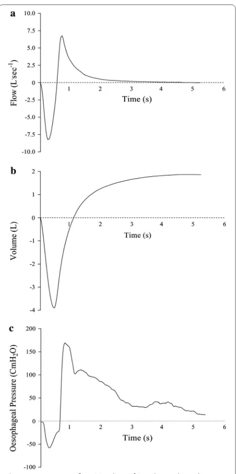
the oesophageal pressure swing (ΔPes) during spirometry was 149 ± 40 cmH2O. Representative data for low,
vol-ume and oesophageal pressure during an FVC manoeu-vre are shown in Fig. 1.
Values are mean ± SD for eight participants. FVC, forced vital capacity; FEV1, forced expiratory volume in
one second; Pesinsp, peak oesophageal pressure recorded
during inspiratory phase of FVC; Pesexp, peak
oesoph-ageal pressure recorded during expiratory phase of
[image:3.595.306.539.84.552.2]FVC; ΔPes, oesophageal pressure swing (peak-to-peak) recorded during FVC manoeuvre; PImax, maximum static inspiratory oesophageal pressure; PEmax, maximum static expiratory oesophageal pressure. Predicted values and z scores for pulmonary volumes and lows are from Quan-jer et al. [6].
Table 1 Baseline (resting) pulmonary function in eight healthy, recreationally-active men
Absolute Relative
FVC, L 5.71 ± 0.51 102 ± 6 %Pred. FEV1, L 4.45 ± 0.47 95 ± 8 %Pred. FEV1/FVC, % 78 ± 5 − 1 ± 0.6 z-score Pesinsp, cmH2O − 47 ± 9 37 ± 10 %PImax Pesexp, cmH2O 102 ± 34 75 ± 17 %PEmax ∆Pes, cmH2O 149 ± 40 – ± –
Fig. 1 Representative flow (a), volume (b), and oesophageal pressure (c) traces from a single subject performing an FVC manoeuvre. Peak flow = 6.78 L s−1; volume
[image:3.595.56.290.631.724.2]Discussion
he aim of this report was to characterise the intra-thoracic pressure-response to spirometry performed by healthy men. Our main indings were that an FVC manoeuvre resulted in a peak inspiratory oesophageal pressure of − 47 cmH2O (37% PImax), and a peak
expira-tory oesophageal pressure of 102 cmH2O (75% PEmax).
hese large intra-thoracic pressures may have implica-tions for respiratory health in vulnerable patients.
A forced vital capacity manoeuvre can be split into two distinct phases: (i) inspiration to total lung capacity; (ii) forced expiration to residual volume. he typical, healthy response to deep inspiration is bronchodilation [7]. In mild-to-moderate asthma, however, bronchodilation following a deep inspiration is inhibited, and in severe asthma, a deep inspiration may induce bronchoconstric-tion [8]. It has been suggested that spirometry-induced bronchoconstriction is caused, at least in part, by an increase in airway wall oedema, secondary to an increase in intra-thoracic pressure across the airway capillaries [8]. Moreover, cardiac complications, including myo-cardial infarction, aortic aneurysm, hypertension and angina, are among the most common contraindications for lung function testing [9], and may be caused by large changes in intra-thoracic pressures during spirometry, and a subsequently elevated blood pressure [9]. Indeed, arrhythmia during spirometry was observed in 10% of patients referred for cardio-pulmonary exercise testing; notably the authors report the onset of arrhythmia dur-ing the inspiratory phase of the manoeuvre [3].
The large positive intra-thoracic pressures we observed during forced expiration may contribute to chronic deleterious consequences in susceptible individuals. Spirometry has been proposed to induce gastro-oesophageal reflux in approximately half of individuals referred for outpatient gastro-oesopha-geal reflux assessment [4]. While the exact mecha-nism of spirometry-induced gastro-oesophageal reflex is unknown, it is likely attributable to an increased intra-abdominal pressure, resulting in upward vec-torial forces on gastric contents. Moreover, during activities that increase intra-abdominal pressure (e.g., deep inspiration, forced expiration, trunk flexion), the right crus of the diaphragm contracts to increase pres-sure on the lower oesophageal sphincter, thereby pre-venting gastric-oesophageal reflux [10]. As such, it is possible that reflux during forced expiration may be symptomatic of diaphragm weakness.
Limitations
here are two limitations that should be considered when interpreting the data presented in this study. First, data were collected in a healthy cohort; i.e., participants free
from cardiorespiratory disease, and the intra-thoracic pressures exhibited may not be representative of a clini-cal population. Further studies are needed to elucidate the typical response in, for example, chronic obstruc-tive pulmonary disease (COPD) and asthma. Second, we recorded intra-thoracic pressures using oesophageal bal-loon-tipped catheters. While balloon catheters are widely used and exhibit excellent reliability, other common tech-niques involve multi-pair oesophageal electrode cath-eters, or pneumotachographs for the measurement of mouth-pressure. here is a lack of consistency in the lit-erature with respect to the technique used; consequently, we urge caution when comparing among studies.
To conclude, this is the irst report to characterise the intra-thoracic pressure-response to spirometry. We observed near maximal oesophageal pressures during expiration, and large peak-to-peak oesophageal pres-sure swings during an FVC manoeuvre which may part-explain some of the deleterious efects of pulmonary function testing. Future studies should aim to clarify cau-sation, and comment on the mechanistic basis.
Abbreviations
FVC: forced vital capacity; TLC: total lung capacity; RV: residual volume; FEV1: forced expiratory volume in one second; Pes: oesophageal pressure; ∆Pes: oesophageal pressure swing (peak-to-peak) recorded during FVC manoeuvre; Pesinsp: peak oesophageal pressure recorded during inspiratory phase of FVC; Pesexp: peak oesophageal pressure recorded during expiratory phase of FVC; PImax: maximum static inspiratory oesophageal pressure; PEmax: maximum static expiratory oesophageal pressure; COPD: chronic obstructive pulmonary disease.
Authors’ contributions
Conception and design of research (NBT, AJS); performed experiments (NBT); analyzed data (NBT); interpreted results of experiments (NBT, AJS); prepared figures (NBT); drafted manuscript (NBT, AJS); edited and revised manuscript (NBT, AJS); approved final version of manuscript (NBT, AJS). Both authors read and approved the final maunscript.
Author details
1 Academy of Sport and Physical Activity, Sheffield Hallam University, Shef-field S10 2BP, UK. 2 Centre for Human Performance, Exercise and Rehabilitation, Brunel University, London, UK. 3 Department of Sport, Health and Exercise Science, University of Hull, Hull, UK.
Acknowledgements Not applicable.
Competing interests
The authors declare that they have no competing interests.
Availability of data and materials
The datasets used and/or analysed during the current study are available from the corresponding author on reasonable request.
Consent for publication Not applicable.
Ethics approval and consent to participate
Page 4 of 4 Tiller and Simpson BMC Res Notes (2018) 11:110
• We accept pre-submission inquiries
• Our selector tool helps you to find the most relevant journal
• We provide round the clock customer support • Convenient online submission
• Thorough peer review
• Inclusion in PubMed and all major indexing services • Maximum visibility for your research
Submit your manuscript at www.biomedcentral.com/submit
Submit your next manuscript to BioMed Central
and we will help you at every step:
Funding Not applicable.
Publisher’s Note
Springer Nature remains neutral with regard to jurisdictional claims in pub-lished maps and institutional affiliations.
Received: 15 January 2018 Accepted: 1 February 2018
References
1. Miller MR, Crapo RO, Hankinson JL, et al. General considerations for lung function testing. Eur Respir J. 2005;26:153–61.
2. Mackay AD, Mustchin CP, Sterling GM. The response of asthmatic patients and normal subjects to maximum respiratory manoeuvres. Spirometry— induced bronchoconstriction. Eur J Respir Dis Suppl. 1980;106:35–40.
3. Araújo CG, Vianna LC. How often does spirometry testing induce cardiac arrhythmias? Prim Care Respir J. 2009;18:185–8.
4. Teo MY, Zhou J, Ho V, Brannan J. Gastro-oesophageal reflux (GOR) during spirometry: prevalence and effects. Eur Respir J. 2017;50:PA2499. 5. Miller MR, Hankinson JL, Brusasco V, et al. Standardisation of spirometry.
Eur Respir J. 2005;26:319–38.
6. Quanjer PH, Stanojevic S, Cole TJ, et al. Multi-ethnic reference values for spirometry for the 3–95-year age range: the global lung function 2012 equations. Eur Respir J. 2012;40:1324–43.
7. Pliss LB, Ingenito EP, Ingram RH. Responsiveness, inflammation, and effects of deep breaths on obstruction in mild asthma. J Appl Physiol. 1989;66:2298–304.
8. Burns GP, Gibson GJ. A novel hypothesis to explain the bronchconstrictor effect of deep inspiration in asthma. Thorax. 2002;57:116–9.
9. Cooper BG. An update on contraindications for lung function testing. Thorax. 2011;66:714–23.