ORIGINAL INVESTIGATION
Canagliflozin attenuates the progression
of atherosclerosis and inflammation process
in APOE knockout mice
Νarjes Nasiri‑Ansari
1†, Georgios K. Dimitriadis
2,3,4†, Georgios Agrogiannis
5, Despoina Perrea
6,
Ioannis D. Kostakis
7, Gregory Kaltsas
4, Athanasios G. Papavassiliou
1, Harpal S. Randeva
2,4,8*‡and Eva Kassi
1,9*‡Abstract
Background: Sodium glucose co‑transporter2 inhibitors reduce the incidence of cardiovascular events in patients with type 2 diabetes mellitus based on the results of recent cardiovascular outcome studies. Herein, we investigated the effects of long‑term treatment with canagliflozin on biochemical and immunohistochemical markers related to atherosclerosis and atherosclerosis development in the aorta of apolipoprotein E knockout (Apo‑E(−/−)) mice.
Methods: At the age of 5 weeks, mice were switched from normal to a high‑fat diet. After 5 weeks, Apo‑E(−/−) mice were divided into control‑group (6 mice) treated with 0.5% hydroxypropyl methylcellulose and Cana‑group (7 mice) treated with canagliflozin (10 mg/kg per day) per os. After 5 weeks of intervention, animals were sacrificed, and heart and aorta were removed. Sections stained with hematoxylin–eosin (H&E) were used for histomorphometry whereas Masson’s stained tissues were used to quantify the collagen content. Immunohistochemistry to assess MCP‑1, CD68, a‑smooth muscle actin, MMP‑2, MMP‑9, TIMP‑1 and TIMP‑2 expression was carried out and q‑PCR experiments were performed to quantify mRNA expression.
Results: Canagliflozin‑group mice had lower total‑cholesterol, triglycerides and glucose levels (P < 0.01), while heart rate was significantly lower (P < 0.05). Histomorphometry revealed that one in seven Cana‑group mice versus four in six control mice developed atheromatosis, while aortic root plaque was significantly less, and collagen was 1.6 times more intense in canagliflozin‑group suggesting increased plaque stability. Immunohistochemistry revealed that MCP‑1 was significantly less expressed (P < 0.05) in the aortic root of canagliflozin‑group while reduced expression of a‑actin and CD68 was not reaching significance (P = 0.15). VCAM‑1 and MCP‑1 mRNA levels were lower (P = 0.02 and
P= 0.07, respectively), while TIMP‑1/MMP‑2 ratio expression was higher in canagliflozin‑group approaching statistical significance (P = 0.07).
Conclusions: Canagliflozin attenuates the progression of atherosclerosis, reducing (1) hyperlipidemia and hyper‑ glycemia, and (2) inflammatory process, by lowering the expression of inflammatory molecules such as MCP‑1 and VCAM‑1. Moreover, canagliflozin was found to increase the atherosclerotic plaque stability via increasing TIMP‑1/ MMP‑2 ratio expression.
Keywords: Canagliflozin, SGLT2i, Atherosclerosis, Inflammation, APOE knockout mice
© The Author(s) 2018. This article is distributed under the terms of the Creative Commons Attribution 4.0 International License (http://creat iveco mmons .org/licen ses/by/4.0/), which permits unrestricted use, distribution, and reproduction in any medium, provided you give appropriate credit to the original author(s) and the source, provide a link to the Creative Commons license,
and indicate if changes were made. The Creative Commons Public Domain Dedication waiver (http://creat iveco mmons .org/
publi cdoma in/zero/1.0/) applies to the data made available in this article, unless otherwise stated.
Open Access
*Correspondence: harpal.randeva@warwick.ac.uk; ekassi@med.uoa.gr
†Νarjes Nasiri‑Ansari and Georgios K. Dimitriadis are joint first authors ‡Harpal S. Randeva and Eva Kassi are joint last authors
1 Department of Biological Chemistry, National and Kapodistrian
University of Athens Medical School, Athens, Greece
2 Division of Translational and Experimental Medicine‑Metabolic
and Vascular Health, Warwick Medical School, University of Warwick, Coventry CV4 7AL, UK
Background
According to data from the World Health Organization (WHO), over 3 million people die worldwide from dia-betes and its related complications every year, mainly due to cardiovascular disease (CVD) [1]. Despite paucity of information regarding the aetiopathogenesis of T2DM related cardiovascular complications, the toxicity of high blood glucose to the endothelium and other cells of the vessels seem to play an important role in the development of atherosclerosis and subsequent CVD. Atherosclerosis represents a systemic inflammatory process which impli-cates both cells of immune system and those of vessel wall. The basic pathologic lesion is atheromatous plaque. The atherogenic process evolves in different stages, start-ing from the endothelium activation/dysfunction and resulting in plaque vulnerability and rupture [2]. At the earlier stages of the atheromatous process, endothelial dysfunction/activation is characterized among others by increased expression of adhesion molecules and inflam-matory molecules such as VCAM-1, ICAM-1 and MCP-1 and IL-6 by the endothelial and vascular smooth mus-cle cells. During the later stages of plaque rupture and/ or erosion, among other factors, the metalloproteinases MMP-2, MMP-9 as well as their inhibitors TIMP-1 and TIMP-2; both expressed in endothelial cells and vascu-lar smooth muscle cells, seem to play a critical role, since they regulate the collagen degradation of the extracellular matrix (ECM) [3]. Monocyte chemoattractant protein-1 (MCP-1) has been postulated to be a direct mediator of plaque instability [4].
SGLT2 inhibitors (SGLT2i) are a new class of oral anti-diabetic drugs, targeting the sodium-glucose co-transporter 2 which is the main glucose co-transporter of the kidney, and is responsible for reabsorption of 90% of glucose from primary urine. SGLT2 inhibition reduces the reabsorption of glucose and therefore enhances uri-nary glucose excretion, consequently decreasing both fasting and postprandial hyperglycemia and preventing glucotoxicity, and consequently hyperglycemia-induced damage. However, pleiotropic effects of these agents have already emerged [5].
Recent clinical trials amongst them CVD-REAL Nordi, EMPA-REG OUTCOME and canagliflozin CANVAS program have shown that SGLT2 inhibitors (dapagliflo-zin, empagliflo(dapagliflo-zin, canagliflozin) use is associated with reduced cardiovascular disease and cardiovascular mor-tality compared with use of other glucose-lowering drugs in patients with T2DM, even though hemoglobin A1c (HbA1c) difference between randomized groups was marginal [6–8]. This suggests direct beneficial effects of SGLT2i on CVD risk besides the indirect effects attrib-uted to better glycemic control, blood pressure or actions on extra-cardiovascular tissues such as adipose tissue.
Interestingly, recent studies have shown that SGLT-2 inhibitors can reduce pro-inflammatory IL-6, MCP-1 and ICAM-1 expression in blood vessels of rodent dia-betic models, yet the molecular mechanisms remain largely unknown. To this direction, Mancini and col-laborators reported recently that canagliflozin, but not empagliflozin or dapagliflozin can activate AMPK and inhibit IL-1β-stimulated secretion of IL-6 and mono-cyte chemoattractant protein-1 (MCP-1) in cultured human endothelial cells, while AMPK-independent mechanisms were also recognized [9]. Another research group has currently investigated the anti-inflammatory effects of SGLT-2 inhibitors in immune cells such as macrophages/monocytes both involved in the athero-genic process. According to their results, canagliflo-zin at clinically-relevant concentrations exerted the most potent inhibition-compared to other SGLT-2 inhibitors-of production and release of inflammatory factors IL1a, IL-6 and TNF-α [10]. These effects medi-ated by inhibiting intracellular glycolysis, enhancing autophagy, and promoting p62-mediated IL-1 degrada-tion. Of note, enhanced autophagy and p62 levels might be mediated by increasing AMPK and NFκB activities, respectively; whether the above anti-inflammatory effects of canagliflozin were associated with SGLT2 should be further investigated [10].
Paradoxically, today there are more clinical than experimental data regarding the beneficial effects of SGLT2i on CVD, evaluating endothelial function, arte-rial stiffness, atherogenic cholesterols in patients with DM type 2 etc. [11–13]; however, although various SGLT2i such as empagliflozin, dapagliflozin, ipragliflo-zin and luseoglifloipragliflo-zin have been evaluated in animal experimental studies regarding their the anti-ather-ogenic effects, there is paucity of evidence regarding canagliflozin.
To this context, we investigated for the first time the effects of long-term treatment with canagliflozin on atherosclerosis development in the aorta of APOE(−/−)
mice as well as on biochemical and immunohistochem-ical markers linked to atherosclerosis.
Materials and methods Animals
APOE(−/−) mice (on the genetic background C57BL/6)
Experimental protocols
Thirteen male APOE−/− mice were kept on a standard
rodent chow. At the age of 5 weeks mice were switched to high fat diet (20–23% by weight; 40–45% kcal from fat) containing cholesterol (0.2% total).
After 5 weeks, mice were randomly divided into two groups (1) canagliflozin-group 10 mg/kg/day (n = 7) administered orally by gavage, and (2) control-group (n = 6) administered the same volume of 0.5% hydroxy-propyl methylcellulose/day (vehicle), via gavage. After 5 weeks of oral treatment with canagliflozin or vehicle, mice were sacrificed under isoflurane anesthesia by tran-section of the diaphragm and, the aorta along with heart were rapidly excised. Food intake and body weight were measured once weekly over a period of 10 weeks. Blood glucose levels were also measured after 8–10 h fast via tail puncture at baseline, before canagliflozin/vehicle oral administration, once during experiment (3 week) and before experiment endpoint. Canagliflozin was pur-chased from Selleck Chemand dissolved in 0.5% hydroxy-propyl methylcellulose.
Mice blood pressure measurement
Blood pressure was measured once at baseline, before canagliflozin oral administration began and once before sacrificing animals. Mice were acclimatized to the restrainer on a warming pad for 2 consecutive days before final measurements. Measurements were per-formed in quiet environment to avoid causing mice anxiety. Blood pressure measurements were performed (15–25 measurements/mouse) using a computerized non-invasive tail-cuff system (CODAs, Kent Scientific, USA). All measurements are expressed as mean value of heart rate, systolic and diastolic blood pressure.
Serum analysis of biochemical parameters
Blood was drawn once before the onset of canagliflozin administration from the facial vein and once by heart
puncturing after sacrificing mice. Serum glucose, choles-terol, triglycerides, and HDL- and LDL-cholesterol levels were determined using a dedicated autoanalyzer.
RNA isolation and real time PCR
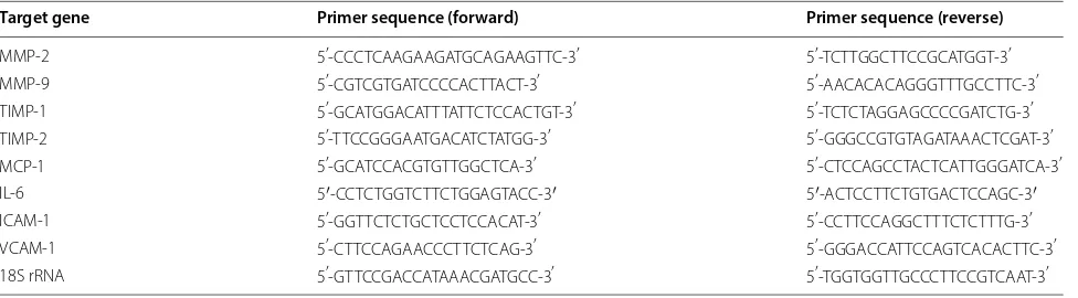
Total RNA was extracted from fresh frozen aorta using RNeasy kit (Qiagen). Extracted mRNA was then reverse transcribed into cDNA using the iScript cDNA synthesis kit (Bio-Rad). Real-time PCR analysis was performed as described previously [14]. The expression of Matrix Met-alloproteinase (MMP-2 and MMP-9) and their inhibi-tors (TIMP-1 and TIMP-2), IL-6, intercellular adhesion molecule 1 (ICAM-1), vascular cell adhesion molecule 1 (VCAM-1) and monocyte chemotaxis protein (MCP-1) was measured using Luna® Universal qPCR Master Mix (New England Biolabs) on a CFX96 (Bio-RAD). The sequences of primers used for RT-PCR analysis in this study are listed in Table 1. A melting curve analysis was performed to confirm the specificity of qPCR products. Fold-changes were calculated using the 2−∆∆Ct method
and were normalized against 18s rRNA expression. All reactions were performed in triplicates and repeated three times.
Histochemistry and immunohistochemistry
Quantification of atherosclerotic lesion area
Aortic tissues were fixed and embedded in paraffin. The 4-μm-thick sections were stained with hematoxy-lin–eosin (H&E) and used for histopathological analysis whereas Masson’s trichrome stained sections were used to quantify tissue section’s collagen content. The degree of pathological changes was evaluated microscopically by measuring the area of atheromatous plaques. Results are reported as the percentage of the neointima area con-taining the lesion. Threshold was set and the positively stained area for each histochemical stain was automati-cally calculated and then the percent of the positively stained area to the total cross-sectional vessel wall area
Table 1 qPCR primer sequences used in this study
Target gene Primer sequence (forward) Primer sequence (reverse)
MMP‑2 5′‑CCC TCA AGA AGA TGC AGA AGTTC‑3′ 5′‑TCT TGG CTT CCG CAT GGT ‑3′
MMP‑9 5′‑CGT CGT GAT CCC CAC TTA CT‑3′ 5′‑AAC ACA CAG GGT TTG CCT TC‑3′
TIMP‑1 5′‑GCA TGG ACA TTT ATT CTC CAC TGT ‑3′ 5′‑TCT CTA GGA GCC CCG ATC TG‑3′
TIMP‑2 5′‑TTC CGG GAA TGA CAT CTA TGG‑3′ 5′‑GGG CCG TGT AGA TAA ACT CGAT‑3′
MCP‑1 5′‑GCA TCC ACG TGT TGG CTC A‑3′ 5′‑CTC CAG CCT ACT CAT TGG GATCA‑3′
IL‑6 5′‑CCT CTG GTC TTC TGG AGT ACC‑3′ 5′‑ACT CCT TCT GTG ACT CCA GC‑3′
ICAM‑1 5′‑GGT TCT CTG CTC CTC CAC AT‑3′ 5′‑CCT TCC AGG CTT TCT CTT TG‑3′
VCAM‑1 5′‑CTT CCA GAA CCC TTC TCA G‑3′ 5′‑GGG ACC ATT CCA GTC ACA CTTC‑3′
[image:3.595.57.545.590.725.2]or intimal plaque lesion area was reported. Plaque area analysis was carried out using Image Pro Plus software version 5.1 (Media Cybernetics, Inc.).
Immunohistochemistry
For immunohistochemistry, all sections were deparaffi-nized at 60 °C. Antigen retrieval was performed using citrate buffer (PH.6.0) for 7 min at 100 °C followed by blocking with normal goat serum (CST, 5425S) for 1 h. Slides were then incubated with appropriate concentra-tion of primary antibodies against CD68 (ZYTOMED, MSK055), α-smooth muscle actin (ZYTOMED, MSK030), MCP-1 (ACRIS, AM32136PU-N), MMP-2 (Proteintech Group, 103732-AP), MMP-9 (Proteintech Group, 10375-2-AP) and their inhibitors TIMP-1 (Santa Cruz Biotechnology, sc-21734) and TIMP-2 (Santa Cruz Biotechnology, sc-21735) followed by incubation with corresponding secondary antibody conjugated to horse-radish peroxidase (ZYTOMED, ZUC053-100) and visu-alized by applying DAB (CST.8059P). All slides were counterstained with hematoxylin and integral absorbance was examined under light microscope and results were quantified using Image Pro Plus software version 5.1 (Media Cybernetics, Inc.). A positive tissue control was used to ensure the specificity of antibodies used in this study.
Statistical analysis
Normality of quantitative data distribution was assessed using Shapiro–Wilk test. Student’s t-test, Welch’s t-test or Mann–Whitney U test were used for comparisons between two groups with quantitative data as appro-priate. Chi square test or Fisher’s exact test were used for comparisons among groups with qualitative data as appropriate. Correlations between quantitative param-eters were tested with Pearson’s correlation coefficient or Spearman’s rank correlation coefficient as appropri-ate. All tests were two-tailed, and results were considered statistically significant if the P-value was < 0.05. Statistical analysis was performed using the 23rd edition of statisti-cal package for social sciences (SPSS) (IBM Corporation, Armonk, NY, USA).
Results
Oral administration of canagliflozin for 5 weeks improved heart rate and biochemical/metabolic parameters associated with atherosclerosis
No significant difference in daily food intake was observed between the two groups. Nevertheless, body weight was significantly increased in both groups fol-lowing HFD and 5 weeks of oral canagliflozin/vehicle administration as compared to the value measured at experiment baseline. No significant difference in weight
gain was observed between canagliflozin and control group (Additional file 1: Figure S1).
Fasting blood glucose (8 h of fasting) and serum lipid levels were measured before canagliflozin/vehicle oral administration as well as at the end of intervention period. A significant reduction in glucose, total choles-terol, LDL-cholesterol and triglyceride levels (P < 0.01) were observed in canagliflozin group (Fig. 1). After treat-ment with canagliflozin, glucose levels returned to nor-mal range, contrary to placebo group where glucose increased significantly above normal range with glucose level progression to diabetes range (P < 0.001).
At the end of intervention period, total cholesterol, glucose and triglyceride levels were significantly lower in Cana-group (P = 0.01, P = 0.001, P = 0.02 respectively). Moreover, diastolic blood pressure values was signifi-cantly higher in the control group at experiment end-point (P = 0.05) (Additional file 2: Table S1).
At the end of canagliflozin/placebo oral treatment, there was a significant difference from baseline in fasting glucose (P < 0.001), triglycerides (P < 0.01), and total cho-lesterol (P < 0.05) between groups. Mean ± SD changes in LDL-, HDL-cholesterol and creatinine levels from base-line were similar in both groups (Fig. 2).
Canagliflozin significantly reduced heart rate (**P ≤0.01) (Fig. 3), whereas no significant change was observed in the control group (Fig. 3a). This finding was confirmed by comparing heart rate changes from base-line (value measured before onset of canagliflozin/vehicle oral administration) between the two groups. (*P ≤ 0.05) (Fig. 3b). As it is shown in Additional file 2: Table S1 post-treatment heart rate was reduced in Cana-group compared to control-group approaching statistical sig-nificance (P = 0.076).
Canagliflozin reduces atherosclerosis lesion formation and increases collagen content
0 500 1000 1500 2000 2500 3000 3500 4000
Before After Before After Total Blood Cholesterol
Mg/d
l
Cana-Group Control-Group
0 50 100 150 200 250 300 350 400
Before After Before After Serum Triglycerides
Cana-Group Control-Group
** **
0 500 1000 1500 2000 2500 3000 3500 4000
Before After Before After
**
Cana-Group Control-Group
Mg/d
l
Serum LDL-Cholesterol
0 50 100 150 200 250 300 350 400 450 500
Before After Before After
Mg/d
lM
g/dl
Fasng Blood Glucose
Cana-Group Control-Group
**
**
Fig. 1 Serum lipid and fasting blood glucose levels in Cana‑ and control‑groups after 5 weeks of canagliflozin/vehicle oral administration. Significant reduction in total cholesterol, triglyceride, LDL‑cholesterol and fasting blood glucose levels was observed in Cana‑group at the end of experimental procedure compared to baseline. Fasting glucose was the only significantly increased parameter observed in the control group at the end of intervention. Data are shown as mean ± SD (***P ≤ 0.001, **P ≤ 0.01)
-100 -50 0 50 100 150 200
Control Group Cana-Group
Fasng Blood Glucose changes from baseline
***
Mg/dl
-1200 -1000 -800 -600 -400 -200 0 200 400 600
Control Group Cana-Group
Mg/dl
Total Serum Cholesterol change From baseline
*
-200 -150 -100 -50 0 50 100
Control Group Cana-Group
Total Serum Cholesterol change From baseline
**
Mg/dl
[image:5.595.55.542.87.379.2] [image:5.595.56.539.436.704.2]Canagliflozin reduced the expression of inflammatory molecules and improved metalloproteinase profile
To evaluate the effect of canagliflozin treatment on the expression of inflammatory (IL-6, MCP-1), adhe-sion molecules (ICAM-1, V-CAM-1), matrix metal-loproteinases (MMP-2, MMP-9) and their inhibitors (TIMP-1, TIMP-2), total RNAs were isolated from the thoracic aorta and analyzed using real-time quantita-tive RT-PCR.
We demonstrate that oral canagliflozin administration significantly reduces VCAM-1 mRNA levels (P = 0.01) while marginally induces TIMP-1 and decreases MCP-1 mRNA expression levels (P = 0.07). Canagliflozin treat-ment causes no significant alteration in IL-6, ICAM-1, MMP-2, MMP-9 and TIMP-2 mRNA levels compared to controls (Fig. 5a). A balance between MMPs and TIMPs is known as an indicator of MMPS overall collagenolytic activity. To this end, TIMP-1/MMP-2 ratio mRNA levels
0 200 400 600 800 1000 1200 1400
Before Aer Before Aer
**
Control-Group Cana-Group
Heart Rate
Beats
per
mi
nute
-250 -200 -150 -100 -50 0 50 100 150 200 250
Control Group Cana-Group
Beats
per
mi
nute
Heart rate change From baseline
*
a b
Fig. 3 Canagliflozin/vehicle oral administration effect on heart rate of APOE(−/−) mice. a 5 weeks of canagliflozin intervention led to significant
reduction of heart rate (**P < 0.01) while no significant difference was observed in the control group. b Heart rate changes from baseline were significantly different between Cana‑ and control‑groups (*P < 0.05)
Fig. 4 Atherosclerotic plaque extension among APOE(−/−) mice on western diet treated with canagliflozin (Cana‑group) or vehicle (control‑group).
a Selected 4 μm section images from the aortic root stained with H&E and Masson trichrome. Formation of atherosclerotic plaque was examined using H&E staining while histological examination of atherosclerotic plaque collagen content was assessed using Masson trichrome staining. b Quantification of plaque area is shown as a percentage of lumina stenosis by thickened intima. Collagen content was measured using quantification of Masson trichrome positive area over complete plaque area. Values are shown as mean ± SD and *P < 0.05. Original magnification
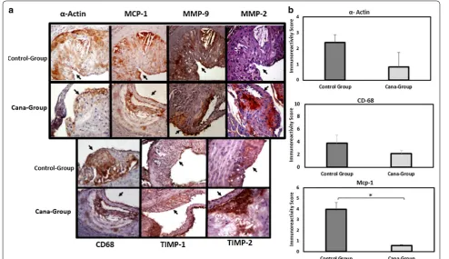
[image:6.595.60.539.88.231.2] [image:6.595.62.539.296.521.2]was measured. Our findings demonstrate that TIMP-1/ MMP-2 ratio mRNA level was higher in Cana-group (Fig. 5b), while approaching significance (P = 0.07). Aor-tic root section immunohistochemistry revealed that smooth muscle (α-actin) and macrophage (CD68) cells content from atherosclerotic plaques were marginally higher in the control group (P < 0.1). Moreover, treatment with canagliflozin led to significant reduction in MCP-1 expression (P < 0.05) and a marginal increase in athero-sclerotic plaque TIMP-1 expression (P < 0.1). Images and quantitative data are shown in Fig. 6.
Discussion
Canagliflozin is an intermediate-acting SGLT2i with proven clinical efficacy regarding glycemic control, blood pressure and weight reduction, in patients with T2DM irrespective of the degree of CVD history or risk factors [15]. In the present study, we investigated the effect of canagliflozin on atherosclerosis formation and demon-strated for the first time, that 5 week canagliflozin admin-istration attenuates atheromatous process in APOE(−/−)
mice fed an atherogenic diet for 10 weeks enough for ath-eroma to be formed [16].
We evaluated the impact of treatment with canagliflo-zin in established atherosclerosis risk factors and were able to demonstrate the anticipated effect on fasting glucose level. In fact, the control-group fed with west-ern diet for 10 weeks, had significantly increased fasting
blood glucose level while canagliflozin administration reversed this effect. In contrast, Terasaki et al. [17] found that western diet fed mice for 4 weeks retained their glu-cose levels in the normal range, an effect that is probably attributed to strain background differences [18]. It should be noted that the majority of available animal data on glucose-lowering effects of SGLT2 inhibitors have used models of streptozotocin-induced diabetic models.
Canagliflozin administration combined with athero-genic diet, did not lead to reduced weight. On the con-trary, there was a statistically significant increase, without differences between the two groups either in weight or in daily food intake. Although weight-loss effects of cana-gliflozin have been demonstrated in clinic trials, with reduction in both subcutaneous and visceral adipose tissue dose-dependently [19, 20], animal studies have yielded conflicting results. Administration of canagliflo-zin 30 mg/kg/day for 4 weeks reduced the weight gain in diet-induced obese mice fed with high-fat diet [21]. In a recent study by Ji et al. [22] administration of 60 mg/ kg/day canagliflozin for 4 weeks in mice fed with high-fat diet containing 6% high-fat, reduced significantly the body weight, via reduction of PPRAγ in the liver; interestingly, in line with our results, this effect was not achieved with the lower dose of 15 mg/kg/day. Naznin et al. [23] found that canagliflozin at dose of 30 mg/day attenuated the body weight gain by promoting caloric loss and suppres-sion of obesity-related inflammation in both the nervous
[image:7.595.60.539.87.313.2]0.0 0.5 1.0 1.5 2.0 Control-Group Cana-Group 0.0 0.5 1.0 1.5 2.0 Control-Group Cana-Group VCAM-1 ** ot der ap mo cs asl ev el A NR m evi tal eR an l ort no cl anr et ni Relave mRNA levels as compared to an internal control MCP-1 0 2 4 6 8 10 Control-Group Cana-Group Relave mRNA levels as compared to an internal control TIMP-1 a b 0 1 2 3 4 5 Control-Group Cana-Group ot der ap mo cs asl ev el A NR m evi tal eR an internal control TIMP-2/MMP-9 0 1 2 3 4 Control-Group Cana-Group Relave mRNA levels as compared to an internal control TIMP-1/MMP-2
system and skeletal muscle. Regarding the effect of other SGLT2 is on body weight, administration of dapagliflozin for 12 weeks in non-diabetic APOE(−/−) mice did not
sig-nificantly reduce weight [24]. In another study, dapagli-flozin did not change the weight of APOE(−/−) mice, with
or without diabetes [17] while ipragliflozin 1 mg/kg/day for 4 weeks, reduced significantly the weight of db/db rats [17]. Empagliflozin for 8 weeks decreased body weight and particularly adipose tissue in APOE(−/−) mice, while
did not affect the weight of ZDF rats (type 2 diabetes model) [25]. Among the involved mechanisms, decrease of the subcutaneous fat mass and the size of visceral adi-pocytes as well as enhanced fat utilization and browning, attenuation of obesity-induced inflammation and insu-lin resistance by polarizing M2 macrophages in WAT and liver have been described [26, 27]. Although, the daily food intake and body weight changes did not differ between our two groups, canagliflozin exerted beneficial effects on lipid profile, reducing total cholesterol and tri-glycerides. As with body weight, there are also inconsist-ent results concerning the effects of SGLT-2 inhibitors on
lipids, either reducing or not affecting total cholesterol, LDL-cholesterol and triglycerides, however data exam-ining the effects specifically of this dose of canagliflozin on lipid profile have not been assessed previously [22, 24,
25, 28–30]. Conflicting results concerning the effects of SGLT-2i on body weight and lipid profile may be due to differences in animal models used (i.e. mice, rats, dia-betic, non-diadia-betic, atherosclerotic, obese etc.), different drugs used (including different doses and durations of treatment), and/or different diets.
Although clinical studies have proved the antihyper-tensive effects (SBP, DBP, pulse pressure and mean arte-rial pressure) of canagliflozin [12], herein we find just a borderline reduction of diastolic pressure. However, canagliflozin reduces significantly heart rate. This could be attributed to a possible decrease in insulin levels as a result of the glucose-lowering effects of canagliflozin since it is well known that insulin increases sympathetic activity [31]. Although we did not measure insulin lev-els, reduction in serum insulin has been reported fol-lowing administration of empagliflozin for 7 weeks in a
Fig. 6 Atherosclerotic lesion characteristics in APOE(−/−) mice fed with HFD and treated with canagliflozin (Cana‑group) or vehicle (control‑group).
[image:8.595.54.549.88.373.2]metabolic syndrome model rat [26]. It should be men-tioned that Terasaki et al. [17], showed no differences in heart rate following administration of dapagliflozin for 4 weeks, however their APOE(−/−) mouse model feeding
western diet did not increase blood glucose levels, thus there were no changes in blood glucose with dapagliflo-zin administration.
It is well established that inflammatory cytokines and adhesion molecules play a crucial role in the initiation and progression of atherosclerotic process. Since we found that the majority of the mice in Cana-group did not form atherosclerotic plaque in contrast to control-group, we investigated the expression of ICAM-1, VCAM-1, IL-6 and MCP-1 between groups and found lower mRNA expression of MCP-1 and VCAM-1. A study by Oelze et al. [32] showed that empagliflozin for 6 weeks decreased the expression of IL-6 and MCP-1. Moreover, luseogliflozin also reduced the expression of ICAM-1 and IL-6 while did not affect VCAM-1 [30]. In both studies, streptozotocin-induced diabetes models were used.
Interestingly, we additionally confirmed the beneficial effect of canagliflozin on MCP-1 expression at the pro-tein level, in atherosclerotic lesion (plaque). The role of MCP-1 in both initiation and progression of atheroscle-rosis has been well-characterized and various mecha-nisms have been proposed for this [33]. A potential mechanism is by promoting the recruitment of mono-cytes/macrophages in atherosclerotic lesion. In our study, the decreased number of stained macrophages in the plaque could be attributed to, among others, decreased MCP-1 expression. Moreover, it has been reported that MCP-1 induces MMP-2 expression in human endothe-lial cells, as well as the expression of MMP-9 in human smooth muscle cells [34, 35]. Both MMPs are critical fac-tors involved in plaque destabilization, through degrada-tion of collagen-rich extracellular matrix.
According to our data, although canagliflozin admin-istration reduced MCP-1 expression, it did not change the MMP-2, MMP-9, while marginally increased TIMP-1 and TIMP-1/MMP-2 ratio, indicating reduced activity of MMP-2.
Previous studies have shown that high glucose concen-trations decrease the expression of MMPs and increase the expression of their inhibitors (TIMPs) [36]. In our study, by reducing blood glucose at normal levels, cana-gliflozin would be expected to lead to the above-men-tioned profile (increased MMP-2 and MMP-9 expression and decreased TIMP-1 and TIMP-2). However, acting via decreasing either directly, or indirectly-through lower-ing blood glucose [37], MCP-1, not only counterbalances but rather is associated with a favorable TIMP-1/MMP-2 profile in the aortic lesion. Another potential mechanism that could explain this TIMP-1/MMP-2 profile is the
reduction of VCAM-1 by canagliflozin which has been demonstrated to lead to an up-regulation of TIMP-1 [38]. This favorable profile could result, along with other factors, in increased collagen content demonstrated in the plaque of the Cana-group. Of note, another SGLT-2 inhibitor, luseogliflozin, given for 1 week decreased MMP-2 and MMP-9 expression in aorta wall but not in atherosclerotic plaque of streptozotocin-induced diabetic APOE(−/−) mice [30].
Attenuation of plaque formation and decreased num-ber of invasive macrophages has been demonstrated following dapagliflozin administration in streptozo-tocin-induced APOE(−/−) mice, while no effect has been
reported in non-diabetic APOE(−/−) mice [17]. Moreover,
empagliflozin administration for 8 weeks decreased the burden of plaque (plaque area), expression of inflamma-tory molecules TNF, IL-6 and MCP-1, and invasion of plaque by inflammatory cells and this effect was more potent in the empagliflozin mice group compared to glimepiride-group that achieved the same glycemic con-trol, the latter suggesting beneficial effects of the SGLT-2i other than just improved glycemic control [25].
of biochemical parameters, gave us the chance to com-pare their changes and not only the values at the end of intervention, between the two groups.
Limitations of our study is the small number of mice and the fact that the design of this study does not allow to draw conclusions around the possible direct effects of canagliflozin on atherosclerotic process. Furthermore, measurements of serum insulin levels as well as of vis-ceral fat could add substantially to the elucidation of the mechanism via which canagliflozin can reduce atheroma burden. Finally, although the significant difference in the development of atherosclerosis between our two groups substantiates the anti-atherogenic effect of canagliflo-zin, can at the same time make the interpretation of data regarding plaque stability difficult.
Attenuation/inhibition of atherosclerosis in our model is mainly attributed to the glucose and lipid-lowering effects of canagliflozin. Correlation analysis showed that the atherosclerotic area is related to glucose and LDL-cholesterol range after the intervention; however, direct effects of canagliflozin could not be ruled out especially in the light of recent studies demonstrating direct effects of canagliflozin on human endothelial cells and monocyte/ macrophages, both involved in atherogenesis process [9, 10]. Of note, SGLT2 is not detected at mRNA level in human endothelial cells, while it remains uncertain if SGLT2 protein is present [9]. Thus, whether the above anti-inflammatory effects of canagliflozin are associated with SGLT2 or SGLT1 which is expressed in endothelial cells [41], or another facilitative glucose transporter-as it is suggested previously in rat muscle cells-remains unex-plored and of great interest [21].
In summary, our data provide for the first time, evi-dence that canagliflozin attenuates atherosclerosis pro-cess in atherosclerotic mouse model through mechanisms that involve (1) improved glycemic control and decreased cholesterol and triglycerides, and (2) inflammation pro-cess via decreasing the MCP-1 and VCAM-1 expression. Moreover, canagliflozin seems to increase the stability of atherosclerotic plaque and possible mechanisms involve decreased MCP-1 expression and increased TIMP-1/ MMP-2 ratio. Further experimental studies with larger number of mice per group based on power calculation, (including a group of atherosclerotic mice model that do not become diabetic with atherogenic diet), longer dura-tion as well as various doses, will add to current knowl-edge and importantly will delineate possible direct effects of canagliflozin on the atherosclerosis process. Elucida-tion of the precise molecular mechanisms underpinning SGLT2 signalling in cells involved in the atherogenic pro-cess may prove useful in understanding the role of cana-gliflozin in the CVD.
Additional files
Additional file 1: Figure S1. Changes in food intake and weight between groups in response to treatment.
Additional file 2: Table S1. Biochemical parameters and vital signs at the end of five‑week intervention in both groups.
Abbreviations
SGLT2: sodium glucose co‑transporter2; SGLT2i: sodium glucose co‑trans‑ porter2 inhibitor; T2DM: type 2 diabetes mellitus; Apo‑E(−/−): apolipoprotein E
knockout Apo‑E(−/−); CVD: cardiovascular disease; MCP‑1: monocyte chemoat‑
tractant protein 1; CD68: cluster of differentiation 68; MMP‑2: matrix metal‑ loproteinase‑2; MMP‑9: matrix metalloproteinase‑9; TIMP‑1: tissue inhibitor of metalloproteinases‑1; TIMP‑2: tissue inhibitor of metalloproteinases‑2; ICAM‑1: intercellular adhesion molecule 1; VCAM‑1: vascular cell adhesion molecule 1; Cana: canagliflozin; ECM: extracellular matrix; HbA1c: hemoglobin A1c; Il‑6: intrleukin 6; HFD: high fat diet; DBP: diastolic blood pressure; SBP: systolic blood pressure.
Authors’ contributions
NNA performed all the animal experiments, IHC and qPCR and contributed to the writing of the manuscript. GKD conceived the project idea and contrib‑ uted to the writing of the manuscript. GA evaluated the IHC staining. DP was responsible for animal housing condition and animal cares. IDK performed all the statistical analysis. GK and AGP contributed to the interpretation of the results. HSR and EK conceived the project idea, designed and supervised the experiments, interpreted the results and wrote the manuscript with support from NNA and GKD. All authors read and approved the final manuscript.
Author details
1 Department of Biological Chemistry, National and Kapodistrian Univer‑
sity of Athens Medical School, Athens, Greece. 2 Division of Translational
and Experimental Medicine‑Metabolic and Vascular Health, Warwick Medical School, University of Warwick, Coventry CV4 7AL, UK. 3 Division of Endocrinol‑
ogy and Experimental Medicine, Imperial College London, Hammersmith Campus, London, UK. 4 Human Metabolism Research Unit, WISDEM Centre,
University Hospitals Coventry and Warwickshire NHS Trust, Coventry CV2 2DX, UK. 5 Laboratory of Pathological Anatomy, Medical School, National
and Kapodistrian University of Athens, Athens, Greece. 6 Laboratory for Experi‑
mental Surgery and Surgical Research “N.S. Christeas”, Medical School, National and Kapodistrian University of Athens, Athens, Greece. 7 Second Department
of Propedeutic Surgery, National and Kapodistrian University of Athens, Medi‑ cal School, ‘Laiko’ General Hospital, Athens, Greece. 8 Division of Life and Health
Sciences, Aston University, Birmingham B4 7ET, UK. 9 First Department of Inter‑
nal Medicine, Laiko Hospital, National and Kapodistrian University of Athens, Athens, Greece.
Acknowledgements
The authors are grateful to Vasiliki Kalotychou for her technical assistance.
Competing interests
The authors declare that they have no competing interests.
Availability of data and materials
The datasets used and/or analysed during the current study are available from the corresponding author on reasonable request.
Consent for publication
Not applicable.
Ethics approval and consent to participate
Funding
This research did not receive any specific grant from any funding agency in the public, commercial or not‑for‑profit sector.
Publisher’s Note
Springer Nature remains neutral with regard to jurisdictional claims in pub‑ lished maps and institutional affiliations.
Received: 17 April 2018 Accepted: 23 July 2018
References
1. Wholey MH, Wholey M. Current status in cervical carotid artery stent placement. J Cardiovasc Surg (Torino). 2003;44(3):331.
2. Hansson GK, Libby P. The immune response in atherosclerosis: a double‑ edged sword. Nat Rev Immunol. 2006;6(7):508.
3. Ruddy JM, Ikonomidis JS, Jones JA. Multidimensional contribution of matrix metalloproteinases to atherosclerotic plaque vulnerability: multiple mechanisms of inhibition to promote stability. J Vasc Res. 2016;53(1–2):1–6.
4. Li YF, Wang H, Fan Y, Shi HJ, Wang QM, Chen BR, et al. Epigallocatechin‑ 3‑gallate inhibits matrix metalloproteinase‑9 and monocyte chemotactic protein‑1 expression through the 67‑κDa laminin receptor and the TLR4/ MAPK/NF‑κB signalling pathway in lipopolysaccharide‑induced mac‑ rophages. Cell Physiol Biochem. 2017;43(3):926–36.
5. Vallon V, Thomson SC. Targeting renal glucose reabsorption to treat hyperglycaemia: the pleiotropic effects of SGLT2 inhibition. Diabetologia. 2017;60(2):215–25.
6. Birkeland KI, Jørgensen ME, Carstensen B, Persson F, Gulseth HL, Thures‑ son M, et al. Cardiovascular mortality and morbidity in patients with type 2 diabetes following initiation of sodium‑glucose co‑transporter‑2 inhibitors versus other glucose‑lowering drugs (CVD‑REAL Nordic): a multinational observational analysis. Lancet Diabetes Endocrinol. 2017;5(9):709–17.
7. Verma S, Mazer CD, Al‑Omran M, Inzucchi SE, Fitchett D, Hehnke U, et al. Cardiovascular outcomes and safety of empagliflozin in patients with type 2 diabetes mellitus and peripheral artery disease: a subanalysis of EMPA‑REG OUTCOME. Circulation. 2017;137(4):405–7.
8. Doggrell SA. Cardiovascular outcomes with canagliflozin—is it on the CANVAS? Expert Opin Pharmacother. 2017;19(2):163–6.
9. Mancini SJ, Boyd D, Katwan OJ, Strembitska A, Almabrouk TA, Kennedy S, et al. Canagliflozin inhibits interleukin‑1β‑stimulated cytokine and chemokine secretion in vascular endothelial cells by AMP‑activated protein kinase‑dependent and ‑independent mechanisms. Sci Rep. 2018;8(1):5276.
10. Xu C, Wang W, Zhong J, Lei F, Xu N, Zhang Y, et al. Canagliflozin exerts anti‑inflammatory effects by inhibiting intracellular glucose metabo‑ lism and promoting autophagy in immune cells. Biochem Pharmacol. 2018;152:45–59.
11. Shigiyama F, Kumashiro N, Miyagi M, Ikehara K, Kanda E, Uchino H, et al. Effectiveness of dapagliflozin on vascular endothelial function and glycemic control in patients with early‑stage type 2 diabetes mellitus: DEFENCE study. Cardiovasc Diabetol. 2017;16(1):84.
12. Pfeifer M, Townsend RR, Davies MJ, Vijapurkar U, Ren J. Effects of cana‑ gliflozin, a sodium glucose co‑transporter 2 inhibitor, on blood pressure and markers of arterial stiffness in patients with type 2 diabetes mellitus: a post hoc analysis. Cardiovasc Diabetol. 2017;16(1):29.
13. Kutoh E, Wada A, Murayama T, Takizawa Y. Canagliflozin as an initial therapy in drug‑naïve subjects with type 2 diabetes mellitus: a potential involvement of atherogenic lipids in its glycemic efficacy. Drugs R D. 2017;17(2):313–20.
14. Kassi E, Nasiri‑Ansari N, Spilioti E, Kalotychou V, Apostolou PE, Mout‑ satsou P, et al. Vitamin D interferes with glucocorticoid responsiveness in human peripheral blood mononuclear target cells. Cell Mol Life Sci. 2016;73(22):4341–54.
15. Davies MJ, Merton K, Vijapurkar U, Yee J, Qiu R. Efficacy and safety of canagliflozin in patients with type 2 diabetes based on history of
cardiovascular disease or cardiovascular risk factors: a post hoc analysis of pooled data. Cardiovasc Diabetol. 2017;16(1):40.
16. Potteaux S, Ait‑Oufella H, Mallat Z. Mouse models of atherosclerosis. Drug Discov Today Dis Model. 2007;4:165–70.
17. Terasaki M, Hiromura M, Mori Y, Kohashi K, Nagashima M, Kushima H, et al. Amelioration of hyperglycemia with a sodium‑glucose cotransporter 2 inhibitor prevents macrophage‑driven atherosclerosis through mac‑ rophage foam cell formation suppression in type 1 and type 2 diabetic mice. PLoS ONE. 2015;10(11):e0143396.
18. Li J, Wang Q, Chai W, Chen MH, Liu Z, Shi W. Hyperglycemia in apolipopro‑ tein E‑deficient mouse strains with different atherosclerosis susceptibility. Cardiovasc Diabetol. 2011;10(1):117.
19. Meininger G, Canovatchel W, Polidori D, Rosenthal N. Canagliflozin for the treatment of adults with type 2 diabetes. Diabetes Manag. 2015;5(3):183. 20. Cefalu WT, Leiter LA, Yoon KH, Arias P, Niskanen L, Xie J, et al. Efficacy and safety of canagliflozin versus glimepiride in patients with type 2 diabetes inadequately controlled with metformin (CANTATA‑SU): 52 week results from a randomised, double‑blind, phase 3 non‑inferiority trial. Lancet. 2013;382(9896):941–50.
21. Liang Y, Arakawa K, Ueta K, Matsushita Y, Kuriyama C, Martin T, et al. Effect of canagliflozin on renal threshold for glucose, glycemia, and body weight in normal and diabetic animal models. PLoS ONE. 2012;7(2):e30555.
22. Ji W, Zhao M, Wang M, Yan W, Liu Y, Ren S, Lu J, Wang B, Chen L. Effects of canagliflozin on weight loss in highfat diet‑induced obese mice. PLoS ONE. 2017;1:1–11.
23. Naznin F, Sakoda H, Okada T, Tsubouchi H, Waise TMZ, Arakawa K, et al. Canagliflozin, a sodium glucose cotransporter 2 inhibitor, attenuates obesity‑induced inflammation in the nodose ganglion, hypothalamus, and skeletal muscle of mice. Eur J Pharmacol. 2017;794:37–44. 24. Leng W, Ouyang X, Lei X, Wu M, Chen L, Wu Q, et al. The SGLT‑2
inhibitor dapagliflozin has a therapeutic effect on atherosclerosis in diabetic ApoE−/− mice. Mediators Inflamm. 2016. https ://doi. org/10.1155/2016/63057 35.
25. Han JH, Oh TJ, Lee G, Maeng HJ, Lee DH, Kim KM, et al. The benefi‑ cial effects of empagliflozin, an SGLT2 inhibitor, on atherosclerosis in ApoE−/− mice fed a western diet. Diabetologia. 2017;60(2):364–76. 26. Kusaka H, Koibuchi N, Hasegawa Y, Ogawa H, Kim‑Mitsuyama S. Empagli‑
flozin lessened cardiac injury and reduced visceral adipocyte hypertro‑ phy in prediabetic rats with metabolic syndrome. Cardiovasc Diabetol. 2016;15(1):157.
27. Xu L, Nagata N, Nagashimada M, Zhuge F, Ni Y, Chen G, et al. SGLT2 inhibi‑ tion by empagliflozin promotes fat utilization and browning and attenu‑ ates inflammation and insulin resistance by polarizing M2 macrophages in diet‑induced obese mice. EBioMedicine. 2017;20:137–49.
28. Tahara A, Kurosaki E, Yokono M, Yamajuku D, Kihara R, Hayashizaki Y, et al. Effects of SGLT2 selective inhibitor ipragliflozin on hyperglycemia, hyper‑ lipidemia, hepatic steatosis, oxidative stress, inflammation, and obesity in type 2 diabetic mice. Eur J Pharmacol. 2013;715(1–3):246–55.
29. Steven S, Oelze M, Hanf A, Kröller‑Schön S, Kashani F, Roohani S, et al. The SGLT2 inhibitor empagliflozin improves the primary diabetic complica‑ tions in ZDF rats. Redox Biol. 2017;13:370–85.
30. Nakatsu Y, Kokubo H, Bumdelger B, Yoshizumi M, Yamamotoya T, Matsu‑ naga Y, et al. The SGLT2 inhibitor luseogliflozin rapidly normalizes aortic mRNA levels of inflammation‑related but not lipid‑metabolism‑related genes and suppresses atherosclerosis in diabetic ApoE KO mice. Int J Mol Sci. 2017;18(8):1704.
31. Siani A, Strazzullo P, Giorgione N, De Leo A, Mancini M. Insulin‑induced increase in heart rate and its prevention by propranolol. Eur J Clin Phar‑ macol. 1990;38(4):393–5.
32. Matthias O, Swenja KSN, Philipp W, Thomas J, Michael H, Yuliya M, et al. The sodium‑glucose co‑transporter 2 inhibitor empagliflozin improves diabetes‑induced vascular dysfunction in the streptozotocin diabetes rat model by interfering with oxidative stress and glucotoxicity. PLoS ONE. 2014;9(11):e112394.
33. Harrington JR. The Role of MCP‑1 in atherosclerosis. Stem Cells. 2000;18(1):65–6.
•fast, convenient online submission •
thorough peer review by experienced researchers in your field • rapid publication on acceptance
• support for research data, including large and complex data types •
gold Open Access which fosters wider collaboration and increased citations maximum visibility for your research: over 100M website views per year •
At BMC, research is always in progress.
Learn more biomedcentral.com/submissions
Ready to submit your research? Choose BMC and benefit from: 35. Yang CQ, Li W, Li SQ, Li J, Li YW, Kong SX, et al. MCP‑1 Stimulates MMP‑9
expression via ERK 1/2 and p38 MAPK signaling pathways in human aortic smooth muscle cells. Cell Physiol Biochem. 2014;34(2):266–76. 36. McLennan SV, Wang XY, Moreno V, Yue DK, Twigg SM. Connective tissue
growth factor mediates high glucose effects on matrix degradation through tissue inhibitor of matrix metalloproteinase type 1: implications for diabetic nephropathy. Endocrinology. 2004;145(12):5646–55. 37. Li H, Peng W, Jian W, Li Y, Li Q, Li W, et al. ROCK inhibitor fasudil attenuated
high glucose‑induced MCP‑1 and VCAM‑1 expression and monocyte‑ endothelial cell adhesion. Cardiovasc Diabetol. 2012;11(1):65. 38. Deem TL, Cook‑Mills JM. Vascular cell adhesion molecule 1 (VCAM‑1)
activation of endothelial cell matrix metalloproteinases: role of reactive oxygen species. Blood. 2004;104(8):2385–93.
39. Shankman LS, Gomez D, Cherepanova OA, Salmon M, Alencar GF, Haskins RM, et al. KLF4‑dependent phenotypic modulation of smooth muscle cells has a key role in atherosclerotic plaque pathogenesis. Nat Med. 2015;21(6):628.
40. Allahverdian S, Chehroudi AC, McManus BM, Abraham T, Francis GA. Contribution of intimal smooth muscle cells to cholesterol accumula‑ tion and macrophage‑like cells in human atherosclerosis. Circulation. 2014;129(15):1551–9.