Manuscript version: Author’s Accepted Manuscript
The version presented in WRAP is the author’s accepted manuscript and may differ from the published version or Version of Record.
Persistent WRAP URL:
http://wrap.warwick.ac.uk/109382
How to cite:
Please refer to published version for the most recent bibliographic citation information. If a published version is known of, the repository item page linked to above, will contain details on accessing it.
Copyright and reuse:
The Warwick Research Archive Portal (WRAP) makes this work by researchers of the University of Warwick available open access under the following conditions.
© 2018 Elsevier. Licensed under the Creative Commons Attribution-NonCommercial-NoDerivatives 4.0 International http://creativecommons.org/licenses/by-nc-nd/4.0/.
Publisher’s statement:
Please refer to the repository item page, publisher’s statement section, for further information.
Functional connectivity of the precuneus in unmedicated patients with depression
Wei Cheng1,12,#; Edmund T. Rolls2,3,#; Jiang Qiu4,5,#; Deyu Yang8,9,#; Hongtao Ruan1,10; Dongtao Wei5; Libo Zhao8; Jie Meng5; Peng Xie6,7,8,*; Jianfeng Feng1, 2,10,11
1. Institute of Science and Technology for Brain-inspired Intelligence, Fudan University, Shanghai, 200433, China
2. Department of Computer Science, University of Warwick, Coventry CV4 7AL, UK 3. Oxford Centre for Computational Neuroscience, Oxford, UK
4. Key Laboratory of Cognition and Personality (SWU), Ministry of Education, Chongqing, China 5. Department of Psychology, Southwest University, Chongqing, China
6. Institute of Neuroscience, Chongqing Medical University, Chongqing, China 7. Chongqing Key Laboratory of Neurobiology, Chongqing, China
8. Department of Neurology, Yongchuan Hospital of Chongqing Medical University, Chongqing 402160, China
9. Department of Central laboratory, Yongchuan Hospital of Chongqing Medical University, Chongqing 402160, China
10. School of Mathematical Sciences, School of Life Science and the Collaborative Innovation Center for Brain Science, Fudan University, Shanghai, 200433, PR China
11. School of Life Science and the Collaborative Innovation Center for Brain Science, Fudan University, Shanghai, 200433, PR China
12. Department of Statistics, School of Management,
Fudan University, Shanghai, 200433, PR China# These authors contributed equally to this work.
Short title: precuneus and depression
Keywords: depression; cingulate cortex; orbitofrontal cortex; functional connectivity; resting state functional neuroimaging; inferior frontal gyrus; medial temporal lobe; hippocampus
Words in abstract: 249 Words in text: 4208 Tables: 2
Figures: 4
Supplemental material 1.
* Corresponding authors:
Professor Edmund T. Rolls, University of Warwick, Department of Computer Science. Coventry CV4 7AL, UK. Edmund.Rolls@oxcns.org www.oxcns.org
Professor Peng Xie,
Abstract Background
The precuneus has connectivity with brain systems implicated in depression.
Methods. We performed the first fully voxel-level resting state functional-connectivity neuroimaging analysis of depression of the precuneus, with 282 patients with major depressive disorder and 254 controls. Results.
In 125 unmedicated patients, voxels in the precuneus had significantly increased functional connectivity with the lateral orbitofrontal cortex, a region implicated in non-reward and that is thereby implicated in
depression. Functional connectivity was also increased in depression between the precuneus and dorsolateral prefrontal cortex; temporal cortex; and angular and supramarginal areas. In patients receiving medication, the functional connectivity between the lateral orbitofrontal cortex and precuneus was decreased back towards that in the controls. In the 254 controls, parcellation revealed superior anterior, superior posterior, and inferior subdivisions, with the inferior subdivision having high connectivity with the posterior cingulate cortex, parahippocampal gyrus, angular gyrus, and prefrontal cortex. It was the ventral subdivision of the precuneus that had increased connectivity in depression with the lateral orbitofrontal cortex and adjoining inferior frontal gyrus.
Conclusions.
Introduction
Major depressive disorder is ranked by the World Health Organization as the leading cause of years-of-life lived with disability, and in most countries the number of people who suffer from depression during their lives falls within an 8–12% range (1-3). Depression is a major personal burden to sufferers and their families, and is a major economic burden to society (4). Major depressive episodes, found in both major depressive disorder and bipolar disorder are pathological mood states characterized by persistently sad or depressed mood. Major depressive disorders are generally accompanied by: altered incentive and reward processing; impaired modulation of anxiety and worry; inflexibility of thought and behavior; altered integration of sensory and social information; impaired attention and memory; and visceral disturbances (1, 3, 5).
Resting state functional connectivity between brain areas, which reflects correlations of activity, is a fundamental tool in helping to understand the brain regions with altered connectivity and function in mental disorders (6). Some functional connectivity differences of the precuneus have been reported in depression, and make this an area important to understand better in depression. In one study, reduced functional connectivity of the precuneus with the fusiform gyrus, supplementary motor area, and pre- and post-central cortex was reported, together with a correlation between the Hamilton Depression score and increased functional connectivity with the dorsomedial frontal cortex, middle frontal gyrus, and the anterior cingulate cortex (7). In other studies, medicated patients with major depressive disorder had increased functional connectivity between the precuneus and the prefrontal cortex ‘dorsal nexus’ (8, 9). A meta-analysis performed on large-scale resting state brain network showed ‘hypoconnectivity within the frontoparietal network; hyperconnectivity within the default mode network, and hyperconnectivity between frontoparietal control systems and regions of the default mode network’ (10). In previous investigations of functional connectivity differences of the precuneus in depression, much smaller sample sizes with tens of participants were studied, and voxel-to-voxel functional connectivity was not measured. More importantly, most of the patients with depression recruited in these studies were taking medication at the time of the study. In one large-scale previous study with a very different design, areas such as the lateral orbitofrontal cortex, precuneus, and angular gyrus were found to have increased functional connectivity with each other, but this study was performed with an analysis in which the functional connectivities between every pair of voxels in the brain was analysed, and although it had the advantage of being unbiased by prior hypotheses, it was somewhat insensitive to the details of the effects found in each of these regions, and the effects of medication (11). Another study on a completely different group of more than 1000 people from the USA not selected to have depression, showed that the Adult Self Report Depressive Problems score was correlated with increased functional connectivity in areas that included the lateral orbitofrontal cortex and precuneus (12). That provides very useful support for the involvement of the functional connectivity of the precuneus in depression. However, that was a region-based analysis not a voxel-based analysis, and the effects of medication could not be examined. Further background to the present investigation of functional connectivity of the precuneus in depression is that a posterior part of the default mode network that included the precuneus and other parietal areas, and the posterior cingulate cortex, had its increased functional connectivity in depression normalized by medication (13), that in a metanalysis the precuneus was an area with different functional connectivity in depression (10), that in depression there is a reduction in the volume of the precuneus / posterior cingulate cortex region (as well as many other brain areas) (14), and that in depression there is reduced structural connectivity (measured with DTI) between the precuneus and anterior cingulate cortex (15). It is with that background that the present large-scale voxel-level investigation of the functional connectivity of the precuneus and depression was performed.
depression, and how this is different from a precuneus seed-based approach, as set out in the Supplementary Material. Further, we analyse here the effects of medication on the functional connectivity of the precuneus in patients with depression, which also has not been performed with any large patient sample previously.
In the present investigation, we used a dataset from Xinan, as it enabled us to analyse functional connectivity in a large group of unmedicated patients with depression. We relate the discoveries described here on the precuneus to a new theory of depression in which the lateral orbitofrontal cortex has increased sensitivity of a non-reward attractor in depression and with which the precuneus we show here has increased functional connectivity, and the reciprocally related medial orbitofrontal cortex reward system is underactive in depression (5, 16-18).
Methods Participants
There were 282 patients with a diagnosis of major depression, and 254 controls from Xinan (First Affiliated Hospital of Chongqing Medical School in Chongqing, China). All participants were diagnosed according to the Diagnostic and Statistical Manual of Mental Disorder-IV criteria for major depressive disorder. Depression severity and symptomatology were evaluated by the Hamilton Depression Rating Scale (HAMD, 17 items) (19) and the Beck Depression Inventory (BDI) (20). 125 of the patients were not receiving medication at the time of the neuroimaging. Table 1 provides a summary of the demographic information and the psychiatric diagnosis of the participants, and fuller information is provided in the Supplementary Material. The dataset utilised here is a subset of those described in (11), (21) and (22) for which the present different type of analysis could be performed, and the present analysis specifically analyses the precuneus, which was not the focus of any earlier investigation we have performed.
Image Acquisition and Preprocessing
Data for resting state functional connectivity analysis were collected in 3T MRI scanners in an 8 min period in which the participants were awake in the scanner not performing a task using standard protocols described in the Supplementary Material. Data preprocessing was standard, as has been described before (11), and details are provided in the Supplementary Material.
Hypothesis based voxel-wise association studies
In the present study, each resting-state fMRI image included 47,619 voxels. For each pair of voxels, the time series were extracted and their Pearson correlation was calculated for each subject, to provide the measure of functional connectivity, followed by Fisher’s z-transformation. Two-tailed, two-sample t-tests were performed on the Fisher’s z-transformed correlation coefficients to identify significantly altered voxel-wise functional connectivity links in patients with depression compared to controls. The effect of age, gender ratios, education and head motion (mean FD) were regressed out within each dataset in this step. Given that the precuneus had been predefined by prior hypothesis as the region of interest and had 1993 voxels, and that there were 47619
3 × 3 × 3 𝑚𝑚 voxels in the whole automated anatomical labelling atlas (AAL2) brain (23), the number of voxel pairs in this study was approximately (1993×47619), which is much smaller than the 1,133,760,771 (47619×47618/2) voxel pairs in our whole-brain study (11). This enabled highly significant differences in voxel-level functional connectivity of the precuneus with the rest of the brain to be identified in the present study. Finally, a FDR procedure was used to correct for multiple comparisons. In the present study FDR correction for the functional connectivity between any pair of voxels was used, and results are presented based on this statistical test with FDR p<0.05, corresponding to a p threshold of 1.1 × 10⁻⁵ in t-tests.
Visualization of the differences in functional connectivity (FC) for each voxel
disorder. This was defined as: 𝑀𝐴 = ∑𝑁𝛼 𝑇𝑗
𝑗=1 , where 𝑁𝛼 is the number of links between voxel 𝑖 and every
other voxel in the brain that have a p-value of less than 𝛼 (in the present study 1.1 × 10⁻⁵) in t-tests comparing patients with controls; 𝑇𝑗 is the t value of the j ’th significant link in t-tests comparing patients with controls. To distinguish the increased and decreased FCs and avoid positive and negative t values cancelling, we defined the MA of each voxel separately for increased FCs and decreased FCs. A larger value of 𝑀𝐴 implies a more significant difference in the functional connectivity of a voxel.
Results
The fMRI resting state functional connectivity analyses were performed with 282 patients with a diagnosis of major depression, and 254 controls, and this large population was sufficient to allow voxel-level analysis with FDR corrected statistics of the differences of functional connectivity of precuneus voxels with all other voxels in the brain (excluding the cerebellum) in patients vs controls.
Voxel-level differences in functional connectivity in unmedicated patients with depression
Of the 282 patients with major depressive disorder, 125 patients were not receiving medication, and 157 patients were receiving antidepressant medication (Table 1). The patients with medication tended to be longer-term patients than those without medication, who were in many cases first episode patients. When we refer to medicated vs unmedicated in what follows, the longevity of the depression may be a factor. Nevertheless, important comparisons could be made as follows, which apply to unmedicated mainly first-episode patients with major depressive disorder.
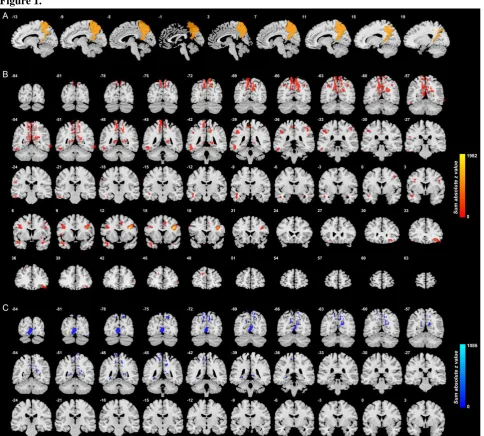
Fig. 1 shows the difference in the functional connectivities of the 125 unmedicated patients from the 254 controls (after FDR correction at p<0.05). This shows that the main differences in unmedicated patients with depression are differences in functional connectivity between the precuneus and the lateral orbitofrontal cortex; the temporal pole; the dorsolateral prefrontal cortex (AAL2 area Frontal_Inf_Tri); some occipital areas; and with voxels on other parts of the precuneus. Table 2 shows the number of voxels in different automated anatomical labelling atlas (AAL2) brain areas with different functional connectivity and the coordinates of the peak voxels. (A list of abbreviations of the AAL2 areas (23) is provided in Table S1.) Table S2 shows which areas have increased and which areas have decreased functional connectivity with the precuneus in depression. Taken together, Tables 2 and S2 show that the areas with increased functional connectivity with the precuneus in depression include the lateral orbitofrontal cortex (AAL2 areas OFClat and Frontal_Inf_Orb_2); the inferior and middle gyri of the prefrontal cortex; temporal cortex; the angular and supramarginal areas; some pre- and postcentral areas with nearby parietal areas; and visual cortical areas. (Some voxels that are mainly lateral orbitofrontal cortex in Fig. 1 appear in Table 2 as medial orbitofrontal cortex AAL2 areas because they just clip these parcels.) The parts of the inferior frontal gyrus shown in Fig. 1 and the coordinates shown in Table 2 are close to those of the inferior frontal gyrus region with connections with the motor laryngeal area (24).
Areas with decreased functional connectivity with the precuneus in depression include the hippocampus and parahippocampal cortex; the fusiform gyrus; and visual cortical areas.
Effect of medication on functional connectivity involving the precuneus
with the precuneus in unmedicated depression, the medication reduces those functional connectivities. An implication is that one way in which antidepressants work is by reducing the FC between the lateral orbitofrontal cortex and the precuneus.
In addition, the medication reduced the functional connectivity of the precuneus with the inferior/middle frontal gyrus (an area implicated in working memory); with the temporal lobe cortex; with the hippocampal / parahippocampal regions (involved in memory); with the ventral insula (an area implicated in autonomic function); and with other precuneus voxels (Fig. 2). A supplementary, ANOVA-based, analysis of the differences in functional connectivity between all AAL2 areas and the precuneus between controls and medicated and unmedicated patients is provided in Fig. S4 and Table S3. The results show that patients with medication have functional connectivities that are altered (mainly decreased) towards the values in the healthy controls.
Analysis of precuneus functional connectivity in healthy controls, and comparison with patients with depression
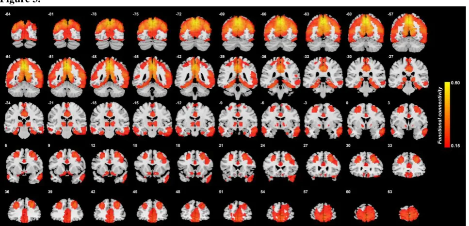
To help interpret the differences in precuneus functional connectivity in depression, Fig. 3 shows the functional connectivity of precuneus voxels with other voxels in the brain in the 254 healthy controls. High functional connectivity is found with the medial and lateral orbitofrontal cortex and anterior cingulate cortex; with the dorsolateral prefrontal cortex; with the olfactory tubercle / ventral striatum; with the inferior temporal gyrus; with the parahippocampal cortex and hippocampus; with the posterior cingulate cortex; and with the lateral and medial parietal cortex.
Fig. 4 shows the main areas with which the precuneus has high functional connectivity in healthy controls; and how for many areas the connectivity is higher with the precuneus in unmedicated patients with depression; and how patients with medication have functional connectivities that are reduced towards the values in the healthy controls. The clear exception is the hippocampus, which has low functional connectivity with the precuneus, which is decreased in depression. The functional connectivity value shown is the mean r (correlation) value between all significant voxels in each of the AAL2 areas indicated. This helps in understanding the functional connectivity of the precuneus in controls. For comparison, the mean r value for the FC of the precuneus with all other brain areas is 0.1434 (std 0.1348). Table S2 shows that in healthy controls the precuneus has moderately high FC with dorsolateral prefrontal cortex, early cortical visual areas, the angular gyrus and the temporal cortex.
Parcellation of the precuneus
To enable a more detailed comparison between patients and controls, we performed a voxel-level parcellation of the precuneus based on the functional connectivity of each precuneus voxel with all 94 AAL2 brain regions (23) in the healthy controls (see Supplementary Material), with the results shown in Fig. S1. An inferior cluster (2, yellow) had especially high functional connectivity with the nearby posterior cingulate cortex but also to a lesser extent with the anterior cingulate cortex; with the parahippocampal gyrus; with prefrontal cortex areas that included Frontal_Med_Orb, the gyrus rectus, and Frontal_Sup_Medial; with the angular gyrus; and with the middle temporal gyrus (Fig. S2). The superior posterior precuneus cluster (1, blue) and the superior anterior cluster (3, red) had somewhat similar functional connectivity to each other, including high functional connectivity with visual areas (calcarine, occipital, lingual and fusiform); with the midcingulate cortex; with other parts of the parietal cortex; with the supramarginal gyrus (Fig. S2). Precuneus cluster 3 (superior anterior) had higher functional connectivity than cluster 1 (superior posterior) with motor-related areas including the precentral gyrus, the paracentral lobule, the midcingulate cortex, and the postcentral gyrus. Precuneus cluster 1 (superior posterior) had higher functional connectivity with the angular gyrus and with some temporal lobe areas than cluster 3 (Fig. S2).
had increased functional connectivity in depression with some motor-related areas (precentral and postcentral gyri), and with temporal lobe cortical areas.
Discussion
The main findings were as follows. Voxels in the precuneus had significantly increased functional connectivity with the lateral orbitofrontal cortex in unmedicated patients with depression. In patients receiving medication, the functional connectivity between the lateral orbitofrontal cortex and precuneus was decreased back towards that in the controls. Functional connectivity was also increased between the precuneus and prefrontal cortex areas involved in short-term working memory. In the 254 controls, it was shown that the precuneus has high functional connectivity with the parahippocampal and dorsolateral prefrontal regions which are involved in memory; and with the parietal cortex. The findings support the theory that the non-reward system in the lateral orbitofrontal cortex has increased effects on memory systems, which contribute to the rumination about sad memories and events in depression (5, 16-18). These new findings provide further evidence that a key target to ameliorate depression is the lateral orbitofrontal cortex (16, 18, 25).
The precuneus and the adjoining retrosplenial cortex (areas 29 and 30) (26-28) (both included in the automated anatomical atlas AAL2 area precuneus used here (23)) are key regions related to spatial function, memory, and navigation (29-31). The retrosplenial cortex provides connections to and receives connections from the hippocampal system, connecting especially with the parahippocampal gyrus areas TF and TH, and with the subiculum (26, 27, 29). The precuneus can be conceptualized as providing access to the hippocampus for spatial and related information from the parietal cortex (given the rich connections between the precuneus and parietal cortex (26, 27, 32) evident in Fig. 4). Further, the precuneus has rich connectivity with the posterior cingulate cortex (32), which provides a pathway into the hippocampal memory system (33, 34), and which also has increased functional connectivity with the lateral orbitofrontal cortex in depression through which sad memories may be facilitated in depression (21). Object information from the temporal lobe connects to and from the hippocampus via the perirhinal cortex (35). This provides a basis for the hippocampus to associate together object and spatial information in the single network in the CA3 region of the hippocampus, to form an episodic memory with object and spatial components (36). However, reward-related / emotional information may also be part of an episodic memory, and connections from the orbitofrontal cortex to the hippocampal system via the perirhinal and entorhinal cortex pathway are likely to be one route (18, 35, 37, 38). Interestingly, the relatively strong functional connectivity between the precuneus and the lateral orbitofrontal cortex described here indicates that reward / punishment-related information also enters this part of the system.
It was of interest that the medication was associated with reduced FC between the precuneus and the lateral orbitofrontal cortex, and also with a number of other areas that have increased functional connectivity with the precuneus in depression (Fig. 2 and Fig. 4).
functional connectivity in depression with a number of areas including the left angular gyrus (involved in language) (11), and with the posterior cingulate cortex involved in memory (21), but they may not have increased connectivity with each other. The common hub to this system is the lateral orbitofrontal cortex OFC47/12. An interesting difference to note is that in healthy controls the precuneus has high functional connectivity with the parahippocampal gyrus but not with the hippocampus per se (Fig. 3), whereas the posterior cingulate cortex has high functional connectivity with both the parahippocampal gyrus and the hippocampus (21), implying that the posterior cingulate cortex is more closely related to hippocampal memory functions (34, 36, 38). The superior anterior precuneus cluster (3, red in Fig. S2) had high functional connectivity with the nearby areas anterior to it involved in motor function. The superior posterior precuneus cluster (1, blue in Fig. S2) had high functional connectivity with the visual areas posterior to it, and in depression had increased functional connectivity with some temporal lobe cortical areas and with the inferior frontal gyrus areas (Fig. S3).
The precuneus is part of the default mode network, which becomes more active when tasks are not being performed in the world, and instead internal thoughts and processing are occurring, and this system may have altered activity in depression (44). The increased functional connectivity between the precuneus and the prefrontal cortex described here, and noted previously in much smaller previous studies (7-9), may relate to increased internal ruminating thoughts in depression. New conceptual contributions of the present research are the links found to the lateral orbitofrontal cortex non-reward system, which links the ruminating thoughts to the negative, sad, thoughts in depression; and the hypothesis that the precuneus may make these sad ruminating thoughts refer especially to the self, and indeed be related to the low self-esteem that is found in depression.
To test some of the implications of the findings and working hypotheses described here, it would be of interest to perform a functional neuroimaging study in which activations in the precuneus and the lateral orbitofrontal cortex to sad vs happy memories are measured. The prediction is that in the precuneus and the lateral orbitofrontal cortex the activations would be higher in depressed people for sad vs happy memories.
The other link that stood out in the unmedicated patients was the increased functional connectivity between the precuneus and the inferior frontal gyrus region shown in Fig. 1 and Table 2, which is probably the inferior frontal gyrus region with connections with the motor laryngeal area (24). It is suggested that this is related to the increased rumination in depression which may produce subliminal speech-related effects. That would be consistent with the increased functional connectivity of the lateral orbitofrontal cortex with both the precuneus and the angular gyrus, a cortical area related to language (11). It is also consistent with the increased functional connectivity of the precuneus with two language-related areas, the angular and supramarginal gyri, as shown in Table 2.
Other links with increased functional connectivity in depression were between the precuneus and the temporal cortical areas (Fig. 1 and Table 2). An investigation of effective (i.e. directed) connectivity in depression showed that it is the forward links from the middle and inferior temporal cortical areas to the precuneus that are increased in depression (45), providing evidence that perceptual and related input may have a greater influence of the precuneus in depression. The effective connectivity in the reverse direction is 20 times smaller, making the precuneus an interesting structure in the nature of its forward and reverse effective connectivities (45).
Some other links with different functional connectivity in depression with the precuneus are with motor areas, and it may be expected that a structure such as the precuneus involved in spatial and related functions has functional connectivity with motor areas. There is also some decreased functional connectivity of the precuneus with the occipital visual areas in depression, as shown in Fig. 1.
Contributors
Edmund T. Rolls, Wei Cheng and Jianfeng Feng contributed to the design of the study. Deyu Yang, Dongtao Wei; Libo Zhao; Jie Meng and Peng Xie contributed to the collection of the data. Wei Cheng, Edmund T. Rolls and Hongtao Ruan contributed to the analysis of the data and the preparation of the manuscript. Edmund T. Rolls and Wei Cheng participated in writing the paper. All collaborators had an opportunity to contribute to the interpretation of the results and to the drafting of the manuscript.
Declaration of interests.
All authors declare no competing interests.
Acknowledgements
Table 1.
A summary of the demographic information and the psychiatric diagnosis in the present
study.
Group Age (years) Sex (male/female) Education (years) Medication
(yes / no) HAMD BDI
Duration of illness First episode (yes / no) Mean FD
Healthy 39.65 ±
15.80 166 / 88 13.01 ± 3.89 / / / / / 0.133 ±0.063
Patient 38.74 ±
13.65 183 / 99 11.91 ± 3.58 157 / 125 20.8 ± 5.87
20.42 ±
9.33 4.16 ± 5.51 209 / 49 0.125 ± 0.054
Statistic (t / p) or (chi-square / p)
0.719 / 0.472 0.013 / 0.911 3.41 / 6.9e-4 / / / / / 1.729 / 0.084
Unmedicated patient
37.60 ±
13.12 84 / 41 12.07 ± 3.72 125 / 0 22.22 ± 4.39
22.51 ±
8.16 2.91 ± 4.44 111 / 14 0.120 ± 0.053
Medicated patient
39.64 ±
14.03 99 / 58 11.78 ± 3.48 0 / 157 19.42 ± 6.73
18.43 ±
9.95 5.33 ± 6.13 98 / 35
0.129 ± 0.054
Statistic (t / p) or (chi-square / p)
-1.250 /
0.212 0.524 / 0.469
0.673 /
0.501 /
3.907 / 1.2e-4
3.520 / 5.1e-4
-3.539 /
4.8e-4 9.570 / 0.002 -1.268 / 0.206
Values are n or mean ± SD.
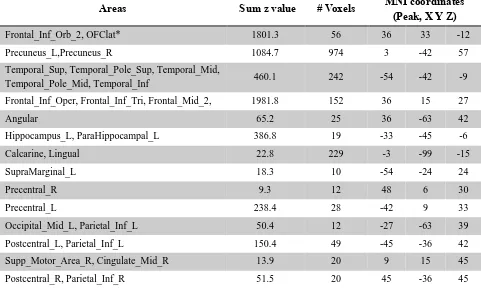
Table 2. Numbers of voxels in different AAL2 areas with significantly different functional connectivity with the precuneus in unmedicated patients with depression. For the precuneus, the table shows the number of precuneus voxels that have different functional connectivity with the whole brain (FDR corrected, p<0.05). The other entries in the table show the numbers of voxels in each of the specified brain regions with different functional connectivity with precuneus voxels (FDR corrected, p<0.05). The z value is the sum across all voxels and FC links of the absolute value of the z score for significant links between pairs of voxels. In the Table, we show clusters with more than 10 voxels.
Areas Sum z value # Voxels MNI coordinates (Peak, X Y Z)
Frontal_Inf_Orb_2, OFClat* 1801.3 56 36 33 -12
Precuneus_L,Precuneus_R 1084.7 974 3 -42 57
Temporal_Sup, Temporal_Pole_Sup, Temporal_Mid,
Temporal_Pole_Mid, Temporal_Inf 460.1 242 -54 -42 -9
Frontal_Inf_Oper, Frontal_Inf_Tri, Frontal_Mid_2, 1981.8 152 36 15 27
Angular 65.2 25 36 -63 42
Hippocampus_L, ParaHippocampal_L 386.8 19 -33 -45 -6
Calcarine, Lingual 22.8 229 -3 -99 -15
SupraMarginal_L 18.3 10 -54 -24 24
Precentral_R 9.3 12 48 6 30
Precentral_L 238.4 28 -42 9 33
Occipital_Mid_L, Parietal_Inf_L 50.4 12 -27 -63 39
Postcentral_L, Parietal_Inf_L 150.4 49 -45 -36 42
Supp_Motor_Area_R, Cingulate_Mid_R 13.9 20 9 15 45
Postcentral_R, Parietal_Inf_R 51.5 20 45 -36 45
Figure legends
Figure 1. A) Voxels of the precuneus defined by the AAL2 atlas. B, C) Anatomical location of voxels with significantly increased (B) and decreased (C) functional connectivity with the precuneus in depression in 125 unmedicated patients vs 254 controls obtained from the voxel-based Association Study (vAS). z values are shown for each voxel, showing the mean difference of functional connectivities for patients with unmedicated depression - controls. Red thus indicates an increase in functional connectivity in depression, and blue a decrease. The right of the brain is on the right of each slice. The Y values are in MNI coordinates. This shows that the main differences in unmedicated depression for the precuneus are an increase in functional connectivity with the lateral orbitofrontal cortex (Y=33), and with a prefrontal area in the inferior frontal gyrus that may be the prefrontal laryngeal cortex (Y=15).
Fig. 2. Anatomical location of voxels with significantly increased (A) and decreased (B) functional connectivity with the precuneus in 125 unmedicated patients vs 157 medicated patients obtained from the voxel-based Association Study (vAS).
z values are shown for each voxel, showing the mean
difference of functional connectivities for patients with medicated depression - unmedicated
depression. Blue thus indicates a lower in functional connectivity in patients with depression who are
medicated than in those that are unmedicated. Here, we only show the voxels with sum absolute z
value larger than 10 and cluster size larger than 20 voxels.
Figure 3.
Functional connectivity of the precuneus in 254 healthy controls. r values are shown,
thresholded at r=0.15.
Figure 3.