Original Article
miR-24-3p stimulates migration, invasion
and proliferation of prostate cancer cells by
targeting suppressor of cytokine signaling 6
Yang Lin1*, Huifang Cao2*, Yuxin Tian1, Xinping Yang1, Changdong Zhou1, Qifu Zhang1
1Department of Urological Surgery, Jilin Province Tumor Hospital, Changchun 130012, Jilin Province, China; 2Departments of Gynecology, Jilin Province Tumor Hospital, Changchun 130012, Jilin Province, China. *Equal contributors.
Received August 11, 2017; Accepted January 8, 2018; Epub March 1, 2018; Published March 15, 2018
Abstract: Prostate cancer is among the most widespread malignancies affecting men in the world. Its aggressive evolution has been associated with altered expression of suppressor of cytokine signaling 6 (SOCS6) but very little is known about the mechanism by which this alteration occurs. The purpose of this study was to explore the role of SOCS6 in prostate cancer cells and the involvement of its regulating microRNA (miR), miR-24-3p. Prostate cancer cell lines were used to determine the transcription level of miR-24-3p and SOCS6 by quantitative reverse-transcrip-tase-polymerase chain reaction (qRT-PCR) and Western blot. Cell proliferation and cell migration assays were done-to determine the effect of miR-24-3p mimics and inhibidone-tors on cell proliferation, invasion and migration. Luciferase
reporter assay with SOCS6 3’-UTR was performed to confirm the control of SOCS6 expression by the miR. The re -sults showed that miR-24-3p was up-regulated in prostate cancer cells whereas SOCS6 protein was downregulated. Overexpression of miR-24-3p in prostate cancer cells promoted cell proliferation, inhibited apoptosis, and increased cell migration and invasion. Luciferase reporter assays showed that SOCS6 is a direct target of its negative regulator miR-24-3p and overexpression of SOCS6 reverses the effects of miR-24-3p on the metastatic phenotype of prostate cancer cells. These results show case miR-24-3p up-regulation in prostate cancer and a mechanism for inhibition of SOCS6 expression. Thus, the miR-24-3p/SOCS6 pathway could be a relevant avenue for prostate cancer treatment.
Keywords: Prostate cancer, SOCS6, miR-24-3p
Introduction
Prostate cancer is one of the most common cancers diagnosed in men. It counts for 15%
of all diagnosed cancers in men and is the
fif-th leading cause of deafif-th from cancer in 2012 [1]. It is a slowly growing cancer without any particular symptoms in the earlier stage andis mostly diagnosed in men above 50 years old [2]. Despite its relatively slow growth, prosta- te cancer cells can migrate and invade other organs such as bones where metastases oc- cur frequently [3, 4]. Molecular mechanisms involved in metastasis are complex and not completely understood [5]. It is widely accept- ed that the cell microenvironment plays a cri- tical role in the induction and silencing of cel- lular signaling pathways that govern cell bio- logy. Among the microenvironment elements involved in this process are cytokines that ha-
ve been associated to the biology of many cancer cells [6, 7]. Cytokine-induced inhibitors of the downstream elements of their recep- tors, such as suppressor of cytokine signaling 6 (SOCS6), could play an important role in the biology of cancer cells [8].
In previous studies, SOCS6 was shown to be
significantly downregulated in colorectal and
prostate cancers [9, 10]. The mechanism by which SOCS6 is down-regulated in cancer cells is not known but several studies point to epi-genetic regulation of its expression by microR-NAs (miRs) [11]. Moreover, the expression
pro-file of miRs is extremely altered in prostate
making them particularly good biomarkers for decision making regarding the appropriate tr- eatment strategy [14]. miRs are also applica-
ble as specific cancer therapeutics [15] and
thus knowing more about the mechanism of
action of miRs is an advantage in defining new
strategies for cancer diagnosis and treatment [16].
The function and mode of action of miR-24- 3p in a diversity of cancers enclosing glioma, hepatocellular carcinoma, lung cancer, breast cancer, bladder cancer and colorectal cancer hasbeen conveyed in previous studies [17-25]. However, despite some studies reporting miR-24-3p as a tumor suppressor while others de- monstrated its role as an oncogene, the pot- ential role of miR-24-3p in prostate cancer is not entirely understood and whether and how the miR-24-3p/SOCS6 axis is involved needs
to be clarified.
Based on the hypothesis that miR-24-3p de- regulation in prostate cancer cells may affect-disease progression, the present study was designed to explore the potential relationship between miR-24-3p and SOCS6 and their ef- fect on prostate cancer cellbiology.
Material and methods
Cell culture
Prostate cancer cell lines LNCaPFGC, PC3 and DU 145 were used in this study and compar- ed to human normal prostate epithelial cell line RWPE1. RWPE-1 cells were cultured in keratinocyte serum-free medium (K-SFM, In- vitrogen, Carlsbad, USA) supplemented with 50 mg/ml bovine pituitary extract, 100 U/ml penicillin and 100 mg/l streptomycin. Other cells were cultured in RPMI 1640 medium (Gibco, USA) supplemented with 10% fetal bovine serum and 1% penicillin/streptomycin antibiotics (Gibco, USA). All cultures were in- cubated at 37°C in 5% CO2, 95% air at- mosphere.
RNA extraction and quantitative real-time RT-PCR
Total RNA was extracted from harvested cells (5 × 10⁴ cells) using TRIzol reagent (Invitro-
gen). The TaqMan miR reverse transcription kit (Applied Biosystems, Foster City, CA) was used on the extracted RNA to generate miR
comple-mentary DNAs that were further amplified and
mRNA expression level was determined by reverse transcription using the SuperScript III
first-strand synthesis system supermix
(Invi-trogen) and a SYBR Green PCR kit (TaKaRa, Dalian, China). The level of the housekeeping gene glyceraldehyde-3-phosphate dehydroge-nase (GAPDH) mRNA was used as a reference
for quantification. The ΔΔCT method was us-ed for quantifications.
Cell transfection
PC-3 Cells in a logarithmic growth phase were transfected with miR-24-3p mimic, miR-24-3p inhibitor (antisense oligonucleotide), or miR-control. Transfection was performed for each cell line with 50 nM of oligonucleotides using riboFECTTM CP transfection reagent (Ribo-bio,
China) according to the user manual. Tran- sfected cells were cultured and forwarded for further experiments.
Cell viability test
Cell lines and transfected cells were seeded in 96-well plates at 104 cells per well. Cell
pro-liferation was measured using Cell Counting Kit-8 (CCK-8) (Dojindo Laboratories, Tokyo, Japan) in accordance with the manufactur- er’s recommendations. Absorbance was mea-sured at 450 nm using a multilableplate read- er. Each experiment was performed with three replicates.
Cell migration and invasion
Transwell assay with FBS in the lower cham- ber was used to determine the migration rate of studied cell lines and transfected cells. Cells were suspended in serum free medium and loaded to the upper chamber (105 cells/
well). The transwell membrane (Millipore, Billerica, MA, USA) with 8 µm pores size was coated with Matrigel (BD Biosciences, San Jose, USA)for tracking cell invasion. Fresh me- dium was loaded in the lower chamber with 10% FBS as the chemoattractant. After 24 h of incubation at 37°C, the invaded cells were
fixed with 100% methanol, stained with 1%
crystal violet for 20 minutes, and counted under a microscope.
In silico analysis
(www.tar-Luciferase reporter assays
Cell lines were seeded in 24 well plates and transfected the following day with expression plasmids constructs harboring the luciferase gene with the SOCS6 3’-UTR or a mutated SOCS6 3’-UTR. Transfection was performed using Lipofectamine 2000 (Invitrogen). Two days after transfection, cells were lysed and analyzed by luciferase reporter assay using the Dual-Luciferase Reporter Assay System (Promega, Madison, WI) according to the ma- nufacturer’s recommendations.
Western blot
The expression levels of proteins were mea-sured by Western blot on all cell lines and tho- se transfected with miR-24-3p mimics or in- hibitors. Cells were harvested 48 h after tr- ansfection, washed twice with cold phospha- te-buffered saline (PBS) and lysed in 50 mM Tris-HCl (pH 7.4), 150 mM NaCl, 1% Nonidet P-40, 10 mM KCl, 1 mM EDTA, 20 mM NaF, 0.25% Na deoxycholate, 5 mM dithiothreitol (DTT). Protease inhibitors (Roche, USA) and phosphatase inhibitors (ThermoFisher Scien-
tific) were added to the mix and total cellular
lysates (20 µg for each sample) were subject- ed to 10% SDS-polyacrylamide gel electro- phoresis. At the end of electrophoresis, pro-teins in the gel were transferred to nitrocellu-lose membrane and blocked with 5% (w/v) skimmed milk for one hour at room tempera-ture. Membranes were then incubated over-night at 4°C with monoclonal primary antibod-iesagainst SOCS6, caspase 3, and cleaved- caspase 3 (Abcam, UK). After incubation with the primary antibody, membranes were wash- ed three times with TBS-T and then incubated
one hour at room temperature with the horse-radish peroxidase-conjugated goat anti-mouse secondary antibody, (Santa Cruz Biotechnolo- gy, USA). Membranes were washed three ti- mes with TBS-T and protein bands were stain- ed using the BM chemiluminescence Blotting Substrate (POD) (Roche, USA) according to the manufacturer’s instructions. Image J soft-ware (NCBI) was used for densitometry an- alysis.
Statistical analysis
Data were expressed as mean ± standard de- viation (SD) of three independent experim- ents performed in triplicate. Statistical analy-ses were performed using one-way or two- way analysis of variance (ANOVA). Values of P
< 0.05 were considered statistically
signifi-cant compared to the respective control.
Results
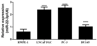
miR24-3p is up-regulated in prostate cancer cells
Prostate cancer cell lines and prostate nor- mal epithelial cells were subjected to whole transcriptome extraction followed by reverse transcription qPCR to quantify the expression level of miR-24-3p. These experiments reveal-
ed a significant increase in the amount of
miR-24-3p expressed in all cancer cell lines in comparison to prostate epithelial RWPE1 cell lines (Figure 1). As shown in Figure 1, miR-24-3p level was the most upregulated in PC-3 cell line. This cell line was therefore selected for further experiments.
miR24-3p overexpression promotes the pro-liferation, invasion and migration of prostate cancer cells
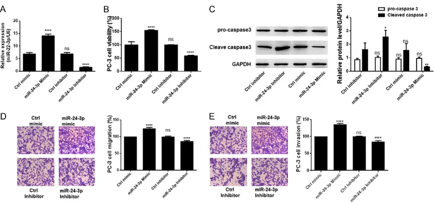
miR-24-3p mimic,the inhibitor, or theirrespec-tive controls were transfected in PC-3 cells
after checking the expression profile of
miR-24-3p in prostate cancer cells by qRT-PCR.
The efficiency of miR-24-3p expression after
[image:3.612.89.287.72.166.2]transfection is reported in Figure 2A. To eva- luate the effect of miR-24-3p on the phenoty- pe of PC-3 cells, the proliferation, apoptosis rate and cell migration and invasion were mea-sured in these cells transfected with miR-24- 3p mimic or inhibitor and compared with con-trol cells. Results of these assays are shown in Figure 1. miR-24-3p is up-regulated in prostate
Figure 2. Overexpression of 3p in prostate cancer cells induces aggressive cancer cell behavior. A.
miR-24-3p expression profile in cells transfected with miR-24-miR-24-3p mimics, the inhibitors, or control. B. miR-24-miR-24-3p overexpres -sion induced the viability of prostate cancer cells. C. Western blot analysis of procaspase 3 and cleaved-caspase 3 indicated that miR-24-3p inhibited cell apoptosis. D. Cell migration rate in transfected cells. E. Rate of cell inva-sion after transfection. All experiments were performed in triplicate. *P < 0.05; **P < 0.01; ****P < 0.0001 and
ns means non-significant when compared to the Ctrl mimic group (Ctrl inhibitor group in the case of western blot
[image:4.612.93.525.372.574.2]analysis).
Figure 3. SOCS6 is a direct target of its inhibitor miR-24-3p. A.Targetscan predicted SOCS6 3’-UTR as seed se-quence for miR-24-3p. B. Expression level of SOCS6 protein in prostate cancer cell lines and RWPE1 cells. C. Lu-ciferase reporter assay indicated miR-24-3p as a negative regulator of its direct target SOCS6. D. Western blot analysis of SOCS6 protein level in PC-3 cells co-transfected with miR-24-3p mimics, the inhibitors, or control and the mutated or wild type 3’-UTR vector. E. SOCS6 mRNA level in PC-3 cells transfected with miR-24-3p mimics, inhibitors or control. All experiments were performed in triplicate. **P < 0.01; **P < 0.01; ****P < 0.0001 and ns means
non-significant when compared to the normal control cells, Ctrl mimic group or wild type (WT) group.
Figure 2B-E. There was a high increase in pro- liferation of PC-3 cells over-expressing miR- 24-3p in comparison to controls (Figure 2B). Furthermore, cells over-expressing the miR
showed a significant reduction in cell
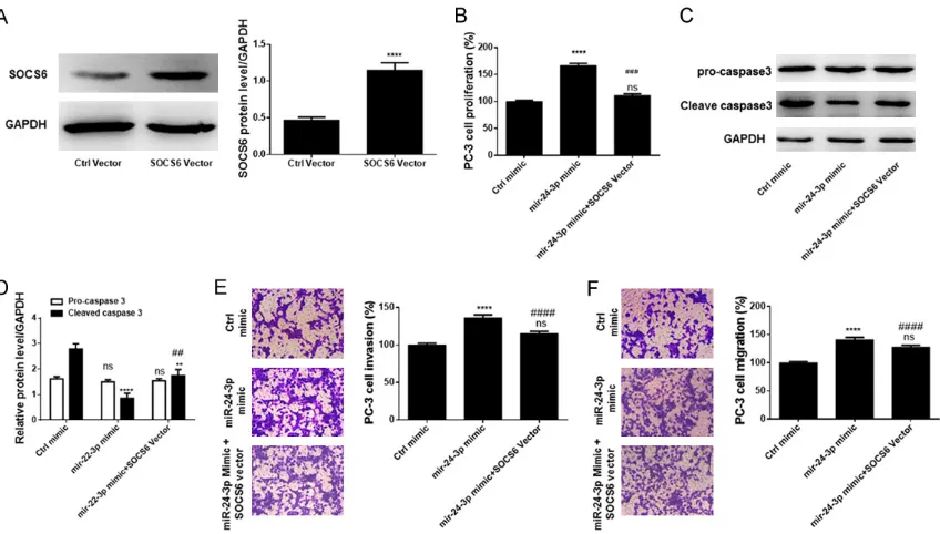
Figure 4. Ectopic expression of SOCS6 inverted the action of miR-24-3p. A. Ectopic SOCS6 expression efficiency.
B. Effect of SOCS6 overexpression on miR-24-3p-induced cell proliferation. C. Western blot analysis of the effect of SOCS6 overexpression on expression of caspase 3 and cleaved caspase 3. D. Densitometry analysis of bands obtained from the Western blot analysis. E. Effect of SOCS6 overexpression on cell migration. F. Effect of SOCS6 overexpression on cell migration. All experiments were performed in triplicate. **P < 0.01; ****P < 0.0001 and
ns means non-significant when compared with Ctrl vector or Ctrl mimic. ##P < 0.01; ###P < 0.001 and ####P <
0.0001 when compared with miR-24-3p mimic group.
and migration (Figure 2D, 2E). In contrast, transfection with miR-24-3p inhibitors in PC-3
cells significantly inhibited cell proliferation,
migration, and invasion but increased the ra- te of apoptosis as measured by the level of cleaved-caspase 3. Altogether, these observa-tions imply that miR-24-3p promotes the meta-static phenotype of prostate cancer cells.
miR-24-3p is a post-transcriptional negative regulator of SOCS6
Targetscan analysis showed in silico that miR-24-3p targets among others the SOCS6 mRNA in the 3’-UTR region (position 3114-3120) as presented in Figure 3A. Western blot analysis of SOCS6 in prostate cancer cell lines and pr-
ostate epithelial cells showed a significantly
decreased level of protein in cancer cell lines in comparison to RWPE1 cells (Figure 3B). The luciferase reporter gene expression assay was performed with the luciferase gene under the control of a SOCS6 3’-UTR fragment (mutated or wild type). In the case of wild-type SOCS6 3’UTR, miR-24-3p drastically decreased the expression level of luciferase whereas the miR
had no significant effect on the expression of
luciferase linked to mutated SOCS6 3’-UTR (Figure 3C). PC-3 cells transfected with miR- 24-3p mimics or inhibitors and vectors harbor-ing SOCS6 gene with either mutated or wild type 3’-UTR were also subjected to Western
blotting and SOCS6 mRNA quantification by
qRT-PCR. The results showed that expression of miR-24-3p or its inhibitor did not affect the level of SOCS6 mRNA while overexpressing
the miR significantly decreased the protein
level (Figure 3D, 3E). These results indicate that miR-24-3p is a post-transcriptional nega-tive regulator of SOCS6.
Overexpression of SOCS6 reverses the effect of miR-24-3p
When PC-3 cells were transfected with a SOC- S6 expression vector, the protein was abun-dantly expressed in the cells despite transf- ection with the miR-24-3p mimics (Figure 4A). These cells were used as models to investi- gate the effect of SOCS6 overexpression on miR-24-3p action. Overexpression of SOCS6
on PC-3 cells as shown in Figure 4B, 4F. Ec- topic overexpression of SOCS6 attenuated the miR-24-3p-induced cell proliferation, migra-tion, and invasion, but increased the rate of apoptosis in cells expressing miR-24-3p. Ove- rall, these results indicate that miR-24-3p accelerates migration, invasion, and prolifera-tion of prostate cancer cells by directly and negatively regulating SOCS6.
Discussion
SOCS6 is one of the understudied members of the cytokine-induced STAT inhibitors family that has been shown to be induced by cell pr- oliferative cytokines and growth factors [26, 27]. As SOCS6 action is to inhibit these cyto-kines and downstream proliferation signaling of cellular growth factors, it makes sense to consider that SOCS6 exerts an anti-prolifera-tive activity as reportedin several previous studies [28-30]. SOCS6 could exert the same anti-proliferative activity in prostate cells in a comparable manner explaining the fact that this protein is highly produced in normal cells but be present in low amounts in prostate can-cer cells as found in this in vitro study. Prosta- te cancer could rise when the protein level of SOCS6 decreases, thus abrogating the control action of this protein on the proliferative sig- nal induced by different cytokines. This obser-vation has already been made clinically and in vitro by other authors [31-33]. One of the un- answered questions in this possible mecha-nism of cell transformation is the mechamecha-nism by which SOCS6 is downregulated in cancer cells. Giving an answer based on experimental data to this question was the central objective in the present study.
A comparison between prostate normal cells and prostate cancer cell lines was performed regarding the expression of miR-24-3p. The results show that the level of miR-24-3p is
significantly high in cancer cell lines.
Mean-while, Western blot results showthat thelevel of SOCS6 protein is decreased in cancer cell lines as compared to prostate epithelial cells. Bioinformatics show that miR-24-3p anneals to SOCS6 mRNA in the 3-’UTR whereby miR- 24-3p directly targets SOCS6 mRNA and do- wn-regulates its translation. The reporter gene
expression assay confirmed that SOCS6 mRNA
is a direct target of the miR. To our knowle-
dge, this is the first time miR-24-3p has been
experimentally shown to downregulate SOCS6 in prostate cancer cell lines. In fact, previous studies demonstrated that SOCS6 expression is under the control of other miRssuch as miR-21, miR-142-3p, miR-183, or miR-494-3p in diverse cancer types [34-36]. miR-24-3p can thus control the expression of SOCS6. The present study could not exclude the action of other miRs but the results in the luciferase
reporter gene assay not only confirm that
SOCS6 3’-UTR is determinant in the expres- sion control but also highlight the importance of miR-24-3p in down-regulating SOCS6 in prostate cancer cell lines.
According to the results of the present study, SOCS6/miR-24-3p regulatory axis plays an ir- revocable role in prostate cancer biology. In
effect, miR-24-3p overexpression was suffi -cient to induce cell proliferation and increase cell migration and invasion. Moreover, ectopic expression of SOCS6 in cancer cells attenuat-ed the proliferation and cell invasion inducattenuat-ed by the miR. miR-24-3p could therefore be responsible for prostate cancer induction, its maintenance and also for the spreading of cancer cells to other organs where metastas- es occur. This explains why SOCS6 downregu- lation is associated with aggressive cancer such as colorectal cancer [9]. Both SOCS6 and miR-24-3p appear to be good markers for prostate cancer prognosis and offer a poten-tially high effective pathway target for prostate cancer treatment.
Overall, the present study identifies SOCS6
and its regulatory miR, miR-24-3p as determi-nant factors in prostate cancer biology. They are central to prostate cancer cell prolifera- tion and metastases. This axis (miR-24-3p/ SOCS6) is a promising therapeutic target for prostate cancer that needs to be more deeply investigated in future studies.
Acknowledgements
This study was financially supported by the
authors’ own funds.
Disclosure of conflict of interest
None.
Tumor Hospital, 1018 Huguang Road, Chaoyang District, Changchun 130012, Jilin Province, China. Tel: +86-431-85873156; E-mail: Zcdjt2199@163. com
References
[1] McGuire S. World cancer report 2014. Geneva, Switzerland: world health organization, inter-national agency for research on cancer, WHO press, 2015. Adv Nutr 2016; 7: 418-419. [2] Adeloye D, David RA, Aderemi AV,
Iseolorunk-anmi A, Oyedokun A, Iweala EE, Omoregbe N and Ayo CK. An estimate of the incidence of prostate cancer in Africa: a systematic review and meta-analysis. PLoS One 2016; 11: e0153496.
[3] Ziaee S, Chu GC, Huang JM, Sieh S and Chung LW. Prostate cancer metastasis: roles of re-cruitment and reprogramming, cell signal net-work and three-dimensional growth character-istics. Transl Androl Urol 2015; 4: 438-454. [4] Kelly K and Yin JJ. Prostate cancer and
metas-tasis initiating stem cells. Cell Res 2008; 18: 528-537.
[5] Tantivejkul K, Kalikin LM and Pienta KJ. Dy-namic process of prostate cancer metastasis to bone. J Cell Biochem 2004; 91: 706-717. [6] Puente Vazquez J, Grande Pulido E and Anton
Aparicio LM. Cytokine and endocrine signaling in prostate cancer. Med Oncol 2012; 29: 1956-1963.
[7] Yoo SY, Lee SY and Yoo NC. Cytokine expres-sion and cancer detection. Med Sci Monit 2009; 15: RA49-56.
[8] Chikuma S, Kanamori M, Mise-Omata S and Yoshimura A. Suppressors of cytokine signal-ing: potential immune checkpoint molecules for cancer immunotherapy. Cancer Sci 2017; 108: 574-580.
[9] Letellier E, Schmitz M, Baig K, Beaume N, Schwartz C, Frasquilho S, Antunes L, Marcon N, Nazarov PV, Vallar L, Even J and Haan S.
Identification of SOCS2 and SOCS6 as bio -markers in human colorectal cancer. Br J Can-cer 2014; 111: 726-735.
[10] Zhu JG, Dai QS, Han ZD, He HC, Mo RJ, Chen G, Chen YF, Wu YD, Yang SB, Jiang FN, Chen WH, Sun ZL and Zhong WD. Expression of SOCSs in human prostate cancer and their association in prognosis. Mol Cell Biochem 2013; 381: 51-59.
[11] Rontauroli S, Norfo R, Pennucci V, Zini R, Ru-berti S, Bianchi E, Salati S, Prudente Z, Rossi C, Rosti V, Guglielmelli P, Barosi G, Vannucchi
A, Tagliafico E and Manfredini R. miR-494-3p
overexpression promotes
megakaryocytopoie-sis in primary myelofibromegakaryocytopoie-sis hematopoietic
stem/progenitor cells by targeting SOCS6. On-cotarget 2017; 8: 21380-21397.
[12] Baumgart SJ and Haendler B. Exploiting epi-genetic alterations in prostate cancer. Int J Mol Sci 2017; 18.
[13] Bucay N, Bhagirath D, Sekhon K, Yang T, Fuku-hara S, Majid S, Shahryari V, Tabatabai Z, Greene KL, Hashimoto Y, Shiina M, Yamamura S, Tanaka Y, Deng G, Dahiya R and Saini S. A novel microRNA regulator of prostate cancer epithelial-mesenchymal transition. Cell Death Differ 2017; 24: 1263-1274.
[14] Cannistraci A, Federici G, Addario A, Di Pace AL, Grassi L, Muto G, Collura D, Signore M, De Salvo L, Sentinelli S, Simone G, Costantini M, Nanni S, Farsetti A, Coppola V, De Maria R and Bonci D. C-Met/miR-130b axis as novel mech-anism and biomarker for castration resistance state acquisition. Oncogene 2017; 36: 3718-3728.
[15] Smith B, Agarwal P and Bhowmick NA. MicroR-NA applications for prostate, ovarian and breast cancer in the era of precision medicine. Endocr Relat Cancer 2017; 24: R157-R172. [16] Armstrong CM, Liu C, Lou W, Lombard AP,
Ev-ans CP and Gao AC. MicroRNA-181a promotes docetaxel resistance in prostate cancer cells. Prostate 2017; 77: 1020-1028.
[17] Gao Y, Liu Y, Du L, Li J, Qu A, Zhang X, Wang L and Wang C. Down-regulation of miR-24-3p in colorectal cancer is associated with malignant behavior. Med Oncol 2015; 32: 362.
[18] Kang H, Rho JG, Kim C, Tak H, Lee H, Ji E, Ahn S, Shin AR, Cho HI, Huh YH, Song WK, Kim W and Lee EK. The miR-24-3p/p130Cas: a novel axis regulating the migration and invasion of cancer cells. Sci Rep 2017; 7: 44847.
[19] Kerimis D, Kontos CK, Christodoulou S, Papa-dopoulos IN and Scorilas A. Elevated expres-sion of miR-24-3p is a potentially adverse prog-nostic factor in colorectal adenocarcinoma. Clin Biochem 2017; 50: 285-292.
[20] Lu K, Wang J, Song Y, Zhao S, Liu H, Tang D, Pan B, Zhao H and Zhang Q. miRNA-24-3p pro-motes cell proliferation and inhibits apoptosis in human breast cancer by targeting p27Kip1. Oncol Rep 2015; 34: 995-1002.
[21] Olbromski M, Grzegrzolka J, Jankowska-Kon-sur A, Witkiewicz W, Podhorska-Okolow M and Dziegiel P. MicroRNAs modulate the expres-sion of the SOX18 transcript in lung squamous cell carcinoma. Oncol Rep 2016; 36: 2884-2892.
[23] Yin Y, Zhong J, Li SW, Li JZ, Zhou M, Chen Y, Sang Y and Liu L. TRIM11, a direct target of miR-24-3p, promotes cell proliferation and in-hibits apoptosis in colon cancer. Oncotarget 2016; 7: 86755-86765.
[24] Yu G, Jia Z and Dou Z. miR-24-3p regulates bladder cancer cell proliferation, migration, in-vasion and autophagy by targeting DEDD. On-col Rep 2017; 37: 1123-1131.
[25] Zhang MX, Zhang J, Zhang H and Tang H. miR-24-3p suppresses malignant behavior of lacri-mal adenoid cystic carcinoma by targeting PRKCH to regulate p53/p21 pathway. PLoS One 2016; 11: e0158433.
[26] Croker BA, Kiu H and Nicholson SE. SOCS reg-ulation of the JAK/STAT signalling pathway. Se-min Cell Dev Biol 2008; 19: 414-422.
[27] Linossi EM, Babon JJ, Hilton DJ and Nicholson SE. Suppression of cytokine signaling: The SOCS perspective. Cytokine Growth Factor Rev 2013; 24: 241-248.
[28] Liu J, Zheng Y, Gao J, Zhu G, Gao K, Zhang W, Shi F and Zhang Q. Expression of SHP-1 and SOCS6 in patients with acute leukemia and their clinical implication. Onco Targets Ther 2017; 10: 1915-1920.
[29] Lai RH, Hsiao YW, Wang MJ, Lin HY, Wu CW, Chi CW, Li AF, Jou YS and Chen JY. SOCS6, down-regulated in gastric cancer, inhibits cell prolif-eration and colony formation. Cancer Lett 2010; 288: 75-85.
[30] Kabir NN, Sun J, Ronnstrand L and Kazi JU. SOCS6 is a selective suppressor of receptor tyrosine kinase signaling. Tumour Biol 2014; 35: 10581-10589.
[31] Qiu X, Zheng J, Guo X, Gao X, Liu H, Tu Y and Zhang Y. Reduced expression of SOCS2 and SOCS6 in hepatocellular carcinoma correlates with aggressive tumor progression and poor prognosis. Mol Cell Biochem 2013; 378: 99-106.
[32] Yoon S, Yi YS, Kim SS, Kim JH, Park WS and Nam SW. SOCS5 and SOCS6 have similar ex-pression patterns in normal and cancer tis-sues. Tumour Biol 2012; 33: 215-221.
[33] Sriram KB, Larsen JE, Savarimuthu Francis SM, Wright CM, Clarke BE, Duhig EE, Brown KM, Hayward NK, Yang IA, Bowman RV and Fong KM. Array-comparative genomic hybrid-ization reveals loss of SOCS6 is associated with poor prognosis in primary lung squamous cell carcinoma. PLoS One 2012; 7: e30398. [34] Wu Q, Luo G, Yang Z, Zhu F, An Y, Shi Y and Fan
D. miR-17-5p promotes proliferation by target-ing SOCS6 in gastric cancer cells. FEBS Lett 2014; 588: 2055-2062.
[35] Qi X, Li J, Zhou C, Lv C and Tian M. MiR-142-3p suppresses SOCS6 expression and promotes cell proliferation in nasopharyngeal carcino-ma. Cell Physiol Biochem 2015; 36: 1743-1752.