Original Article
MicroRNA-130a reduces drug
resistance in breast cancer
Jin Huang, Min Zhao, Hongguang Hu, Jin Wang, Lin Ang, Li Zheng
Department of Pathology, The Second People’s Hospital of Hefei, Hefei 230011, Anhui, China
Received February 8, 2019; Accepted March 26, 2019; Epub July 1, 2019; Published July 15, 2019
Abstract: Objective: Although the advent of chemotherapy has made some progress in the comprehensive treat-ment of breast cancer, drug resistance of tumor cells remains to be one of the main challenges for the treattreat-ment of breast cancers. Several microRNAs have been implicated in the resistant process, but the role of miR-130a in drug resistance in breast cancer remains unclear. The present study aims to investigate the role and mechanisms of miR-130a in drug resistance in breast cancer cells and tissues. Patients and methods: miR-130a mimics was used to up-regulate miR-130a expression in Doxorubicin-resistant MCF-7/Adr breast cancer cell line, followed by MTT assay and colony formation to determine cell viability and relative colony number. The relationship between
the expression of miR-130a and drug resistance was detected by in situ hybridization in the formalin-fixed
paraffin-embedded (FFPE) tissues from 50 breast cancer patients before and after Epirubicin-based neoadjuvant chemo-therapy. Results: Up-regulation of miR-130a level in MCF-7/Adr cells decreased the cell viability and colony number, and reversed Doxorubicin resistance of MCF-7/Adr cells. In breast cancer tissue from patients, the miR-130a level was lower before neoadjuvant chemotherapy than that after neoadjuvant chemotherapy (P < 0.05). Moreover, a
significant increase in the expression of miR-130a was observed in breast tumor tissues from patients sensitive to
neoadjuvant chemotherapy compared to the patients who were resistant to neoadjuvant chemotherapy (P < 0.05). Conclusion: We concluded that miR-130a might weaken drug resistance of human breast cancer cells, and act as an important factor in prediction of therapeutic responses in chemotherapy of breast cancer.
Keywords: MicroRNA, doxorubicin, chemotherapy, resistance, breast cancer
Introduction
With advances in science and technology, there are more innovations in the approach to management of patients with breast cancer. Epirubicin-based neoadjuvant chemotherapy that is designed to be used prior to surgical
removal of tumor has received significant atten
-tion [1, 2]. There is now sufficient evidence that
the Epirubicin-based neoadjuvant chemothera-py leads to complete pathologic response, and the patient will enjoy a better outcome. How- ever, drug resistance has emerged as a major obstacle to successful treatment of breast can-cer [3].
Doxorubicin, a Topoisomerase II catalytic inhibi-tor, is a broad spectrum antibiotic used in the treatment of cancers, including breast cancer [4, 5]. Epirubicin is a semisynthetic L-arabino derivative of Doxorubicin, and an anthracycline drug used for chemotherapy. It can be used in
combination with other medications to treat breast cancer. Epirubicin, like Doxorubicin, ex- erts its antitumor effects by complex with DNA, resulting in damage to DNA and interference with the synthesis of DNA, RNA, and proteins [6]. Epirubicin and Doxorubicin are the two most common chemotherapy drugs used in the treat-ment of breast cancer. Recently, Epirubicin is favored over Doxorubicin, the most popular anthracycline in neoadjuvant chemotherapy regimens of breast cancer as it appears to cause fewer side-effects [7, 8].
MicroRNAs (miRNAs) are non-encoding, single-stranded RNAs of about 18 to 22 nt in length that can regulate target gene expression by
binding to specific mRNAs or by regulating pro
-tein translation processes in specific mRNAs
chemother-apeutic drugs has also been widely recognized [10, 11]. Mutations, abnormal expression, and abnormal processing of miRNA affect the nor-mal function of miRNAs, leading to abnornor-mal expression levels of target genes. If such target genes are associated with the drug sensitivity of tumor cells, the sensitivity of tumor cells to therapeutic drugs will be changed [12, 13]. The miR-130 family includes miR-130a and miR-130b, which are located on chromosomes 11 and 22, respectively, and are highly homolo-gous miRNAs. In recent years, it has been reported that miR-130a plays an important role in the proliferation, apoptosis, metastasis and tumor resistance of various tumor cells, such as osteosarcoma [14], gastric cancer [15], liver cancer [16], and breast cancer [17, 18]. However, the role of miR-130 in chemotherapy resistance of breast cancer remains unclear. In the present study, we investigated the ex- pression of miR-130a in Doxorubicin-resistant MCF-7/Adr breast cancer cell line. Furthermore, we used mimics to up-regulate miR-130a ex- pression level. We found that up-regulation of miR-130a level in MCF-7/Adr cells decreased the cell viability and colony formation. Then, we detected the expression of miR-130a by in situ hybridization using FFPE tissues from 50 breast cancer patients before and after Epirubicin-based neoadjuvant chemotherapy. We found that the miR-130a level before Epirubicin-based neoadjuvant chemotherapy was lower than that after Epirubicin-based neoadjuvant
chemotherapy. A significant increase in the
expression of miR-130a was observed in tumor tissue samples sensitive to Epirubicin-based neoadjuvant chemotherapy compared with those that were resistant to Epirubicin-based neoadjuvant chemotherapy. As a whole, miR-130a might play a role in prediction of thera-peutic responses in Epirubicin-based chemo-therapy of breast cancer.
Materials and methods
Clinical information and tissue specimens
50 paired breast cancer tissues from patients before and after getting Epirubicin-based neo-adjuvant chemotherapy were collected at
the First Affiliated Hospital of Anhui Medical
University (Hefei, Anhui, China) between 2012
and 2017. All tissue diagnoses were confirmed
by permanent histology. All the patients have
received Epirubicin-based neoadjuvant chemo-therapy due to advanced breast cancer. All tis-sue samples were incubated for 10 hours in 10% neutral-buffered formalin before being
embedded in paraffin. A protocol for the use of
tissue samples from patients and follow-up study was approved by the Institutional Review
Boards of the First Affiliated Hospital of Anhui
Medical University. Every patient had signed a consent form.
In situ hybridization
Formalin-fixed (10%) paraffin-embedded sec
-tions are cut onto microscope slides at 4 μm thickness. Sections were deparaffinized in
xylene, rehydrated in a graded series of ethanol solutions, and then incubated with 3% hydro-gen peroxide for 10 min at room temperature, digested with pepsin for 25 min at 37°C, and rinsed with phosphate-buffered saline (PBS)
three times (5 min/wash). A total of 50 μl of
pre-hybridization solution (provided with anti-bodies) was placed on each section, and the sections were incubated at 37°C for 4 h. Subsequently, the pre-hybridization solution
was removed and then replaced with 50 μl of
Scoring was determined by the cytoplasm staining intensity of tumor cells as described: no staining = 0; weakly stained = 1; moderately stained = 2; strong stained = 3. Furthermore, according to the percentage of tumor cell over the sections (< 10% = 1; 10%-25% = 2;
25%-50% = 3; > 25%-50% = 4). The final staining scores
were determined as intensity × percentage to
produce a final score of 0-12. The final staining
scores < 5 were considered to be low
expres-sion, the final staining scores ≥ 5 were consid -ered to be high expression.
Cell lines and cell culture
Human breast cancer cell line MCF-7/ADR was obtained from KeyGENE (Nanjing, China). Doxorubicin was purchased from Beyotime (Shanghai, China). The cell lines were main-tained in RPMI-1640 (Gibco, USA) supplement-ed with 10% fetal bovine serum (Gibco, USA), 1% penicillin sulfate (KeyGENE, China) and 1% streptomycin sulfate (KeyGENE, China), and incubated at 37°C in 5% CO2.
Transfection of miR-130a mimics
MiR-130a level was up-regulated in MCF-7/ADR cells using miR-130a mimics synthesized by Genepharma (Shanghai, China). Cells were plat-ed in six-well plates and culturplat-ed to a density of 60%; Then, Lipofectamine 2000 and miR-130a mimics were added into the culture medium to enhance the transfection according to the
man-ufactures instruction. The transfection efficien
-cy of miR-130a was confirmed using qRT-PCR
after 24 hours.
MTT assay
To measure the cell proliferation, non-transfect-ed MCF-7/Adr cells and miR-130a mimics trans-fected MCF-7/Adr cells were incubated with 0.5 mg/ml MTT buffer (Thermo Fisher, USA) on a 96-well plate at 37°C for 72 hours. The absor-bance at 570 nm was detected at 0 h, 24 h, 48 h, and 72 h using a spectrophotometer (Bio-Rad, USA). Each experiment was repeated at least three times.
The sensitivity of the non-transfected MCF-7/ Adr cells and miR-130a mimics transfected MCF-7/Adr cells to Doxorubicin was determined. Cells were seeded into 96-well plates and
co-cultured with Doxorubicin (2 μM). We measured
the cell activity by using 0.5 mg/ml MTT buffer at 72 h. Each experiment was repeated at least three times.
Soft agar assay for colony formation
Soft agar colony forming assay was used to evaluate cellular transformation in vitro. 500 cells were seeded into 6-well plates and cul-tured in common media (1.5 mL, DMEM medi-um containing 10% FBS and 3% agarose). About 12 days later, colonies that appeared
were fixed with pre-cold methanol and stained
with 2% Giemsa solution. Experiments were repeated three times.
Statistical analysis
Data analysis was performed using the SPSS for Windows (version 18.0; SPSS, Inc., Chicago, IL, USA) statistical software. All data were pre-sented as mean ± standard. For MTT assay and cell colony formation assay, a one-way analysis of variance followed by Bonferroni or Tamhane post hoc tests was used. Pearson’s chi-square test was used to analyze the results of the In situ hybridization. A value of P < 0.05 was
con-sidered statistically significant.
Results
Up-regulation of miR-130a reversed doxorubi-cin resistance of MCF-7/Adr cells and inhibited cell growth
To analyze the function of miR-130a, we over-expressed miR-130a in MCF-7/Adr cells using miR-130a mimics. MTT assay demonstrated that the proliferation ability of MCF-7/Adr cells
was significantly reduced by miR-130a up-regu -lation compared to the negative control cells (Figure 1A). The relative colony number of miR-130a mimics treated MCF-7/Adr cells decreased
significantly than the negative control cells
mimics treated MCF-7/Adr cells decreased
sig-nificantly than the negative control after
co-cultured with Doxorubicin (Figure 1B). The results demonstrated that overexpression of miR-130a inhibited cell growth and reversed Doxorubicin resistance of MCF-7/Adr cells.
neoadjuvant chemotherapy were higher than those who were resistant to epirubicin-based neoadjuvant chemotherapy
Clinical response to epirubicin-based neoadju-vant chemotherapy of 50 patients was classi-Figure 1. A. MTT assay was performed to determine proliferation of
[image:4.612.91.374.70.258.2]miR-130a mimics treated MCF-7/Adr cells compared with negative control before and after co-cultured with Doxorubicin. B. Soft agar colony forming assay was used to evaluate the cellular transformation of miR-130a mim-ics treated MCF-7/Adr cells and negative control cells before and after co-cultured with Doxorubicin.
Figure 2. In situ hybridization was performed to determine the expression level of miR-130a in tissue samples before and after neoadjuvant chemo-therapy. Case 1: the expression level of miR-130a in the tumor tissue before neoadjuvant chemotherapy (A) was higher than the level after neoadjuvant chemotherapy (B). Case 2: the expression level of miR-130a in the tumor tissue before neoadjuvant chemotherapy (C) was lower than the level after neoadjuvant chemotherapy (D).
miR-130a expression levels were up-regulated in breast cancer tissue samples from patients with advanced breast cancer receiving epirubicin-based neoadjuvant chemotherapy
Neoadjuvant chemotherapy, designed to be used prior to surgical removal of a tumor,
has received significant atten -tion. It was applied to treat advanced breast cancer pa- tients usually. We collected 50 paired breast cancer tissu- es from patients before and after getting Epirubicin-based neoadjuvant chemotherapy to measure the miR-130a ex- pression level (Figure 2). We categorized the miR-130a
lev-els as high or low by the final
staining score of in situ hybrid-ization. Before chemotherapy, 12 breast cancer patients (24%) had high tissue levels of miR-130a, 38 breast cancer patients (76%) had low tissue levels of miR-130a. After che-motherapy, 27 breast cancer patients (54%) had high tissue levels of miR-130a, 23 breast cancer patients (46%) had low tissue levels of miR-130a (Table 1). The analysis demon-strated that the expression level of miR-130a were in- creased in the tumor samples of patients after neoadjuvant chemotherapy compared to the samples before treatment (P < 0.05).
[image:4.612.89.373.351.552.2]fied as chemotherapy-resistant (progressive
disease) and chemotherapy-sensitive groups (partial response + stable disease). To further analyze the function of miR-130a in therapeu-tic resistance of breast cancer, we detected the level of miR-130a in the two different groups. In the chemotherapy-sensitive group, 22 breast cancer patients (68.8%) had high tissue levels of miR-130a, and 10 breast cancer patients (31.2%) had low tissue levels of miR-130a. In the chemotherapy-resistant group, 5 breast cancer patients (27.8%) had high tissue levels of miR-130a, and 13 breast cancer patients (72.2%) had low tissue levels of miR-130a (Table 2). These data suggested that miR-130a expression levels in the tumor samples of che-motherapy-sensitive group patients were high-er than the chemothhigh-erapy-resistant group patients (P < 0.05).
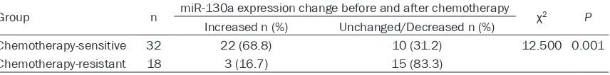
In the chemotherapy-sensitive group, miR-130a tissue expression level of 22 breast can-cer patients (68.8%) post-chemotherapy was increased compared to pre-chemotherapy, and 10 breast cancer patients (31.2%) had un- changed or decreased level. In the chemother-apy-resistant group, miR-130a tissue expres-sion level of 3 breast cancer patients (16.7%) post-chemotherapy was increased compared to pre-chemotherapy, and 15 breast cancer patients (83.3%) had unchanged or decreased
level (Table 3). These data indicated that
miR-130a levels were significantly reduced after
chemotherapy in chemotherapy-resistant gro- up. In contrast, miR-130a levels were increased obviously after chemotherapy in chemothera-py-sensitive group (P < 0.05).
These results verified that miR-130a might play
an important role in resistance to epirubicin-based chemotherapy of breast cancer.
Discussion
Nowadays, neoadjuvant chemotherapy is off- ered to patients with locally advanced breast cancer and also those breast cancer patients
who may benefit from size reduction before
conservation therapy. However, chemotherapy resistance is one of the reasons for the treat-ment failure [1, 3]. Therefore, it is important to identify the mechanism of chemotherapy resis-tance in breast cancer.
More and more evidence indicates that miR-130a is associated with drug resistance and acts as an intermediate in PI3K/Akt/PTEN/
mTOR, Wnt/β-catenin and NF-kB/PTEN drug
[image:5.612.89.519.85.139.2]resistance signaling pathways [19-21]. Yang et al [20] found that upregulation of miR-130a might be associated with MDR1/P-gp-mediated drug resistance in SKOV3/CIS cells and play the role of an intermediate in drug-resistance Table 1. Expression of miR-130a in breast cancer tissues before and after chemotherapy
Group n miR-130a expression χ2 P
High expression n (%) Low expression n (%)
[image:5.612.87.525.177.228.2]Pre-chemotherapy 50 12 (24.0) 38 (76.0) 9.458 0.002 Post-chemotherapy 50 27 (54.0) 23 (46.0)
Table 2.Relationship between miR-130a expression and the clinical response post-chemotherapy
Group n miR-130a expression χ2 P
Low expression n (%) High expression n (%)
Chemotherapy-sensitive 32 10 (31.2) 22 (68.8) 7.785 0.005 Chemotherapy-resistant 18 13 (72.2) 5 (27.8)
Table 3.Differences of miR-130a expression change before and after chemotherapy between chemotherapy-resistant and chemotherapy-sensitive groups
Group n miR-130a expression change before and after chemotherapy χ2 P
Increased n (%) Unchanged/Decreased n (%)
[image:5.612.95.523.276.329.2]pathways in PI3K/Akt/PTEN/mTOR and ABC superfamily drug transporters in ovarian cancer cells. Hu et al [21] reported that colon cancer-associated transcript-1 (CCAT1)/miR-130a axis enhanced cisplatin (DDP) resistance of non-small-cell lung cancer (NSCLC) cells by target-ing sex-determintarget-ing region Y-box 4 (SOX4). Although many studies have reported that miR-130a is related to drug resistance in several cancers, studies on miR-130a of drug resis-tance in breast cancer have not been reported. Miao et al [22] reported that microRNA-130b targets PTEN to mediate drug resistance and proliferation of breast cancer cells via the PI3K/ Akt signaling pathway. New evidence has dem-onstrated that miR-130a plays an important role in diverse physiological processes in breast cancer, including regulating cell growth, metas-tasis and invasion [23-25]. Kong et al [24] found that miR-130a-3p is down-regulated in human breast cancer tissues and exosomes from cir-culating blood, and overexpression of miR-130a-3p in breast cancer stem cells inhibits cellular proliferation, migration, and invasion. Taken together, their results demonstrated that lower levels of exosome-derived miR-130a-3p are associated with lymph node metastasis and advanced TNM stage. Their study revealed a tumor-inhibiting function of miR-130a in breast cancer, and these results are consistent
with our current findings. Our present study
demonstrated that up-regulation of miR-130a reversed Doxorubicin resistance of MCF-7/Adr cells and inhibited cell growth. We also found that miR-130a expression levels were up-regu-lated in breast cancer tissue samples from patients with advanced breast cancer receiving Epirubicin-based chemotherapy. Moreover, our results indicated that miR-130a expression lev-els in breast cancer tissue samples from patients who were sensitive to Epirubicin-based chemotherapy were higher than those who were resistant to Epirubicin-based chemothe- rapy.
In conclusion, we revealed that up-regulated miR-130a expression can enhance the sensitiv-ity of MCF-7/Adr cells to Doxorubicin. Moreover, we found that down-regulation of miR-130a expression might be associated with resistance to Epirubicin-based chemotherapy in breast cancer tissues. More efforts are needed to fur-ther delineate the biofunctional roles of miR-130a for reversing Doxorubicin resistance. It
will provide a potential target to overcome drug
resistance and improve efficacy of chemother -apy for patients with breast cancer.
Acknowledgements
This research was supported by the Fifth Cy- cle Medical Key Specialist Construction Funds of Hefei [grant no. 2016 (256)], Applied Me- dicine Research Project of the Health and Family Planning Commission of Hefei [grant no. hwk2016zd005].
Disclosure of conflict of interest
None.
Address correspondence to: Dr. Min Zhao, Depart- ment of Pathology, The Second People’s Hospital of Hefei, 246 Heping Road, Hefei 230011, Anhui, P. R. China. E-mail: zhaomin3629@163.com
References
[1] Masood S. Neoadjuvant chemotherapy in breast cancers. Womens Health (Lond) 2016; 12: 480-491.
[2] Harbeck N, Gnant M. Breast cancer. Lancet 2017; 389: 1134-1150.
[3] Munzone E, Colleoni M. Clinical overview of metronomic chemotherapy in breast cancer. Nat Rev Clin Oncol 2015; 12: 631-644. [4] Magne N, Largillier R, Marcy PY, Magne J,
Na-mer M. Cardiac toxicity assessment in locally advanced breast cancer treated neoadjuvantly with doxorubicin/paclitaxel regimen. Support Care Cancer 2005; 13: 819-825.
[5] Nitiss JL. Targeting DNA topoisomerase II in cancer chemotherapy. Nat Rev Cancer 2009; 9: 338-350.
[6] Bonadonna G, Gianni L, Santoro A, Bonfante V, Bidoli P, Casali P, Demicheli R, Valagussa P. Drugs ten years later: epirubicin. Ann Oncol 1993; 4: 359-369.
[7] Fan C, Georgiou KR, Morris HA, McKinnon RA, Keefe DMK, Howe PR, Xian CJ. Combination breast cancer chemotherapy with doxorubicin and cyclophosphamide damages bone and bone marrow in a female rat model. Breast Cancer Res Treat 2017; 165: 41-51.
[8] Shin DH, Park SH, Jeong SW, Park CW, Han K, Chung YB. Hepatic uptake of epirubicin by iso-lated rat hepatocytes and its biliary excretion after interavenous infusion in rats. Arch Pharm Res 2014; 37: 1599-1606.
therapeu-tic approaches. Physiol Rev 2016; 96: 1297-1325.
[10] Wu X, Xiao H. MiRNAs modulate the drug re-sponse of tumor cells. Sci China C life Sci 2009; 52: 797-801.
[11] Wu QB, Sheng X, Zhang N, Yang MW, Wang F. Role of microRNAs in the resistance of colorec-tal cancer to chemoradiotherapy. Mol Clin On-col 2018; 8: 523-527.
[12] Fabbri M, Valeri N, Calin GA. MicroRNAs and genomic variations: from proteus tricks to pro-metheus gift. Carcinogenesis 2009; 30: 912-917.
[13] Chen CC, Lee KD, Pai MY, Chu PY, Hsu CC, Chiu CC, Chen LT, Chang JY, Hsiao SH, Leu YW. Changes in DNA methylation are associated with the development of drug resistance in cervical cancer cells. Cancer Cell Int 2015; 15: 98.
[14] Untch M, Loibl S, Konecny GE, Minckwitz GV. Neoadjuvant clinical trials for the treatment of primary breast cancer: the experience of the german study groups. Curr Oncol Rep 2012; 14: 27-34.
[15] Serra KP, Peres RM, Sarian LO, Vassallo J, Pin-to GA, Silva GR, Soares FA,Cunha IW, Espinola J, Bento AM, Corso LM, Derchain S. Cyclooxy-genase-2 (COX2) and p53 protein expression are interdependent in breast cancer but not associated with clinico-pathological surrogate subtypes, tumor aggressiveness and patient survival. Acta Histochemica 2016; 118: 176-182.
[16] Zhu MY, Zhang W, Yang T. Diverse microRNAs with convergent functions regulate tumorigen-esis. Oncol Lett 2016; 11: 915-920.
[17] Arora A, Singh S, Bhatt AN, Pandey S, Sandhir R, Dwarakanath BS. Interplay between metab-olism and oncogenic process: role of microR-NAs. Transl Oncogenomics 2015; 7: 11-27.
[18] Frixa T, Donzelli S, Blandino G. Oncogenic Mi-croRNAs: key players in malignant transforma-tion. Cancers 2015; 7: 2466-2485.
[19] Zhang HD, Jiang LH, Sun DW, Li J, Ji ZL. The role of miR-130a in cancer. Breast Cancer 2017; 24: 521-527.
[20] Yang LY, Li N, Wang HJ, Jia XB, Wang X, Luo J. Altered microRNA expression in cisplatin-resis-tant ovarian cancer cells and upregulation of miR-130a associated with MDR1/P-glyco- protein-mediated drug resistance. Oncol Rep 2012; 28: 592-600.
[21] Hu BL, Zhang HF, Wang ZP, Zhang F, Wei HT, Li Li. LncRNA CCAT1/miR-130a-3p axis increas-es cisplatin rincreas-esistance in non-small-cell lung cancer cell line by targeting SOX4. Cancer Biol Ther 2017; 18: 974-983.
[22] Miao Y, Zheng W, Li N, Su Z, Zhao L, Zhou HM, Jia L. MicroRNA-130b targets PTEN to mediate drug resistance and proliferation of breast can-cer cells via the PI3K/Akt signaling pathway. Sci Rep 2017; 7: 41942.
[23] Chen XW, Zhao M, Huang J, Li YH, Wang SQ, Harrington CA, Qian DZ, Sun XX, Dai MS. Mi-croRNA-130a suppresses breast cancer cell migration and invasion by targeting FOSL1 and upregulating ZO-1. J Cell Biochem 2018; 119: 4945-4956.
[24] Kong XJ, Zhang JF, Li J, Shao JF, Fang L. MiR-130a-3p inhibits migration and invasion by regulating RAB5B in human breast cancer stem cell-like cells. Biochem Biophys Res Com-mun 2018; 501: 486-493.