Case Report
Ovarian endometrioid adenocarcinoma a with yolk
sac tumor in a 41-year-old woman: a case report
Hongyi Li1,3*, Yuping Xie4*, Yangmei Shen2,3
Departments of 1Gynecology and Obstetrics, 2Pathology, West China Second University Hospital, Sichuan
University, Chengdu, Sichuan, China; 3Key Laboratory of Birth Defects and Related Diseases of Women and
Children (Sichuan University), Ministry of Education, Sichuan, China; 4Department of Oncology, Chengdu First
People’s Hospital, Chengdu 610061, Sichuan, China. *Equal contributors and co-first authors.
Received June 11, 2019; Accepted July 22, 2019; Epub September 1, 2019; Published September 15, 2019
Abstract: Background: Yolk sac tumors (YSTs) are the second most common germ cell malignancy of the ovaries generally present in children and young women. YSTs arising in combination with epithelial ovarian carcinoma (EOC) in older women are rarely reported. The YST components in such cases often show a marked morphological and immunophenotypic overlap with epithelial neoplasms, making diagnosis difficult. Case report: A 41-year-old woman presented with irregular vaginal bleeding and a bilateral adnexal mass. The postoperative pathology confirmed a poorly differentiated adenocarcinoma and YST of the left ovary. The short tandem repeat (STR) analysis further in-dicated that the YST component was probably derived from an epithelial precursor neoplasm. Conclusion: This case improves our ability to detect and diagnose YST coexisting with epithelial tumors in older patients by summarizing its histopathologic characteristics and immunohistochemical stains. Molecular analysis should be used to further identify such mixed neoplasms.
Keywords: Ovarian germ cell, epithelial ovarian carcinoma, yolk sac tumor, short tandem repeat, perimenopausal, pathology
Introduction
Yolk sac tumors (YSTs) are the second most common germ cell malignancy of the ovaries generally present in children and young women. While epithelial ovarian carcinoma (EOC) is pre-dominantly a disease of older, postmenopausal women compared with other types of ovarian cancer [1], a few studies have found that YSTs can arise in combination with EOC and result in an elevated serum AFP in older women [2]. Here, we describe an unusual ovarian neo-plasm with components of poorly differentiated adenocarcinoma and YST in a 41-year-old peri-menopausal woman. In addition to highlighting the microscopic features and immunohisto-chemical patterns of this highly unique tumor,
we will briefly discuss the molecular character -istics of the two components in this case, as well as the differential diagnosis.
Case presentation
A 41-year-old woman with a history of irregular vaginal bleeding was admitted to our hospital. An ultrasound scan demonstrated a complex left adnexal mass with cystic and solid compo-nents (6.8 × 5.1 × 6.2 cm) and a cystic mass (4.6 × 4.2 × 4.5 cm) in the right appendage. The preoperative CA125 level was elevated (85.4 U/ml; normal range: 0-34 U/ml), and the serum
α-Fetoprotein (AFP) level was up to 5202.5 ng/
On gross inspection, the left oophorectomy specimen consisted of a 7 cm predominantly solid mass with an irregular-surface associated with the residual left ovary and fallopian tube. The cut surface appeared to be two different structures with solid and cystic areas, while the
solid part showed an extensive rotten fish-like
texture with necrosis and hemorrhage, and the cystic part was multilocular, and many papilla could be seen in the inner wall. The right adnex-al cystic mass contained chocolate-like, clear
inflammatory liquids, but no papillary protru -sions were detected in the inner lining. The uterine and endocervical cavities appeared grossly unremarkable.
Microscopically, the neoplastic areas of the left ovary exhibited two different patterns. Pattern A showed epithelioid differentiation and dis-played visibly irregular adenoid structures of varying size, with dirty necrosis in the glandular cavity (Figure 1A). Some of the glandular epi-theliums resembled an endometriosis cyst, with the cells showing atypical hyperplasia (Figure 1B). The glandular lining was composed of crowded, markedly atypical cells with
fre-quent mitosis, and an infiltrative growth pattern
was noted in the stroma, suggestive of poorly differentiated adenocarcinoma (Figure 1C). Pat-
tern B predominantly showed microcystic and reticular structures made up of primitive cells surrounded by a loose and myxoid background (Figure 1D). A high-power view of this pattern showed primitive nuclei, cytoplasmic vacuola-tion, and a structure similar to Schiller-Duval bodies (Figure 1E). In some areas, pleomorphic tumor cells with hyperchromatic nuclei, little cytoplasms, and conspicuous mitotic activity were also observed (Figure 1F).
[image:2.612.92.528.72.274.2]positive rate of Ki67 staining was about 80% in
[image:3.612.97.529.73.304.2]both patterns, but the remainder of the immu- nohistochemical stains listed above were nega-tive in both tumor components. Figure 2. The epithelial tumor markers EMA, CK7, CA125, and PAX8 were positive in the adenocarcinoma compo-nents but completely negative in YST.
[image:3.612.95.532.355.659.2]Since there are few reports about YST in elderly patients, we wanted to carry out some further
molecular investigation to confirm our suspi -cion. The preserved tumor specimen and a nor-mal tissue underwent a genetic comparison to determine the tumor’s origin. DNA was extract-ed from the tumor specimen and some nor- mal tissue using an AllPrep® DNA/RNA kit (Qiagen, Valencia, CA, USA) following the manu-facturer’s instructions. An AGCU EX22 PCR
amplification system (AGCU ScienTech Inc.,
Wu-xi City, China) comprising 21 autosomal STRs and one gender determination gene of the two samples was used in this study. Electrophoresis
[image:4.612.91.372.75.487.2]ond most prevalent malignant ovarian germ cell tumor (MOGCTs) histologic subtype after dys-germinoma. They occur most commonly before age 30 and usually in pure form, sometimes in association with other types of germ cell neo-plasms like teratoma, choriocarcinoma, embry-onal carcinoma, or polyembryoma [3]. YSTs coexisting with a variety of histologic patterns have been described, but those with an epithe-lial malignant component are extremely rare, especially in older women. Among all the ovari-an epithelial neoplasms associated with YST, it seems that endometrioid carcinoma is the most common epithelial component, occurring Figure 4. Short tandem repeat (STR)-based concordance study of the 22
microsatellite markers tested, two different alleles (A, D16S539 and B, CSF1PO) were found between the tumor and the patient’s normal ovarian tissue.
was performed using an ABI 3130XL Genetic Analyzer (App- lied Biosystems). The fragment sizes were automatically deter-mined using Genemapper ID software (Version 3.2.1; App- lied Biosystems). The STR re- sults showed that, of the 22 microsatellite markers tested, differences were found betwe- en the tumor cells and the pa- tient’s normal tissue (Figure 4A and 4B). We presumed that the mixed-type tumor was a microsatellite instability and the YST component was prob-ably derived from the epithelial precursor neoplasm.
Based on the morphologic, IHC features and STR results, an extremely unusual combinati- on of poorly differentiated ade-nocarcinoma with malignant YST arising in the left ovary was diagnosed.
The patient recently finished 6
cycles of intraperitoneal BEP (bleomycin + etoposide + cis-platin) chemotherapy and de- veloped neutropenia and oth- er tolerant side effects. Her serum AFP level is normal, and she is currently alive with no evidence of disease.
Discussion
sec-in 12 cases (67%), and 6 of these were associ-ated with an endometriotic cyst reported in a previous case report [4]. Here, we describe a rare case of ovarian endometrioid adenocarci-noma with a YST component occurring in a 41-year-old woman. Most of the cases of this type have occurred in postmenopausal or peri-menopausal women, but the age distribution contrasts with the much younger age of pa- tients with typical YST [5]. The bimodal age dis-tribution of patients with ovarian YST suggests differing histogenetic mechanisms of tumori-genesis in the 2 age groups. As germ cells are
not identified histologically in the ovaries of
postmenopausal women, a direct origin of ma- lignant neoplasms from germ cells is highly unlikely at that age. We highly suspected that this rare mixed tumor represents an adenocar-cinoma with aberrant differentiation because it occurs in the same age range as EOC and shows an aggressive behavior, but the molecu-lar events involved in this transformation have not yet been elucidated.
It is likely that the YST component can show transitional forms and a wide variety of pat-terns that frequently occur in combination. Some of these patterns are rare and can be confused with other types of tumors. Patho- logically, the endometrioid-like variant of YST simulated primary endometrioid
adenocarcino-ma sometimes adenocarcino-makes diagnosis difficult. The endometrioid-like pattern reflects an unusual
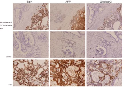
form of endodermal differentiation within yolk sac tumors that should be distinguished from endometrioid carcinoma, the recognition of which should be facilitated by a panel of im- munohistochemical stains that are often ex- pressed differentially in these two neoplasms— endometrioid-like yolk sac tumors are frequent-ly reactive for AFP, SALL4, and Gfrequent-ly-3, but CK7 and EMA are usually negative. Endometrioid adenocarcinomas of the ovaries tend to have opposite patterns of immunoreactivity [6, 7].
The identification of the specific morphologic
features of mixed ovarian tumors is vitally important since it is very easy for the patholo-gist to misdiagnose these groups of tumors as having two different origins [8]. In recent years, STR (also called microsatellite) analysis has been used extensively as a diagnostic tool to
compare specific loci on DNA from two or more samples for the detection and classification of
tumors [9]. Loci that show alterations in frag-ment length between normal and tumor tissue are considered to be unstable, but loci that exhibit no length variations between normal and tumor tissue are considered stable. Mi-
crosatellite instability in tumor DNA is defined
as the presence of alternate sized repetitive DNA sequences that are not present in the cor-responding germline DNA [10]. Therefore, STR analysis was used to determine the tumor ori-gin in this case. The results showed that, com-pared with the normal tissue, there were muta-tions of 2 alleles in the mixed tumor genotypes, which means the tumor existed with unstable microsatellites. In other reported studies of endometrioid adenocarcinoma with a compo-nent of ovarian YST, the authors thought that somatic carcinomas have the ability to acquire a germ cell differentiation, and the germ cell component is thought to derive from somatic mesodermal cells and not from germ cells [11-13]. Although the exact explanation for this bio-logic behavior is not known, our case along with other reported reports have raised the possibil-ity that the YST does not always have a germ cell origin and can be derived from epithelial tumors, perhaps through one of the four theo-ries including the teratoma theory, retrodiffer-entiation, collision theory, and neometaplasia [14]. Additionally, the term “somatically derived YSTs” should be used to categorize these neo-plasms. Further molecular analysis may also provide information about better therapeutic approaches for the treatment of these types of rare tumors in the future.
postoperative treatment consisting of 6 cours-es of the BEP regimen, and the patient is cur-rently alive with no evidence of disease for 8 months. We hope that other authors describe
their experiences to define the most appropri -ate approach to this rare tumor.
Disclosure of conflict of interest
None.
Address correspondence to: Yangmei Shen, De- partment of Pathology, West China Second Uni- versity Hospital, Sichuan University, Chengdu 610041, Sichuan, China. Tel: 0086-13036664276; E-mail: sym.julia@163.com
References
[1] Ledermann JA, Raja FA, Fotopoulou C, Gonza-lez-Martin A, Colombo N, Sessa C; ESMO Guidelines Working Group. Newly diagnosed and relapsed epithelial ovarian carcinoma: ESMO clinical practice guidelines for diagno-sis, treatment and follow-up. Ann Oncol 2018; 29 Suppl: iv259.
[2] McNamee T, Damato S, McCluggage WG. Yolk sac tumours of the female genital tract in older adults derive commonly from somatic epitheli-al neoplasms: somaticepitheli-ally derived yolk sac tu-mours. Histopathology 2016; 69: 739-751. [3] Goyal LD, Kaur S, Kawatra K. Malignant mixed
germ cell tumour of ovary-an unusual combi-nation and review of literature. J Ovarian Res 2014; 7: 91.
[4] Roth LM, Talerman A, Levy T, Sukmanov O, Cz-ernobilsky B. Ovarian yolk sac tumors in older women arising from epithelial ovarian tumors or with no detectable epithelial component. Int J Gynecol Pathol 2011; 30: 442-451.
[5] Nasioudis D, Chapman-Davis E, Frey MK, Ca-puto TA, Holcomb K. Management and progno-sis of ovarian yolk sac tumors; an analyprogno-sis of the National Cancer Data Base. Gynecol Oncol 2017; 147: 296-301.
[6] Ulbright TM. Gonadoblastoma and hepatoid and endometrioid-like yolk sac tumor: an up-date. Int J Gynecol Pathol 2014; 33: 365-373. [7] Ramalingam P, Malpica A, Silva EG,
Gershen-son DM, Liu JL, Deavers MT. The use of cyto-keratin 7 and EMA in differentiating ovarian yolk sac tumors from endometrioid and clear cell carcinomas. Am J Surg Pathol 2004; 28: 1499-1505.
[8] Sadlecki P, Walentowicz-Sadlecka M, Grabiec M. Molecular diagnosis in type I epithelial ovar-ian cancer. Ginekol Pol 2017; 88: 692-697. [9] Hile SE, Shabashev S, Eckert KA.
Tumor-specif-ic mTumor-specif-icrosatellite instability: do distinct mecha-nisms underlie the MSI-L and EMAST pheno-types? Mutat Res 2013; 743-744: 67-77. [10] Nojadeh JN, Behrouz Sharif S, Sakhinia E.
Mic-rosatellite instability in colorectal cancer. EX-CLI J 2018; 17: 159-168.
[11] Hong DG, Chong GO, Seong WJ, Lee YS, Cho YL, Park JY, Chae JM, Park IS. A case of ovarian endometrioid adenocarcinoma with yolk sac tumor in a 35-year-old woman. Eur J Gynaecol Oncol 2010; 31: 471-474.
[12] McBee WC Jr, Brainard J, Sawady J, Rose PG. Yolk sac tumor of the ovary associated with en-dometrioid carcinoma with metastasis to the vagina: a case report. Gynecol Oncol 2007; 105: 244-247.
[13] Rutgers JL, Young RH, Scully RE. Ovarian yolk sac tumor arising from an endometrioid carci-noma. Hum Pathol 1987; 18: 1296-1299. [14] Giuliani J, Marzola M, Pizzutilo P, Martinello R,
Marzola A, Indelli M, Frassoldati A. Ovarian en-dometrioid adenocarcinoma with a yolk sac tumor component in a postmenopausal wom-an: case report and review of the literature. Clinical Ovarian and Other Gynecologic Cancer 2012; 5: 31-32.