Case Report
Intimal sarcoma of the abdominal
aorta and common iliac arteries presenting
as epithelioid angiosarcoma of the skin: a case report
Shogo Tajima1, Katsuyuki Hoshina2, Tets Oushik1, Kunihiro Shigematsu2, Masashi Fukayama1
1Department of Pathology, Graduate School of Medicine, The University of Tokyo, Tokyo, Japan; 2Division of
Vascular Surgery, Department of Surgery, Graduate School of Medicine, The University of Tokyo, Tokyo, Japan
Received March 1, 2015; Accepted April 15, 2015;Epub May 1, 2015; Published May 15, 2015
Abstract: Intimal sarcoma (IS) is the most common type of sarcoma of the aorta. IS tumor emboli can involve various organs, including the skin. However, a limited number of IS cases with an initial presentation of skin metastasis has been reported. Cutaneous metastasis as a form of epithelioid angiosarcoma (EAS) has not been well described. Herein, we present a 61-year-old Japanese man with an initial presentation of EAS of the skin, followed by multiple metastases to the skin as a form of EAS prior to detection of IS of the infrarenal aorta and common iliac arteries. In our case, the IS was CD31 and cytokeratin positive but did not express CD34 and factor VIII-related antigen. The EASs in our case exhibited diffuse CD31 expression, and focal factor VIII-related antigen and cytokeratin expression
were observed throughout the tumor, including the neoplastic vascular structure; CD34 expression was not identifi -able. IS metastasis to the skin has been documented as a form of angiosarcoma. However, IS metastasis has not been well described as a form of EAS. Our case could prove a morphological change from IS to EAS. Given the rarity of primary cutaneous EAS, it is recommended that primary sites other than the skin should be thoroughly investi-gated when EAS of the skin is encountered.
Keywords: Intimal sarcoma, aorta, common iliac artery, metastasis, skin, epithelioid angiosarcoma
Introduction
Two types of aortic sarcoma, intimal and mural, have been reported [1]. The intimal type extends along the lumen and often forms intra-luminal polyps that cause aortic obstruction or peripheral emboli; the mural type develops in the media or adventitia and usually grows extramurally to para-aortic tissues [2-4]. The luminal-type intimal sarcoma (IS) is the most common sarcoma of the aorta [5]. IS often involves the abdominal aorta among other large systemic arteries, and it is occasionally observed to span both the abdominal aorta and the iliac artery [5, 6]. Tumor emboli of IS involve various organs, including the skin, brain, lungs, bowels, kidneys, liver, and bones [7]. However, only a few cases of IS presenting ini-tially as skin metastasis have been reported [7, 8]. IS metastasis to the skin has been docu-mented as a form of angiosarcoma [5, 8].
However, IS metastasis as a form of epithelioid angiosarcoma (EAS) has not been well described.
Herein, we present a 61-year-old Japanese man who presented initially with EAS of the skin, fol-lowed by detection of IS of the infrarenal aorta and common iliac arteries.
Clinical summary
A 61-year-old Japanese man was seen at an outpatient clinic because of a painful nodule on the penile skin. The patient reported that he had noticed the nodule approximately 1 month before presentation and had not experienced with any recent trauma. At the outpatient clinic,
he reported no fever, fatigue, or other specific
Intimal sarcoma presenting as cutaneous epithelioid angiosarcoma
5857 Int J Clin Exp Pathol 2015;8(5):5856-5862
nodes were observed. Subsequently, excision of the penile skin nodule was performed. The pathological diagnosis was angiosarcoma. An enhanced computed tomography (CT) scan revealed a few nodules in the lungs; these con-sidered metastases of the cutaneous angiosar-coma to the lung. The patient was referred to our hospital 1 month after presentation at the clinic for intensive examination and treatment. He then complained of another painful nodule on his left abdomen. Another skin excision was performed, and the biopsy results were consis-tent with angiosarcoma. Positron emission tomography (PET) demonstrated increased tracer uptake in the L5 vertebral body, the left side and S3 level of the sacrum, and the left calcaneus. At that time, primary cutaneous angiosarcoma and metastasis to multiple sites were not considered. Extradermal primary sites
were investigated but could not be confirmed.
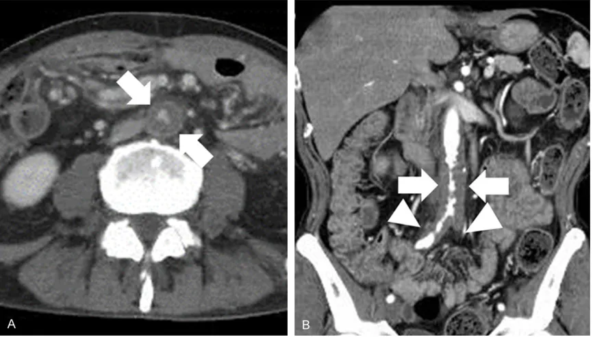
The patient received 5 cycles of docetaxel-based (60 mg/m2) chemotherapy. Seven months after the initial diagnosis, he developed metastatic disease on his back, groin, and penis. These lesions were excised and he began to receive pazopanib (200 mg/day) ther-apy. Two months after the excision, he began to exhibit intermittent claudication, and contrast-enhanced CT showed an exacerbating
thick-ened abdominal aortic wall from below the level of the renal arteries to the bilateral common iliac arteries (Figure 1A, 1B). The patient was admitted for vascular surgery. A transplant from the infrarenal aorta to the bilateral com-mon iliac arteries was performed. Pathological
findings revealed an IS. Proximal and distal sur -gical margins were all positive. Besides pain
from the left fibula metastasis, the patient
recovered well from this major surgical intervention.
Pathological findings
[image:2.612.91.524.72.318.2]Intimal sarcoma presenting as cutaneous epithelioid angiosarcoma
5859 Int J Clin Exp Pathol 2015;8(5):5856-5862
lar formation (AE1/AE3, 1:100; Dako) (Figure 2F); however, the samples were negative for CD34 (QBEnd 10, 1:100; Dako). A diagnosis of angiosarcoma was rendered for each excised specimen.
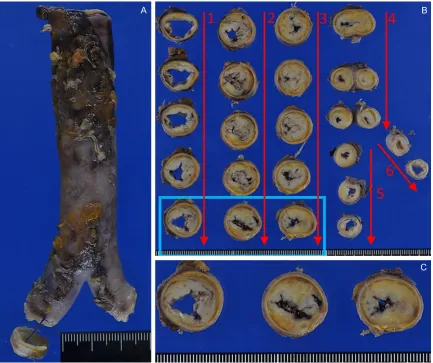
The surgically resected abdominal aorta and common iliac arteries included 9 cm of the
[image:4.612.91.524.161.524.2]infrarenal aorta, 3 cm of the right common iliac artery, 0.5 cm of additionally resected tissue, and 2.5 cm of the left common iliac artery (Figure 3A). The cut surface revealed the for-mation of a whitish thrombus along the entire length of the resected aorta and common iliac arteries; this thrombus nearly occluded the lumen in the center of the resected aorta Figure 2. Microscopic findings of skin excision specimens. A. Representative specimen showed a tumor in the der
-mis with a hemorrhagic appearance (1.25× magnification). B. Constituent tumor cells were nearly identical among
the specimens and formed focal vascular structures (400×). C. Tumor cells exhibited an epithelioid morphology with enlarged nuclei and prominent nucleoli; some cells had intracytoplasmic lumina (600×). D. Tumor cells exhib-ited strong immunostaining for CD31 (400×). E. Tumor cells exhibexhib-ited focal immunostaining for factor VIII-related antigen (400×). F. Tumor cells exhibited strong immunostaining for pan-cytokeratin, including the area of vascular formation (400×).
Intimal sarcoma presenting as cutaneous epithelioid angiosarcoma
5861 Int J Clin Exp Pathol 2015;8(5):5856-5862
(Figure 3B). Yellowish atherosclerosis was observed beneath the whitish thrombus, and no mass was apparent (Figure 3C). His- topathological examination of the surgically resected specimen revealed a large circumfer-ential thrombus, particularly at the central part of the infrarenal aorta (Figure 4A). A tumor cell layer was present on the thrombus surface (Figure 4B). At the proximal and distal margins, the thrombus was smaller and patchily distrib-uted, resulting in areas without thrombus (Figure 4C); these areas showed direct contact between the tumor cells and the intima (Figure 4D). Tumor cells with a short-spindled to epithe-lioid morphology formed a layer with polygonal cells at the luminal side and short-spindled cells at the opposite side (Figure 4E). These cells had enlarged nuclei with prominent nucle-oli (Figure 4E, inset). IHC revealed that these cells were strongly positive for CD31 (Figure 4F), negative for factor VIII-related antigen (Figure 4G) and CD34, and positive for pan-cytokeratin (Figure 4H). A diagnosis of IS was rendered.
Upon reviewing the biopsy specimens, we con-sidered that this angiosarcoma might be better
classified as EAS, given the epithelioid mor -phology and pan-cytokeratin positivity.
Discussion
Reports of IHC-detected CD31 and cytokeratin expression in IS vary among studies. CD31 expression, including focal positivity, was previ-ously reported in 13 of 13 cases (100%) [5], 0 of 42 cases (0%) [9], and 13 of 26 cases (50%) [6]; cytokeratin expression was observed in 2 of 13 cases (15%) [5], 6 of 42 cases (14%) [9],
and 10 of 26 (38%) [6]. Some authors define
CD31-positive IS as angiosarcoma and CD31- and cytokeratin double-positive IS as EAS even if the morphological features of vascular differ-entiation are not apparent [6]. However, we did
not want to describe a case of IS as angiosar-coma if CD31 expression did not accompany morphological vascular differentiation. This viewpoint is supported by a study in which IS was shown to express at least focal CD31 [5]. In our case, although the IS strongly expressed CD31 and cytokeratin, we did not diagnose it as EAS because of its lack of morphological vas-cular differentiation.
In contrast, the skin metastases of IS in our case expressed these markers along with vas-cular formation and epithelioid morphology in more than 90% of the constituent tumor cells. The term EAS should be strictly applied to tumors in which more than 80-90% of constitu--90% of constitu-90% of constitu-ent cells exhibit epithelioid morphology [10]. It was therefore valid to designate the metastatic skin lesions in our case as EAS.
Both primary and metastatic cutaneous angio-sarcomas are rare among malignant tumors of the skin. Primary cutaneous angiosarcomas arise in three main settings [11-13]: the head and neck of elderly patients; post-radiotherapy; and in association with lymphedema. Focal epi-thelioid changes are frequently observed in angiosarcomas arising in these three main set-tings; however, EAS is more often encountered outside of these settings. EAS is most com-monly observed in deep, soft tissue and is rare-ly encountered as a primary skin tumor [10]. Therefore, when encountering EAS of the skin, it would be wise to consider the possibility that it might be a cutaneous metastasis rather than primary cutaneous tumor. Although cases of cutaneous metastasis of EAS originating in a non-skin site are exceptional [10], cutaneous metastases from cardiac and mediastinal EAS have been documented [14, 15]. In cases such as ours involving multiple skin and internal organ lesions, it would be impossible to deter-mine whether the skin lesion was a primary cutaneous or metastatic lesion [10].
Figure 4. Microscopic findings of the surgically resected infrarenal aorta and common iliac arteries. (A) A large circumferential thrombus, especially at the central part of the infrarenal aorta (1.25× magnification). (B) Higher magnification of the boxed area in (A). The thrombus surface features a tumor cell layer (arrows) (40×). (C) The
In conclusion, at the metastatic site, IS could change to EAS with vascular formation. Given the rarity of primary cutaneous EAS, primary sites other than the skin should be thoroughly evaluated when encountering EAS of the skin.
Disclosure of conflict of interest
None.
Address correspondence to: Dr. Shogo Tajima, Department of Pathology, Graduate School of Medicine, the University of Tokyo, 7-3-1 Hongo, Bun- kyo-ku, Tokyo 113-0033, Japan. Tel: +81-3-5841-3341; Fax: +81-3-3815-8379; E-mail: stajima-tky@ umin.ac.jp
References
[1] Wright EP, Glick AD, Virmani R and Page DL. Aortic intimal sarcoma with embolic metasta-ses. Am J Surg Pathol 1985; 9: 890-897. [2] Shirani S, Soleymanzadeh-Ardabili M and
Arami M. Intimal sarcoma of the descending aorta. Arch Iran Med 2007; 10: 253-254. [3] Salm R. Primary fibrosarcoma of aorta. Cancer
1972; 29: 73-83.
[4] le Rochais JP, Icard P, Coffin O, Galateau F,
Khalil M and Maiza D. Intimal sarcoma of the thoracic aorta: a case report. Eur J Vasc Endovasc Surg 1999; 18: 181-182.
[5] Sebenik M, Ricci A Jr, DiPasquale B, Mody K, Pytel P, Jee KJ, Knuutila S and Scholes J. Undifferentiated intimal sarcoma of large sys-temic blood vessels: report of 14 cases with
immunohistochemical profile and review of the
literature. Am J Surg Pathol 2005; 29: 1184-1193.
[6] Staats P, Tavora F and Burke AP. Intimal sarco-mas of the aorta and iliofemoral arteries: a clinicopathological study of 26 cases. Pathology 2014; 46: 596-603.
[7] Sparsa A, Liozon E, Wechsler J, Soria P, Delage-Core M, Loustaud V, Bedane C, Bonnetblanc JM and Vidal E. Aortic angiosarcoma clinically mimicking polyarteritis nodosa. Scand J Rheumatol 2006; 35: 237-240.
[8] Rudd RJ, Fair KP and Patterson JW. Aortic an-giosarcoma presenting with cutaneous metas-tasis: case report and review of the literature. J Am Acad Dermatol 2000; 43: 930-933. [9] Neuville A, Collin F, Bruneval P, Parrens M,
Thivolet F, Gomez-Brouchet A, Terrier P, de Montpreville VT, Le Gall F, Hostein I, Lagarde P, Chibon F and Coindre JM. Intimal sarcoma is the most frequent primary cardiac sarcoma: clinicopathologic and molecular retrospective analysis of 100 primary cardiac sarcomas. Am J Surg Pathol 2014; 38: 461-469.
[10] Suchak R, Thway K, Zelger B, Fisher C and Calonje E. Primary cutaneous epithelioid an-giosarcoma: a clinicopathologic study of 13 cases of a rare neoplasm occurring outside the setting of conventional angiosarcomas and with predilection for the limbs. Am J Surg Pathol 2011; 35: 60-69.
[11] Alessi E, Sala F and Berti E. Angiosarcomas in lymphedematous limbs. Am J Dermatopathol 1986; 8: 371-378.
[12] Billings SD, McKenney JK, Folpe AL, Hardacre MC and Weiss SW. Cutaneous angiosarcoma following breast-conserving surgery and radia-tion: an analysis of 27 cases. Am J Surg Pathol 2004; 28: 781-788.
[13] Mark RJ, Tran LM, Sercarz J, Fu YS, Calcaterra TC and Juillard GF. Angiosarcoma of the head and neck. The UCLA experience 1955 through 1990. Arch Otolaryngol Head Neck Surg 1993; 119: 973-978.
[14] Val-Bernal JF, Figols J, Arce FP and Sanz-Ortiz J. Cardiac epithelioid angiosarcoma presenting as cutaneous metastases. J Cutan Pathol 2001; 28: 265-270.
[15] Chen TJ, Chiou CC, Chen CH, Kuo TT and Hong HS. Metastasis of mediastinal epithelioid
an-giosarcoma to the finger. Am J Clin Dermatol