Original Article
Decrease in prosaposin in spermatozoon is associated
with polychlorinated biphenyl exposure
Jia-Li Cai1, Ling-Bin Sun1, Zhi-Zhun Guo2, Xiao-Ming Jiang1, Guan-Chao Zheng2, Hui-Ling Qiu1, Ai-Guo Sha1,
Chong-Gang Wang2, Jian-Zhi Ren1, Zheng-Hong Zuo2
1Reproductive Medicine Center, The Affiliated Chenggong Hospital of Xiamen University, Xiamen, Fujian 361002, China; 2State Key Laboratory of Cellular Stress Biology, School of Life Sciences, Xiamen University, Xiamen
361005, China
Received January 11, 2015; Accepted February 28, 2015; Epub March 1, 2015; Published March 15, 2015
Abstract: Polychlorinated biphenyls (PCBs) are a class of ubiquitous persistent organic pollutants and they have been associated with declining male fertility. In the present study, we aimed to determine the responsiveness of prosaposin (Psap) expression to PCB exposure. Male C57 mice were exposed to PCB mixture (Aroclor 1254) of environmental related doses by oral gavage. After exposure for 50 days, the expression of Psap was significantly decreased by PCB exposure in epididymides and epydidymal spermatozoa, but not in testis. The Psap abundance in sperm was decreased in a dose-dependent manner. Benchmark dose modeling revealed the 95% lower confidence limit on the benchmark dose (BMDL) and Benchmark Dose (BMD) for Psap reduction were 1.25 and 8.89 μg/kg Aroclor 1254, and for sperm motility reduction were 11.85 and 61.9 μg/kg Aroclor 1254. The depressed Psap level also showed a significant correlation (P<0.01, r=-0.531) with PCB accumulation in liver. In men with detectable PCB exposure in semen, Psap expression in sperm was significantly decreased whereas the semen parameters were unaffected. Linear regression showed that a negative association between total PCB level in seminal plasma and Psap level in ejaculated spermatozoa (P<0.05, r=-0.396). In conclusion, our data suggested that the abundance of Psap in sperm sample may be a sensitive endpoint to predict PCB exposure.
Keywords: Polychlorinated biphenlys, prosaposin, sperm, human, mice
Introduction
Polychlorinated biphenyls (PCBs) are a class of legacy chemicals with high environmental per-sistency. Despite the ban on their production, PCBs remain to be a global environmental problem due to their high resistance against electrical, thermal, chemical, or biological breakdown and ongoing “leaking” to the envi-ronment from existing applications and waste. Accumulated evidence have associated expo-sure to PCBs with many adverse health effects, including effects on reproduction [30] and development [34], thyroid system [36], nervous system [27], immune system [28], cardiovascu-lar system [22], and metabolism [18].
The effects of PCBs on male reproductive sys-tem have long been of concerned. PCB conge-ners have shown estrogenic, antiestrogenic, or anti-androgenic effects [16] which might
dis-rupt the male reproductive functions and hypo-thyroidism induced by PCB exposure in early life also results in declines in reproductive end-points. A number of animal studies have report-ed adverse effects in relation to PCBs following both developmental exposures and exposure in adulthood, including reduced organ weights, impaired spermatogenesis, and disrupted ste-roid hormone function [1, 5, 6, 11]. Epide- miological studies have associated exposure of human to PCBs with declining semen parame-ters, sperm DNA integrity, and circulating repro-ductive hormone levels [30]. Impaired male reproductive functions are undoubtedly impact
final reproductive outcomes. A recent study has
demonstrated that exposures of PCBs in male partner might contribute to decreased human fecundity, as measured by a prolonged time to
In toxicological studies, biomarkers of effect are used to associate the xenobiotic exposures with their harmful downstream health conse-quences. They are typically physiological indi-cators which presence after physiologically rel-evant doses and response to xenobiotic of
interest in a specific and sensitive manner.
Furthermore, they should be facile in measure-ment and validatable in humans exposed to the toxin. Semen parameters are the most fre-quently used biomarkers to investigate the xenobiotic-related effects on male fertility. They are correlated directly to the fertility and can be measured objectively with a relative simple tool [33]. However, semen quality parameters pro-vide less information about the mechanism of the action and the organ targeted. Toxicants can affect the male reproductive system at one of several sites, or at multiplesites, as well as physiological functions. There is so far no single all-encompassing biomarker of reproductive capacity in men. Identifying novel biomarkers
reflecting have no doubt enabled us to better
evaluate the effect of the exposure on the male reproductive system.
The epididymis is known to play an important role in providing the microenvironment for sperm maturation and storage of sperm [13]. Numerous proteins synthesized and secreted by the epididymal epithelia cells are thought to be involved in male reproductive activities including the initiation of sperm maturation, sperm-oocyte recognition, the acrosome reac-tion, interaction with the zona pellucida, and binding to and fuse with plasma membrane of the oocyte. The caput epididymis is the most metabolically active region, accounting for 70-80% of the total overall protein secretion in the epididymal lumen [13]. Characterizing glob-al transcript responses to PCBs in the caput epididymis might yield a clue concerning the potential mechanisms for their toxicological effects and might provide candidate markers for fertility and toxicologic evaluations.
In our previous studies, we have demonstrated
that low doses of PCB exposure up to 500 μg/
kg impaired sperm count, sperm motility and sperm morphology without signiciantly affect-ing the weight or morphology of the organs [10, 11]. The exposure also induced a global alter-nation in the gene expression of caput epididy-mis [10]. Amongst the dysregulated genes, the
expression of prosaposin (Psap) mRNA is dose-dependently down-regulated in the microarray data. Psap is a 65-70 kilodalton glycoprotein that is targeted to the lysosome, as well as is
secreted to extracellular fluids (Hiraiwa et al,
1992) and a role of Psap in sperm-oocyte inter-action has been suggested [29] (Magargee et al. 2000). The goals of this study were to vali-date the effects of PCB exposure on the protein expression of Psap in male reproductive tract and to determine whether PCB-induced diminu-tions in Psap were present on epididymal sperm.
Materials and methods
Animals and treatment
All animal experiments were conducted accord-ing to the research protocols approved by the Xiamen University Institutional Animal Care and Use Committee. Male C57 mice, aged 21 days and weighing 12-14 g, were purchased from Fujian Medical University, China, housed at 24±1°C under a 12:12 h light-dark cycle, with free access to food and water. After a quaran-tine period, mice with adequate weight gain and without clinical signs were divided random-ly into three experimental groups. Aroclor 1254 of analytical grade purity (lot LB38310; Supelco, Bellefonte, PA) was dissolved in a 10% ethanol saline solution as previously described [10, 11] and serially diluted with the same solu-tion for exposure experiments. The mice in each treatment were administered with Aroclor 1254 by oral gavage once every three days
(either 5 or 500 μg/kg doses); control mice received an equal volume of vehicle (5 μL/g). The mice were sacrificed after 50 days expo -sure under slight ether anesthesia. The epididy-mis was removed, cleared of adhering connec-tive tissue and stored at -80°C for further anal-ysis or placed in 4% formaldehyde for 24 h, rinsed in water, and stored in 70% ethanol until further processing for immunohistochemical examination.
The epididymis of each mouse was excised and rinsed with prewarmed PBS. The isolated Cauda epididymis was placed in 1 mL of human
tuba fluid (Chemicon, Temecula, CA), cut finely
evaluation. Evaluation of sperm motility was carried out as previously described [40]. RNA isolation and Real-time PCR
Total RNA was extracted with Trizol (Invitrogen, Carlsbad, CA) according to the manufacturer’s instructions. One milligram of RNA was reverse-transcribed using the Quantitect Reverse Transcription kit (QIAGEN, Valencia, CA). Quantitative RT-PCR was performed using
SYBR green fluorescence and the Qiagen Rotor
Gene 3000 Real-Time PCR cycler (QIAGEN, Valencia, CA). Each reaction was run in triupli-cate and consisted of 20 ng of cDNA, Fast Power SYBR Green PCR System (Applied Bio-
systems) and 300 nM of the gene-specific prim -ers. The fold change in gene expression was calculated using the comparative cycle thresh-old method with the housekeeping genes. The expression of L32, RPL11 and Cyclophilin were
used as internal control in the quantification.
PCR was performed using the following prim-ers: Psap: 5’-GCCAGAGGGCAGGAGCATT and 5’-CTGACCCAGGGACAGCAACA, L32: 5’-TCCAC- AATGTCAAGGAGCTG and 5’-ACTCATTTTCTTC- GCTGCGT, RPS11: 5’-CGTGACGAAGATGAAGA- TGC and 5’-GCACATTGAATCGCACAGTC, Cyclo- philin: 5’-ACACGCCATAATGGCACTGG and 5’- ATTTGCCATGGACAAGATGCC.
Western blotting assay
Protein was extracted from frozen tissues using
homogenization, and fraction samples (40 μg
proteins) were mixed with 2× Coomassie bril-liant blue, heated to 100°C in a water bath for 5 min. Next, 10% sodium dodecyl sulfate poly-acrylamide gel electrophoresis (SDS-PAGE) was performed at a constant voltage of 100 V for 1.5 h. After SDS-PAGE, the proteins were transferred to a nitrocellulose membrane and blocked at room temperature for 2 h in PBS buffer containing 5% nonfat dried milk or 5%
bovine serum albumin to prevent nonspecific
binding of reagents, and then incubated over-night at 4°C with a primary antibody. Anti-Psap antibody was obtained from Abcam (Cambridge, MA). After this, the membrane was washed three times in PBST for 15 min and incubated with secondary antibody (1:10000 dilution; Pierce, USA) for 1 h at room temperature. Then the membrane was washed three times in PBST and chemiluminescence detection (Sigma, UK) was performed. The intensity of
bands was quantified using Quantity One soft -ware (Bio-Rad).
Immunohistochemistry
Fixed tissues were embedded in paraffin and cut into 6 μm sections. The sections were depa
-raffinized, rehydrated, and stained following the
previously described methods [11]. To optimize immunohistochemical staining, an antigen retri- eval protocol was carried out by immersing the sections in 10 mM citrate buffer (pH 6.0) and heating in a microwave. The sections were incu-bated with anti-Psap antibodies overnight at
4°C in a humidified chamber and the negative
control was incubated in the presence of irrelev- ant IgG instead of the primary antibodies. Psap measurement in human spermatozoa and seminal plasma
A total of 36 male counterparts of women
visit-ing the Affiliated Chenggong Hospital of Xiamen
University for in vitro fertilization (IVF) treat-ment were enrolled in the study and donated the semen samples. The study was approved by the Institutional Review Board and informed consent was obtained from participants. All couples were diagnosed as female factor infer-tility and the cases with known causes of male infertility such as a history of orchitis, cryptor-chidism, varicocele, vas deferens obstruction, and Y chromosome microdeletions were exclud-ed. Semen parameters were analyzed accord-ing to WHO criteria (WHO 2010). Semen sam-ples were centrifuged to separate the sperm from seminal plasma. For the resulting sperm, sperm proteins were isolated as previously described [7]. The sperm lysate and the semi-nal plasma were stored at -80°C for following Psap measurement.
Psap was determined using an ELISA kit (Uscn Life Science Inc., Wuhan, China) according to the manufacturer’s instructions. The enzyme-substrate reaction is terminated by the addition of a sulphuric acid solution and the color change is measured spectrophotometrically at a wavelength of 450 nm. The concentration of Psap in the samples is then determined by comparing the O.D. of the samples to the stan-dard curve. The minimum detectable level of Psap detected is less than 8.2 pg/ml.
Determination of PCBs
Briefly, 1 g portion sample was weighted and
25 mL acetone-n-hexane (1:1, v/v) was used as the extractant. The extraction was carried out using MARS-X microwave-accelerated reaction system model (CEM, USA) under 100°C, 35 psi for 15 min at 300 W power output. The MAE solution were cooled to room temperature and dried through a short column packed with anhy-drous Na2SO4. A cleanup on a SiO2-Al2O3 col-umn was followed and the colcol-umns were eluted
with n-hexane. The eluate was finally concen -trated to 1 ml under nitrogen for Gas chromato-graphic (GC) analysis.
For semen samples from human participants,
1 ml seminal plasma was fortified with 20 μl
internal standard solution of 4, 4’-Dib-
romooctafluorobiphenyl (1000 ppb). The sam -ple was thereafter mixed with 1 ml formic acid and equilibrated by ultrasonic treatment for 30 min. The resulting solution was extracted with solid phase extraction (SPE) cartridges. Prior to the sample application, the cartridges were washed with dichloromethane and activated with methanol and water. After conditioning, the cartridges were not allowed to dry. After sample loading, the SPE cartridge was rinsed with 1 ml water. The sorbent bed was dried by centrifugation (15 min, 3000 rotations/min). A SiO2-Al2O3 column was placed under the SPE cartridges. The SPE cartridge was eluted with
2×500 μl n-hexane and 2×500 μl dichlorometh -ane directly into the clean-up cartridge. Further, PCBs were eluted with 3 ml n-hexane and 3 ml dichloromethane from the clean-up cartridge.
The final eluate was concentrated and stored
as described above for liver samples.
An Agilent 6890N with electron capture
detec-tion (ECD) was used for PCB quantificadetec-tion
(Agilent, USA). The column employed was a
DB-5 (Agilent, USA). An aliquot of 1 μl of stan -dard solution or sample solution was injected for each GC-ECD analysis. The number of deter-minations for each sample was three. Nitrogen
was both the carrier gas at a flow-rate of 1.5
ml/min and the make-up gas at 60 ml/min. The temperature of the injector and the detector were maintained at 280 and 300°C, respec-tively. The column was programmed from 85°C, held for 2 min, to 180°C at 30°C/min, held for 2 min, to 200°C at 20°C/min, held for 3 min, to 230°C at 3°C/min, to 250°C at 5°C/min and held for 8 min.
Quantification of PCBs was performed using
external standard calibration. To determine
Aroclor 1254 accumulation in exposed mice, Aroclor 1254 was used as external standard and eleven major peaks were selected for
quantification. To determine environmental
exposure of PCBs in seminal plasma, twelve PCB congeners (PCB 8, PCB 18, PCB 28, PCB 44, PCB 66, PCB 101, PCB 103, PCB 153, PCB 138, PCB 187, PCB 128 and PCB 206) were used as standard. Peak area ratios (analyte response/internal standard response) were plotted against the concentration ratios (ana-lyte concentration/internal standard
concen-tration). Method limits of quantification (LOQ)
were calculated as 3× standard deviation of the analyte values in procedural blanks. Method LOQs for individual PCBs ranged between were 2 to 6 ng/g LW. Recoveries ranged between 66% PCBs in semen.
Statistical analysis
All statistics were performed using SPSS 19.0. Results were reported as mean ± S.E. The data were statistically analyzed with one-way
analy-sis of variance (ANOVA) and the significance
level between data was examined using least
significantly difference (Duncan; P=0.05) post hoc tests. Linear regression analysis was used to determine statistically if a measurement was dose-dependent or time-dependent, while a Pearson correlation was used to relate Psap expression to other endpoints.
Benchmark dose modeling
The United States EPA Benchmark Dose (BMD) Software Version 2.4 was utilized to compare benchmark doses (BMD). One standard devia-tion from the control mean was used to deter-mine the benchmark dose response. For each
endpoint, the best-fitting model was chosen
using the methodology recommended in the Benchmark Dose Technical Guidance [15], which includes visual inspection of the model
fit to the data and analysis of the chi-square
value, p-value, Akaike’s Information Criterion
(AIC), and the 95% lower confidence limit on the
benchmark dose (BMDL).
Results
PCBs dose-dependently decreased Psap in mouse epididymis and sperm
days, which decreased the sperm parameters in a dose-dependent manner whereas the body weight remained unchanged. Real-time PCR
analysis at 5 μg/Kg group and 500 μg/Kg
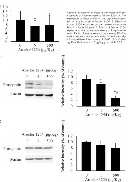
revealed a trend toward decreased Psap mRNA in epididymis (Figure 1A), which validated our
[image:5.612.97.529.56.668.2]previous finding in microarray [10]. The western blot analysis confirmed the trend (Figure 1B)
and showed the protein expression responded to PCB exposure in a dose-dependent manner.
In the 500 μg/Kg group, the protein expression
decreased 70% over control.
Sertoli cells are important source of Psap in male reproductive tract and the expression of Psap in testes was also investigated by western blot. In contrast to the epididymides, the testes showed no sign of reduction of Psap abun-dance, even in the highest dose tested (Figure 1C).
We further investigate the alteration of Psap distribution in testes and epididymides after PCB exposure. In coincidence with previous reports, an intense staining was found in testes and a moderate staining present in the caput epididymides (Figure 2). Neither the intense
nor the pattern of the staining was observably
changed in testes after 500 μg/Kg Aroclor
1254 exposure. In the caput epididymis, Psap staining present in the supernuclear region of epithelia cells (arrowhead), as well as the apical surface of the epithelium (arrow). In the lumen, staining presented associated with epididymal sperm (curved arrow), which suggested a role of the protein in the epididymal maturation. As
the mice exposed to 500 μg/Kg Aroclor 1254,
the decrease in the staining was observed in the epithelium and the lumen.
[image:6.612.91.522.72.398.2]Western blot was performed on the isolated epididymal spermatozoa to determine the responsiveness of Psap to PCB exposure. The decrease of Psap associated with epididymal sperm showed a dose-dependent manner (P<0.001, r=0.674). In mice exposed to 50 μg/
Kg and 500 μg/Kg Aroclor 1254, sperm Psap
was decreased by 42 percent and 58 percent relative to control value (Figure 3).
Benchmark dose modeling for Psap and sperm parameters
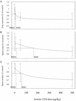
To evaluate dose-response effect of Aroclor 1254 on Psap expression, BMD modeling was utilized. The dose-response effect on Psap was compared with epididymal sperm concentra-tion, sperm motility and sperm abnormality, which are frequently used as toxicological end-points in animal and epidemiological studies [30]. In coincidence with our previous studies [10, 11], the sperm concentration and sperm
motility were significantly reduced at 500 μg/
Kg group, whereas the sperm abnormality was
significantly increased in 50 μg/Kg group and 500 μg/Kg group. For Psap expression, sperm count and sperm motility, the Hill Model fit the
data best based on global Goodness-of-Fit cri-teria, AIC, Chi-squared residuals and graphical
verification (Figure 4). For sperm abnormality,
however, all available models are a poor fit to
the present data, which suggested that no cer-tain estimation on BMD can be derived from the data.
Benchmark dose modeling indicated that BMD using percent reduction in sperm Psap was
[image:7.612.91.521.68.479.2]8.89 μg/kg while BMD for sperm count and sperm motility was 164.67 μg/kg and 61.9 μg/
Figure 4. Comparison of dose-response effects of Aroclor 1254 on endpoint. The curve is calculated using the Hill Model. A. Decrease incidence of Psap expression in sperm in response to PCB exposure. B. Decrease incidence of epididymal sperm count in response to PCB exposure. C. Decrease incidence of epididymal sperm motility in response to PCB exposure. Data are presented as percent alternations relative to the respective control. The BMD is the concen-tration that elicits a response of one standard deviation over control mean. The BMDL is the concentration corresponding to the lower 95% confidence interval.
kg. The BMDLs for Psap expression, sperm
count and sperm motility was 1.26 μg/kg, 15.73 μg/kg and 11.85 μg/kg, respectively.
Correlation between PCB accumulation and endpoints
Internal exposure of PCBs in mice was estimat-ed by determining the hepatic Aroclor 1254 accumulation and was correlated with sperm Psap expression by linear regression. For com-parison purpose, epididymal sperm count, sp-
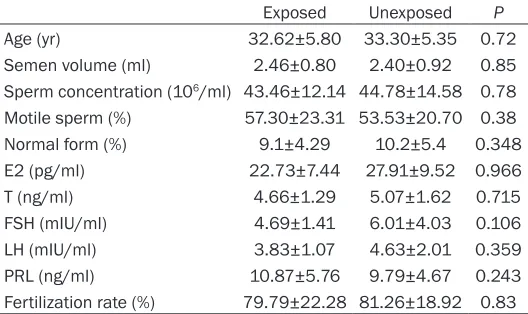
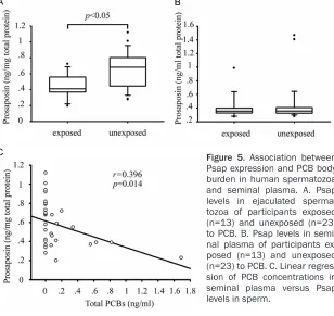
pants were comparable (Table 2). For exposed participants, the level of Psap in sperm was 0.45±0.16 ng/mg total protein, which was
sig-nificantly different to the value of 0.65±0.24
ng/mg total protein in unexposed participants. In contrast, comparison of the Psap level in (Figure 5) seminal plasma showed no signifi -cant difference between exposed and unex-posed participants. Linear regression suggest-ed that a negative association between total PCB level in seminal plasma and Psap associ-ated with sperm (P<0.05, r=-0.396).
erm motility and sperm abnormality were also cor-related to hepatic PCB lev-els. The sperm Psap expre- ssion, epididymal sperm co unt, sperm motility and sperm abnormality were sig-
nificantly correlated to
hep-atic PCB accumulation (Ta- ble 1). The correlation
coef-ficient between Psap
exp-ression and PCB accumula-tion was higher than that between PCB accumulation and sperm motility or sperm count. In addition, correlatio- ns between alternations in endpoints were investigat-ed. Relative expression of
Psap in sperm was signifi -cantly associated with sper- m abnormality (Table 1). However, no other signif- icant correlation between Psap expression and other endpoints was observed. Depressed Psap in human spermatozoon is associ-ated with PCB exposure
partici-Discussion
Male reproductive endpoints, including sperm concentration, motility, and morphology, sperm DNA integrity and serum reproductive hormone levels have been associated with PCB exposure [30]. In the present study, we reported that Psap, a glycoprotein detectable in sperm, were dose-dependently reduced by PCBs. The expo-sure doses suppressing the Psap expression were comparable to the reported daily intakes in human [25, 41], when the dose translation from animal to human is taken into consider-ation [35]. Within the dose-range tested in the present study, the PCB induced repression of sperm Psap might precede other male repro-ductive endpoints, including sperm motility. In epidemiological studies, sperm motility is the most consistent reproductive endpoint associ-ated with PCB exposure in men and data show a lack of exposure threshold for a PCB-related effect on sperm motility [30]. However, in the present study, the BMD and BMDL were lower
and 500 μg/kg, it could be clearly distinguished
from the response-curves that the response of Psap precedes sperm count and motility. The correlation between Psap reduction and the increase of sperm abnormality might sug-gest a functional association. The role of Psap in male reproductive system has been suggest-ed in knock-out mouse model. Ablation of Psap gene in mice results in several abnormalities, such as a decrease in testis size and an involu-tion of the prostate, seminal vesicle, and epi-didymis, although levels of testosterone in blood remain normal. The spermatogenesis of
Psap deficient mice is impaired and late sper -matids were particularly affected. It appears that Psap is involved in the development and maintenance of the male reproductive organs, as well as, in cellular differentiation in male reproductive system [32].
For ejaculated sperm, Psap might also be essential for fertilization. The seminal Psap for Psap reduction than percent reduction in sperm motility, which might suggested that the former might be a more sensitive predictor.
This point was further fortified by
the pilot study based on male par-ticipants enrolled from an IVF pro-gram, in which Psap levels in sperm were associated with semen PCB concentrations in semen.
Benchmark dose approach is use to derive a point of departure (POD) in estimating the dose-response effect. The method is less depen-dent on dose selection and spacing, and it takes into account the shape of the dose-response curve [14]). It provides a useful tool for analyzing data with limited dose setting and small sample size, because it is
dif-ficult for the
No-Observed-Adverse-Effect-Level approach to distinguish differences in sensitivity to toxic exposure in such data set. In the present study, BMD modeling was used to allow a comparison of dose-response effects between end-points. Although it might call for a more intense dose spacing and larger data set to determine the
[image:9.612.91.357.105.216.2]‘true’ BMDs lie between 50 μg/kg
Table 2. Characteristics of participants exposed and unex-posed to PCBs
Exposed Unexposed P
Age (yr) 32.62±5.80 33.30±5.35 0.72
Semen volume (ml) 2.46±0.80 2.40±0.92 0.85 Sperm concentration (106/ml) 43.46±12.14 44.78±14.58 0.78 Motile sperm (%) 57.30±23.31 53.53±20.70 0.38
Normal form (%) 9.1±4.29 10.2±5.4 0.348
E2 (pg/ml) 22.73±7.44 27.91±9.52 0.966
T (ng/ml) 4.66±1.29 5.07±1.62 0.715
FSH (mIU/ml) 4.69±1.41 6.01±4.03 0.106
LH (mIU/ml) 3.83±1.07 4.63±2.01 0.359
PRL (ng/ml) 10.87±5.76 9.79±4.67 0.243
[image:9.612.91.355.275.432.2]Fertilization rate (%) 79.79±22.28 81.26±18.92 0.83
Table 1. Correlation coefficient between Psap expression,
hepatic Aroclor 1254 concentration, sperm motility and abnormality
Endpoint P correlation
PCB accumulation versus sperm abnormality 0.001 0.557** PCB accumulation versus Psap 0.003 -0.531** PCB accumulation versus sperm motility 0.008 -0.478** PCB accumulation versus sperm count 0.023 -0.413*
Psap versus abnormality 0.029 -0.4*
Psap versus sperm count 0.054 0.355
Psap versus sperm motility 0.13 0.283
ization rate. However, due to the limited size of study cohort and inca-pability to adjust for potential confounders, it was unable to con-clude that depressed Psap levels in sperm without major changes in semen parameters have no effect on fertil-ization rate.
Psap in male reproduc-tive tract is secreted by Sertoli cells as well as epididymal principal ce- lls [31]. In testis, Psap exists as a secretory protein as well as a lyso-somal protein [24]. The Psap secreted by Sertoli cells binds to the tail of late spermatids and spermatozoa and is present in a relatively facilitates the sperm-egg interaction through a
60-amino acid sequence fragment, which is conserved among human and animals [2-4, 17, 29]. It is demonstrated that the pregnancy rate for thawed sperm from bulls is increased when the sperm is exposed to the fragment [3]. It appears that Psap itself is equally as good as the fragment in restoring sperm binding capa-bility [17]. Although the mechanism of action is unknown, these studies suggest that the role of Psap was increasing the number of sperm bind-ing to the outer egg layer, zona pellucida. It is
postulated that insufficient amount of the pro -tein essential for the initial binding of a sperma-tozoon to an egg investment might result in poor fertilizing potential of sperm, representing either a substantial proportion of the cells in a semen sample from certain subfertile individu-als or most samples of frozen-thawed sperm [4]. Previous study has demonstrated that
sperm-oocyte interaction is significantly
reduc-ed in PCB treatreduc-ed male rats [21]. If the Psap levels in sperm are sensitive to environmental exposure, would it possible that depressed Psap levels contribute to the prolonged TTP associated with PCB exposure? Our data failed
to show any significant difference between
exposed and unexposed participants in
fertil-high concentration in luminal fluids from the rat
rete testis, efferent ducts [23]. On the other hand, the fact that Psap is also synthesized in the epithelial cells in the extra testicular tract suggest that this protein might play an integral role through the process of sperm maturation [20]. In the efferent ducts, the Psap from testes is endocytosed by the nonciliated cells and the abundance decreased dramatically [23], which might suggest the differential role of Psap in testis and epididymis. In the present study, the expression of Psap in testis and epididymis is differentially regulated. Within the dose-range
tested in the present study, no significant alter -nation of Psap expression is observed in tes-tes. In contrast, the caput epididymides showed a decrease in Psap expression at both mRNA and protein levels after PCB exposure, which might suggest dysfunction in epididymides might be response for the declining Psap levels in sperm. Once again, it suggested that epididy-mis is a sensitive target organ contributing to PCB induced male reproductive toxicity, as it have been indicated in epidemiological studies [8].
[image:10.612.93.401.74.365.2]The Psap level is regulated in vivo and in vitro as a result of steroid hormone actions. In the
endometrial epithelium, Psap expression increased in response to elevated levels of estrogen during the oestrous cycle [37].
Supplement of 17β-estradiol in breast cancer
culture dose-dependently increased secretion of Psap into medium [12]. Using prostate cell line as an in vitro model, Koochekpour and col-leagues show that the expression of Psap is under regulation of androgen receptor [26]. The toxic effects of PCBs on male reproductive sys-tem are associated with their ability to disrupt both the expression/activity of enzymes required for sex steroid synthesis/catabolism, and the expression/ability of hormone recep-tors to bind endogenous ligands. Thus, it is rea-sonable to postulate that the expression of Psap is responsive to endocrine disrupting activities of PCBs. However, there is a report indicating that the immunohistological staining of Psap in epdididymal principal cells, as well as in narrow, clear, and basal cells, remain unchanged after orchidectomy with or without testosterone supplementation, efferent duct ligation, or hypophysectomy [19], which sug-gests an alternative mode of regulation on
Psap. To clarify the specific effects of PCBs on
Psap expression, additional experiments with PCB congeners in different mode of action are still required.
In summary, we have described the dose-dependent reduction of Psap in male reproduc-tive tract after PCB exposure. The Psap levels in sperm were adversely associated with exter-nal and interexter-nal PCB exposure, in mice and men. The PCB induced depression of Psap
pre-ceded significant effects on sperm motility and
concentration. Adversely association between Psap relative expression and sperm abnormal-ity suggested a correlation between Psap lev-els and sperm function. Taken together, our data suggested that determining the expres-sion of Psap in sperm might provide a link between PCB exposure and exposure-mediat-ed damage to target organs, and have the potential to become a useful predictor of PCB related decrease in fertility.
Acknowledgements
The study was supported by and National Key Technology R&D Program of China (2007BAc- 27B02), the National Funds for the Natural Sciences (81302454) and the Major Science
and Technology Platform in Xiamen City (No. 3502z20111006).
Disclosure of conflict of interest
None.
Address correspondence to: Dr. Zheng-Hong Zuo, School of Life Sciences, Xiamen University, 422 Siming South Road, Xiamen 361005, Fujian Province, China. Tel: 86-592-2187353; E-mail: [email protected]
References
[1] Aly HA, Domenech O and Abdel-Naim AB. Aroclor 1254 impairs spermatogenesis and in-duces oxidative stress in rat testicular mito-chondria. Food Chem Toxicol 2009; 47: 1733-1738.
[2] Amann RP, Hammerstedt RH and Shabanowitz RB. Exposure of human, boar, or bull sperm to a synthetic peptide increases binding to an egg-membrane substrate. J Androl 1999; 20: 34-41.
[3] Amann RP, Seidel GE Jr and Brink ZA. Exposure of thawed frozen bull sperm to a synthetic pep-tide before artificial insemination increases fertility. J Androl 1999; 20: 42-46.
[4] Amann RP, Shabanowitz RB, Huszar G and Broder SJ. Increased in vitro binding of fresh and frozen-thawed human sperm exposed to a synthetic peptide. J Androl 1999; 20: 655-660.
[5] Anbalagan J, Kanagaraj P, Srinivasan N, Aruldhas MM and Arunakaran J. Effect of poly-chlorinated biphenyl, Aroclor 1254 on rat epi-didymis. Indian J Med Res 2003; 118: 236-242.
[6] Andric NL, Andric SA, Zoric SN, Kostic TS, Stojilkovic SS and Kovacevic RZ. Parallelism and dissociation in the actions of an Aroclor 1260-based transformer fluid on testicular an -drogenesis and antioxidant enzymes.Toxico- logy 2003; 194: 65-75.
[7] Bains R, Adeghe J and Carson RJ. Human sperm cells express CD44. Fertil Steril 2002; 78: 307-312.
[9] Buck Louis GM, Sundaram R, Schisterman EF, Sweeney AM, Lynch CD, Gore-Langton RE, Maisog J, Kim S, Chen Z and Barr DB. Persistent environmental pollutants and couple fecundi-ty: the LIFE study. Environ Health Perspect 2013; 121: 231-236.
[10] Cai J, Wang C, Huang L, Chen M and Zuo Z. A novel effect of polychlorinated biphenyls: im-pairment of the tight junctions in the mouse epididymis. Toxicol Sci 2013; 134: 382-90. [11] Cai J, Wang C, Wu T, Moreno JM, Zhong Y,
Huang X, Chen Y and Zuo Z. Disruption of sper-matogenesis and differential regulation of tes-ticular estrogen receptor expression in mice after polychlorinated biphenyl exposure. Toxicology 2011; 287: 21-28.
[12] Campana WM, O’Brien JS, Hiraiwa M and Patton S. Secretion of prosaposin, a multifunc-tional protein, by breast cancer cells. Biochim Biophys Acta 1999; 1427: 392-400.
[13] Cornwall GA. New insights into epididymal biol-ogy and function. Human Reprod Update 2009; 15: 213-227.
[14] Davis JA, Gift JS and Zhao QJ. Introduction to benchmark dose methods and U.S. EPA’s benchmark dose software (BMDS) version 2.1.1. Toxicol Appl Pharmacol 2011; 254: 181-191.
[15] EPA. Benchmark Dose Technical Guidance. Washington, D.C: Risk Assessment Forum; 2012.
[16] Hamers T, Kamstra JH, Cenijn PH, Pencikova K, Palkova L, Simeckova P, Vondracek J, Andersson PL, Stenberg M and Machala M. In vitro toxicity profiling of ultrapure non-dioxin-like polychlorinated biphenyl congeners and their relative toxic contribution to PCB mixtures in humans. Toxicol Sci 2011; 121: 88-100. [17] Hammerstedt RH, Cramer PG, Barbato GF,
Amann RP, O’Brien JS and Griswold MD. A frag-ment of prosaposin (SGP-1) from rooster sperm promotes sperm-egg binding and im-proves fertility in chickens. J Androl 2001; 22: 361-375.
[18] Hatch EE, Nelson JW, Stahlhut RW and Webster TF. Association of endocrine disruptors and obesity: perspectives from epidemiological studies. Int J Androl 2010; 33: 324-332. [19] Hermo L and Andonian S. Regulation of
sulfat-ed glycoprotein-1 and cathepsin D expression in adult rat epididymis. J Androl 2003; 24: 408-422.
[20] Hermo L, Morales C and Oko R. Immunocyto- chemical localization of sulfated glycoprotein-1 (SGP-1) and identification of its transcripts in epithelial cells of the extratesticular duct sys-tem of the rat. Anat Rec 1992; 232: 401-422. [21] Hsu PC, Guo YL and Li MH. Effects of acute
postnatal exposure to
3,3’,4,4’-tetrachlorobi-phenyl on sperm function and hormone levels in adult rats. Chemosphere 2004; 54: 611-618.
[22] Humblet O, Birnbaum L, Rimm E, Mittleman MA and Hauser R. Dioxins and cardiovascular disease mortality. Environ Health Perspect 2008; 116: 1443-1448.
[23] Igdoura SA, Hermo L, Rosenthal A and Morales CR. Nonciliated cells of the rat efferent ducts endocytose testicular sulfated glycoprotein-1 (SGP-1) and synthesize SGP-1 derived sapo-sins. Anat Rec 1993; 235: 411-424.
[24] Igdoura SA, Rasky A and Morales CR. Trafficking of sulfated glycoprotein-1 (prosaposin) to lyso-somes or to the extracellular space in rat Sertoli cells. Cell Tissue Res 1996; 283: 385-394.
[25] Kannan K, Tanabe S, Quynh HT, Hue ND and Tatsukawa R. Residue pattern and dietary in-take of persistent organochlorine compounds in foodstuffs from Vietnam. Arch Environ Contam Toxicol 1992; 22: 367-374.
[26] Koochekpour S, Lee TJ, Sun Y, Hu S, Grabowski G, Liu Z and Garay J. Prosaposin is an AR-target gene and its neurotrophic domain upregulates AR expression and activity in prostate stromal cells. J Cell Biochem 2008; 104: 2272-2285. [27] Korrick SA and Sagiv SK. Polychlorinated
bi-phenyls, organochlorine pesticides and neuro-development. Curr Opin Pediatr 2008; 20: 198-204.
[28] Kramer S, Hikel SM, Adams K, Hinds D and Moon K. Current status of the epidemiologic evidence linking polychlorinated biphenyls and non-hodgkin lymphoma, and the role of im-mune dysregulation. Environ Health Perspect 2012; 120: 1067-1075.
[29] Magargee SF, Cramer PG and Hammerstedt RH. Increased in vitro binding and fertilizing ability of mouse sperm exposed to a synthetic peptide. Mol Reprod Dev 2000; 57: 406-411. [30] Meeker JD and Hauser R. Exposure to
poly-chlorinated biphenyls (PCBs) and male repro-duction. Syst Biol Reprod Med 2010; 56: 122-131.
[31] Morales CR, Hay N, El-Alfy M and Zhao Q. Distribution of mouse sulfated glycoprotein-1 (prosaposin) in the testis and other tissues. J Androl 1998; 19: 156-164.
[32] Morales CR, Zhao Q, Lefrancois S and Ham D. Role of prosaposin in the male reproductive system: effect of prosaposin inactivation on the testis, epididymis, prostate, and seminal vesicles. Arch Androl 2000; 44: 173-186. [33] Ong CN, Shen HM and Chia SE. Biomarkers for
male reproductive health hazards: are they available? Toxicol Lett 2002; 134: 17-30. [34] Parent AS, Naveau E, Gerard A, Bourguignon
ac-tions of endocrine disruptors on the hypothala-mus, hippocampus, and cerebral cortex. J Toxicol Environ Health B Crit Rev 2011; 14: 328-345.
[35] Reagan-Shaw S, Nihal M and Ahmad N. Dose translation from animal to human studies re-visited. FASEB J 2008; 22: 659-661.
[36] Salay E and Garabrant D. Polychlorinated bi-phenyls and thyroid hormones in adults: a sys-tematic review appraisal of epidemiological studies. Chemosphere 2009; 74: 1413-1419. [37] Spencer TE, Graf GH and Bazer FW. Sulfated
glycoprotein-1 (SGP-1) expression in ovine en-dometrium during the oestrous cycle and early pregnancy. Reprod Fertil Dev 1995; 7: 1053-1060.
[38] WHO. WHO Laboratory Manual for The Examin- ation and Processing of Human Semen. Geneva: World Health Organization.
[39] Xiong G, He X and Zhang Z. Microwave-assisted extraction or saponification combined with mi -crowave-assisted decomposition applied in pretreatment of soil or mussel samples for the determination of polychlorinated biphenyls. Analytica Chimica Acta 2000; 413: 49-56. [40] Yan F, Chen Y, Zuo Z, Yang Z and Wang C.
Effects of tributyltin on epididymal function and sperm maturation in mice. Environ Toxicol Pharmacol 2009; 28: 19-24.