Original Article
The role and function of matrix metalloproteinase-8
in rhegmatogenous retinal detachment
Hong Pan1, Zhiming Zheng2
Departments of 1Ophthalmology, 2Neurosurgery, Shandong Provincial Hospital Affiliated to Shandong University, Jinan 250021, Shandong, People’s Republic of China
Received September 7, 2016; Accepted November 18, 2016; Epub July 1, 2017; Published July 15, 2017
Abstract: Rhegmatogenous retinal detachment (RRD) is one blinding disease, and has pathological features cor-related with migration of retinal pigment epithelium (RPE) cells to viscous body. Matrix metalloproteinase-8 (MMP-8) participates in eye diseases including xerophthalmia and retinal disease. Its role in RRD, however, has not been illustrated with the functional mechanism. RPE cells from RRD model mice and normal mice were separated and cultured. MMP-8 expression plasmid was transfected into RPE cell in model group. Real time PCR and Western blot were employed to test expression level of MMP-8, whilst MTT method was used to test proliferation activity of RPE
cells. Caspase 3 activity was quantified by test kit. Transwell migration assay was adopted to measure invasion ability of RPE cells. ELISA method was used to test expression level of inflammatory factors interleukin-1β (IL-1β) and tumor necrosis factor-α (TNF-α). MMP-8 expression level was significantly decreased in RPE cells of RRD group, which also had enhanced cell proliferation and migration, accompanied with higher IL-1β and TNF-α levels (P<0.05
compared to control group). After MMP-8 transfection and over-expression, RPE cell proliferation and migration
were inhibited, along with higher Caspase 3 activity, plus lower IL-1β and TNF-α expression (P<0.05 compared to
model group). RRD caused decreased expression of MMP-8 in RPE cells. MMP-8 can facilitate RPE cells proliferation
and migration via modulating cell apoptotic activity and secretion of inflammatory factor, thus participating in RRD
pathogenesis and progression.
Keywords: Rhegmatogenous retinal detachment, retinal pigment epithelium, matrix metalloproteinase-8, cell
apoptosis, inflammatory factor
Introduction
Rhegmatogenous retinal detachment (RRD) frequently occurs in middle and aged popula-tions, especially in male patients. It normally develops sequentially in both eyes, and com-monly happens in people with severe myopia refractive errors [1, 2]. RRD is manifested as the detachment of neural layer from retinal pig-ment epithelium (RPE) layer of retina, and severely affects patient’s life quality as one blinding disease [3]. RRD is also featured as liquidation of viscous body following forma-
tion of cleft on retina, making the detachment
between retina and RPE cells [4]. RPE is formed by single layers of epithelial cells with regular arrangement. Polygonal RPE cells can be divid-ed into apical, soma and basal compartments [5]. There are over one million RPE cells in each human eye. However, due to the inability to
regenerate, RPE cells cannot be replaced after death, but are occupied by adjacent cells via sliding [6, 7]. Occurrence of RRD causes sepa-ration of retinal neural epithelium and RPE, depriving the nutrient supply of outer retina via choroid plexus, further aggravating post-macu-lar area injury and retinal damage. Without timely treatment or relocation, detached retina may develop atrophy and denature, leading to vision damage [8]. RRD in macular region can cause sharply decreased vision function, and irreversible loss of vision without timely treat-ment [9, 10].
of retina and viscous body, causing retinal detachment [11, 12]. Matrix metalloproteinase (MMPs) can degrade effective component of ECM and basal membrane, and regulate cell adhesion, thus playing a critical role in ECM dynamic balance [13, 14]. Previous study showed the involvement of MMPs in various diseases including tumor, rheumatoid arthritis and proliferative vitreoretinopathy [15, 16]. The function and mechanism of MMP-8, which is one important member of MMPs family, in RRD, however, have not been elucidated.
Materials and methods
Experimental animals
Healthy male Wistar rats (2 months old, SPF
grade, body weight 250±20 g) were purchased from laboratory animal center of Shandong
University and were kept in an SPF grade facili
-ty with fixed temperature (21±1°C), fixed humid
-ity (50-70%) and 12 h light/dark cycle. Rats
were used for all experiments, and all proce-dures were approved by the Animal Ethics
Committee of Provincial Hospital Affiliated to
Shandong University.
Major materials and equipment
0.1% atropine eye dropping, 0.3% ofloxacin eye
dropping and Mydrin-P were purchased from
Merck (US). Pentobarbital sodium and lidocaine
were purchased from Zhaohui Pharm (China).
PVDF membrane was purchased from Pall Life
Sciences (US). Western blotting reagents were purchased from Beyotime (China). ECL reagent was purchased from Amersham Biosciences (US). Rabbit anti-mouse MMP-8 monoclonal antibody and goat anti-rabbit horseradish per-oxidase (HRP)-labelled IgG secondary antibo- dy were purchased from Cell Signaling (US).
Β-actin (MAB8929) was purchased from R&D (US). IL-β and TNF-α ELISA kits were purchased from R&D (US). Caspase 3 activity assay kit
was purchased from Cell signaling (US). RNA
extraction kit, pcDNA3.1 vector, and reverse transcription kit were purchased from Axygen
(US). Transwell chamber was purchased from Corning (US). pcDNA3.1 empty plasmid and pcDNA3.1-MMP-8 plasmid were synthesized by Gimma (China). Surgical microscope was pur-chased from Suzhou Instrument (China). ABI
2000 fluorescent quantitative PCR cycler was
purchased from ABI (US). Microplate reader
was purchased from BD (US). Other common reagents were purchased from Sangon (China).
Animal grouping and treatment
Healthy male Wistar rats were randomly divid-ed into two groups (N=20), including control and RRD model group.
RRD model preparation
Rats were anesthetized by 30 mg/kg pentobar
-bital sodium, and were fixed in a supine posi
-tion. 0.1% atropine eye dropping, 0.3% ofloxa -cin eye droppings were applied 3 d before surgery. Mydrin-P was used to dilate the pupil.
Focal anesthesia was performed by sub-con -junctival injection of 2% lidocaine and 0.75% levobupivacaine. Bulbar conjunctiva was open- ed, and an incision was made 3 mm posterior of corneoscleral junction to prepare channels
for incision, perfusion, and optical fiber lighting.
The scaffold of corneal contact lens was placed in, along with surgical microscope. Partial vis-cous body was removed under sterile condi-tion. Retina was scratched gently to form 2~3 optic disc holes. Incisions of both cornea and bulbar conjunctiva were closed. 10000 U of gentamicin and dexamethasone were injected underneath the bulbar conjunctiva, along with post-op application of chloramphenicol eye dropping to prevent infection. Optical coher-ence tomography can observe interruption of
retinal neural epithelium, in addition to signifi
-cant uplift of highly-reflective pigmented epi -thelium to form detachment with certain
angles. These features plus liquid dark region
indicated successful generation of the model [17].
Culture of RPE cells
0.25% trypsin digestion for 30 min at 37°C,
cells suspensions were then centrifuged at 800 rpm for 10 min. the supernatant was dis-carded and cultured medium containing 100 U/
ml penicillin and 100 μg/ml streptomycin was added for incubation at 37°C with 5% CO2. Culture medium was changed every changed every other day. Cells were passed every 2-3 days. RPE cells at log-growth phase at 2nd to 8th
generation were used for experiments. Model group was randomly divided into three groups: model group; empty transfection group, which was transfected by empty pcDNA3.1 plasmid; and MMP-8 group, which was transfected with pcDNA3.1-MMP8 plasmid.
Liposome transfection of MMP8 plasmid into RPE cells
pcDNA3.1 and MMP8 plasmid were transfect-ed into RPE cells, which were culturtransfect-ed in 6-well
plate until 70-80% confluence. Liposome
rea-gent containing pcDNA3.1 and MMP8 plasmid
were mixed well with 200 μl serum-free culture
medium for 15 min room temperature incuba-tion. Lipo2000 reagent was then mixed with pcDNA3.1 or pcDNA3.1-MMP8 plasmid dilu-tions for 30 min continuous incubation at room temperature. Serum was removed from cul-tured cells, which were then rinsed gently in PBS. 1.6 ml serum-free medium was added for
6 h incubation at 37°C with 5% CO2. Normal culture medium was then added for 48 h incu-bation in further experiments.
Real-time PCR for MMP8 mRNA expression in RPE cells
Trizol reagent was used to extract mRNA from
all RPE cells. Following manual instruction of the test kit, reverse transcription kit was per -formed to synthesize DNA. Primers were de- signed by Primer 6.0 based on target gene sequence, and were synthesized by Invitrogen (China) as shown in Table 1. Real-time PCR was then performed to detect target gene
expres-sion under the following conditions: 56°C 1
min, followed by 35 cycles each containing
92°C 30 s, 58°C 45 s and 72°C 35 s; Data
were collected by PCR cycler. CT value was
Western blot for MMP8 protein expression
Proteins of RPE cells were extracted by RIPA lysis buffer containing proteinase inhibitor. In brief, cells were mixed with lysis buffer for 15~30 min iced incubation. Using ultrasonic rupture (5 s, 4 times) and centrifugation (10000
g, 15 min at 4°C), proteins were quantified from the supernatant and were kept at -20°C for
Western blotting. Proteins were separated in
10% SDS-PAGE, and were transferred to PVDF membrane by semi-dry method. Non-specific binding sites were blocked by 5% defatted milk
powders for 2 hours. Anti-MMP8 monoclonal
antibody (1:1000) or β-actin antibody (1:2000) was applied for 4°C overnight incubation. Goat
anti-rabbit IgG (1:2000) was then added for 30-min incubation after PBST rinsing. After PBST washing and ECL development for 1 min, the membrane was exposed under X-ray. An imaging analyzing system and Quantity one
software were then used to scan X-ray films and
to detect the density of bands with repeated measures (N=4).
MTT assay for cell proliferation
RPE cells at log-phase were seeded into 96- well plate at 5000 cells per well containing
DMEM medium with 10% FBS. After 24 h incu -bation, the supernatant was discarded, cells were then randomly divided into control, model, empty transfection, and MMP8 groups as abovementioned. After 48-hour incubation, 20
μl sterile MTT solution was then added into
each test well in triplicates. With 4 h continu-ous culture, the supernatant was completely
removed, with the addition of 150 μl DMSO
for 10 min vortex until the complete resolving of crystal violet. Absorbance (A) values was measured at 570 nm in a microplate reader. The proliferation rate was calculated in each group. Each experiment was repeated in tripli-cates for statistical analysis.
Transwell chamber assay for cell migration Following instruction of test kit, cells were
changed from serum-free culture medium. After 24 h, 1:5 50 mg/L Matrigel dilution was Table 1. Primer sequence
Target gene Forward primer 5’-3’ Reverse primer 5’-3’ GAPDH AGTACCAGTCTGTTGCTGG TAATAGACCCGGATGTCTGGT MMP8 ACCCTTCCCTCTAGTGAATC TAGATGGACCTCTGTTTAAT
used to coat bottom and upper membrane of
the chamber, which was then air-dried at 4°C. 500 μl DMEM medium containing 10% FBS and 100 μl serum-free RPE cell suspensions were
added into the inner and outer side of the chamber, respectively. Chambers were placed into 24-well plate in triplicates, in parallel with control group using Transwell chamber without Matrigel. After 48 h incubation, the lower chamber was rinsed by PBS. Cells on the mem-brane were removed, followed by cold ethanol
fixation. The membrane was stained by crystal
violet. Under the microscope, number of cells on the lower membrane was counted in triplicates.
Caspase 3 activity assay
Caspase 3 activity in cells was measured
fol-lowing manual instruction of test kit. In brief,
cells were digested in trypsin, and were
centri-fuged at 600 g for 5 min under 4°C. The super -natant was discarded, followed by the addition of cell lysis buffer and iced incubation for 15 min. The mixture was then centrifuged at
20000 g for 5 min under 4°C, followed by the
addition of 2 mM Ac-DECD-pNA. Optical density (OD) values at 400 nm wavelength were mea-sured to calculate Caspase 3 activity.
ELISA for expression of inflammatory factor IL-1β and TNF-α
Cell culture supernatant was collected and tested following the manual instruction of ELISA
kits. Absorbance (A) values at 450 nm wave
length were measured in all wells using a micro-plate reader within 15 min of adding the quen- ching buffer. Linear regression model was then
plotted based on the concentration of standard samples and respective A values. Sample con-centration was further deduced based on A value and regression function.
Statistical analysis
All data were expressed as mean ± standard deviation (SD). Student t-test was used to com-pare means between two groups. SPSS 11.5 software was used to analyze all data. Analysis of variance (ANOVA) was used to compare
means across groups. A statistical significance was defined when P<0.05.
Results
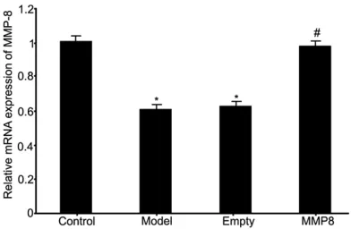
MMP-8 mRNA expression in RPE cells from RRD rats
Real time PCR was used to test mRNA expre- ssion of MMP-8 in RPE cells collected from RRD rat model and the effect of MMP-8 plas-mid transfection on mRNA expression. Results
showed significantly decreased MMP-8 expres
-sion in RRD group (P<0.05 compared to control
group). The transfection of MMP-8 plasmid into
RPE cells significantly facilitated MMP-8 mRNA expression in model cells (P<0.05 compared to
[image:4.612.91.287.73.201.2]model group). The transfection of empty plas-mid into model RPE cells did not affect MMP-8 mRNA expression compared to model group (Figure 1).
MMP-8 protein expression in RPE cells of RRD rats
Western blot was used to test protein expres-sion of MMP-8 in RPE cells collected from RRD rat model and the effect of MMP-8 plasmid transfection on MMP-8 protein expression. Results showed similar results as those from
mRNA levels, as shown by significantly
decre-ased MMP-8 protein expression in RRD group
(P<0.05 compared to control group). The trans
-fection of MMP-8 plasmid into RPE cells signifi -cantly facilitated MMP-8 protein expression in
model cells (P<0.05 compared to model group,
Figure 2).
Proliferation of RPE cells by MMP-8 in RRD model
MTT assay was used to analyze the prolifera-tion of RPE cells after MMP-8 plasmid
transfec-tion. Results showed significantly enhanced
Figure 1. MMP-8 mRNA expression in RPE cells of
RRD. *P<0.05 compared to control group. #P<0.05
RPE cell proliferation in RRD group (P<0.05
compared to control group). The transfection
of MMP-8 plasmid into RPE cells significantly
facilitated MMP-8 expression and inhibited cell
proliferation (P<0.05 compared to model
gro-up, Figure 3). These results suggested altered MMP-8 expression in RRD cells helped to regu-late abnormal proliferation of RPE cells.
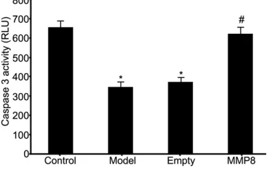
Caspase 3 activity of RPE cells under regulat-ing MMP-8 level in RRD model
Caspase 3 activity assay kit was used to ana -lyze Caspase 3 activity of RPE cells in RRD model after MMP-8 plasmid transfection.
Results showed significantly lower Caspase 3 activity in RPE cells of RRD group (P<0.05 com -pared to control group). The transfection of
MMP-8 plasmid into RPE cells significantly facil
-itated Caspase 3 activity (P<0.05 compared to
model group, Figure 4). These results suggest-ed elevatsuggest-ed MMP-8 expression in RPE cells of RRD model helped to enhance Caspase 3 activ-ity and further RPE cell apoptosis.
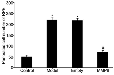
RPE cell migration ability under MMP-8 over-expression
Transwell chamber assay kit was used to ana -lyze the effect of MMP-8 on migration ability of RPE cells in RRD model. Results showed en- hanced migration of RPE cells in RRD group
(P<0.05 compared to control group). The trans
-fection of MMP-8 plasmid into RPE cells signifi -cantly facilitated MMP-8 expression and
inhib-ited cell migration ability (P<0.05 compared to
model group, Figures 5 and 6). These results suggested elevated MMP-8 expression in RPE cells of RRD model enhanced migration ability of RPE cells.
Expression of inflammatory factors in RPE cells by MMP-8 expression
ELISA was used to analyze the effect of MMP-8
on expression of inflammatory factors of RPE
cells in RRD model. Results showed elevated
IL-1β and TNF-α expressions in RPE cells of RRD group (P<0.05 compared to control group).
The transfection of MMP-8 plasmid into RPE
[image:5.612.90.290.67.237.2]cells significantly facilitated MMP-8 expression and inhibited IL-1β and TNF-α expressions (P<0.05 compared to model group, Figure 7).
Figure 2. MMP-8 protein expression in RPE cells of RRD. A. MMP-8 protein expression in RPE cells from RRD model. A. Control group; B. Model group; C. Empty plasmid transfection group; D. MMP-8 group.
B. Analysis of MMP-8 protein expression. *P<0.05 compared to control group; #P<0.05 compared to
[image:5.612.90.288.338.466.2]model group.
Figure 3. Effects on RPE cell proliferation by
MMP-8 expression in RRD model. *P<0.05 compared to control group; #P<0.05 compared to model group.
Figure 4. Effects on caspas3 activity of RPE cells
by MMP-8 expression in RRD model. *P<0.05 com
-pared to control group; #P<0.05 com-pared to model
[image:5.612.92.287.531.655.2]Discussion
The early phase manifestation of RRD is the migration of RPE cells into viscous body, accom-panied with abnormal hyperplasia of RPE cells.
Further migration causes aggregation on the
lower surface of retina or viscous-retina
inter-face to secrete inflammatory factors, causing
abundant secretion of ECM and its remodeling, eventually forming pathology precipitation, stretching retina and further detachment [18,
19]. RRD is one inflammatory process and can
affect ECM synthesis or degradation [20]. MMPs as the only proteinase that can hydrolyze
fibrous collagen, can degrade ECM protein,
maintain physiological renewal of ECM, and prevents ECM from over-precipitation [21]. MMPs can participate in various pathology-physiology processes including traumatic re- pair, tissue model regeneration and embryonic development, and regulate cell adhesion, plus regulation on extracellular components or other proteins [22]. Besides degrading ECM and maintaining homeostasis, MMPs can also par-ticipate in regulating various functions
includ-ing cytokine secretion, cell surface protein and
proteinase inhibitor [23].
As one important member of MMPs family, MMP-8 participates in pathogenesis of various diseases including corneal disease and sclera
inflammation [24]. This study therefore estab -lished an RRD rat model whose RPE cells were separated and cultured to analyze the effect and mechanism of MMP-8 in RRD. Results
showed significantly decreased MMP-8 expre
ssion in RPE cells of RRD model, plus elevat- ed cell proliferation/migration potency, lower Caspase 3 activity, and elevated expression of
inflammatory factors IL-1β and TNF-α. These
results suggested that during pathogenesis of RRD, lower MMP-8 expression causes inhibi-tion of RPE apoptosis, plus abnormal hyperpla-sia, further accelerating migration ability of RPE
cells, and stimulating secretion of inflammatory
factors. By transfection of MMP-8 plasmid into RPE cells of RRD model, the over-expression of MMP-8 facilitated RPE apoptosis, inhibited abnormal hyperplasia of RPE, and further sup-pressed RPE migration in addition to the
inhibi-tion of secretin of inflammatory factors. Such
process might be correlated with ECM
[image:6.612.90.289.70.291.2]degrada-Figure 5. Effects of MMP-8 regulation on RPE cell mi-gration of RRD group.
Figure 6. Analysis of the effects on RPE cell
migra-tion by MMP-8 expression in RRD model. *P<0.05 compared to control group; #P<0.05 compared to
[image:6.612.326.521.74.203.2]model group.
Figure 7. Effects on inflammatory factor expression
in RPE cells by MMP-8 expression in RRD model.
[image:6.612.89.288.341.473.2]tion by MMP-8, which further inhibited inflam -matory stimulus and decrease RPE hyperplasia [25].
Conclusion
RRD causes decreased MMP-8 expression in RPE cells. MMP-8 can facilitate proliferation and migration of RPE cells via modulating cell
apoptosis and secretion of inflammatory fac -tors, thus participating in occurrence and pro-gression of RRD.
Acknowledgements
This work was supported by
Science-Techno-logy Development Project of Shandong Province
(2013G0021809); National Science
Founda-tion for Young Scientists of China (81500698)
and Promotive Research Fund for Excellent
Young and Middle-Aged Scientisits of Shandong Province (BS2013YY044).
Disclosure of conflict of interest None.
Address correspondence to: Dr. Zhiming Zheng, Department of Neurosurgery, Shandong Provincial
Hospital Affiliated to Shandong University, #324 Jing
Wu Road, Jinan 250021, Shandong, People’s
Republic of China. Tel: +86-18663766527; Fax:
+86-18663766527; E-mail: XingYQzxc@163.com
References
[1] Huang C, Zhang T, Liu J, Ji Q and Tan R.
Chang-es in axial length, central cornea thicknChang-ess,
and anterior chamber depth after rhegmatog-enous retinal detachment repair. BMC Oph-thalmol 2016; 16: 121.
[2] Yumusak E, Ornek K and Ozkal F. Bilateral si -multaneous rhegmatogenous retinal
detach-ment following laser in situ keratomileusis.
Case Rep Ophthalmol 2016; 7: 341-5.
[3] Gotzaridis S, Liazos E, Petrou P and Georgalas I. 25-gauge vitrectomy and incomplete
drain-age of subretinal fluid for the treatment of pri -mary rhegmatogenous retinal detachment. Ophthalmic Surg Lasers Imaging Retina 2016; 47: 333-5.
[4] Kominami A, Ueno S, Kominami T, Nakanishi
A, Piao CH, Ra E, Yasuda S, Asami T and
Tera-saki H. Restoration of cone interdigitation zone
associated with improvement of focal macular ERG after fovea-off rhegmatogenous retinal re-attachment. Invest Ophthalmol Vis Sci 2016; 57: 1604-11.
[5] Heriot WJ. Thermofusion of the retina with the RPE to seal tears during retinal detachment repair. Graefes Arch Clin Exp Ophthalmol 2016; 254: 691-6.
[6] Koutsandrea C, Kanakis M, Papaconstantinou
D, Brouzas D, Ladas I, Petrou P and Georgalas
I. Scleral buckling versus vitrectomy for retinal detachment repair: comparison of visual fields and nerve fiber layer thickness. Ophthalmo -logica 2016; 235: 10-7.
[7] Takahashi E, Fukushima A, Haga A, Inomata Y, Ito Y, Fukushima M and Tanihara H. Effects of
mechanical stress and vitreous samples in retinal pigment epithelial cells. Biochem Bio-phys Res Commun 2016; 470: 569-74. [8] Sukseree S, Chen YT, Laggner M, Gruber F, Pe
-tit V, Nagelreiter IM, Mlitz V, Rossiter H, Pollre-isz A, Schmidt-Erfurth U, Larue L, Tschachler E
and Eckhart L. Tyrosinase-cre-mediated dele -tion of the autophagy gene Atg7 leads to ac-cumulation of the RPE65 variant M450 in the retinal pigment epithelium of C57BL/6 mice. PLoS One 2016; 11: e0161640.
[9] Knickelbein JE, Liu B, Arakelyan A, Zicari S,
Hannes S, Chen P, Li Z, Grivel JC, Chaigne-Delalande B, Sen HN, Margolis L and Nussen-blatt RB. Modulation of immune responses by extracellular vesicles from retinal pigment epi-thelium. Invest Ophthalmol Vis Sci 2016; 57: 4101-7.
[10] Mones J, Leiva M, Pena T, Martinez G, Biarnes
M, Garcia M, Serrano A and Fernandez E. A
swine model of selective geographic atrophy of
outer retinal layers mimicking atrophic AMD: a
Phase I escalating dose of subretinal sodium iodate. Invest Ophthalmol Vis Sci 2016; 57: 3974-83.
[11] Shaw PX, Stiles T, Douglas C, Ho D, Fan W, Du
H and Xiao X. Oxidative stress, innate immuni-ty, and age-related macular degeneration. AIMS Mol Sci 2016; 3: 196-221.
[12] Moreira EF, Cai H, Tezel TH, Fields MA and
Del Priore LV. Reengineering human Bruch’s Membrane increases rod outer segment phagocytosis by human retinal pigment epithe-lium. Transl Vis Sci Technol 2015; 4: 10. [13] Fernandez-Godino R, Pierce EA and Garland
DL. Extracellular matrix alterations and depos-it formation in AMD. Adv Exp Med Biol 2016; 854: 53-8.
[14] Fields MA, Cai H, Bowrey HE, Moreira EF, Beck Gooz M, Kunchithapautham K, Gong J, Vought E and Del Priore LV. Nitrite modification of ex -tracellular matrix alters CD46 expression and
VEGF release in human retinal pigment epithe -lium. Invest Ophthalmol Vis Sci 2015; 56: 4231-8.
-tion by female sex hormones of collagen gel contraction mediated by retinal pigment epi-thelial cells. Invest Ophthalmol Vis Sci 2014; 55: 2621-30.
[16] Lee HS, Jun JH, Jung EH, Koo BA and Kim YS.
Epigalloccatechin-3-gallate inhibits ocular neo-vascularization and vascular permeability in human retinal pigment epithelial and human retinal microvascular endothelial cells via
sup-pression of MMP-9 and VEGF activation. Mol -ecules 2014; 19: 12150-72.
[17] Cederlund M, Ghosh F, Arner K, Andreasson S and Akerstrom B. Vitreous levels of oxidative stress biomarkers and the radical-scavenger
alpha1-microglobulin/A1M in human rheg-matogenous retinal detachment. Graefes Arch Clin Exp Ophthalmol 2013; 251: 725-32. [18] Hou X, Han QH, Hu D, Tian L, Guo CM, Du HJ,
Zhang P, Wang YS and Hui YN. Mechanical force enhances MMP-2 activation via p38 sig-naling pathway in human retinal pigment epi-thelial cells. Graefes Arch Clin Exp Ophthalmol 2009; 247: 1477-86.
[19] Coral K, Angayarkanni N, Madhavan J, Bharath
-selvi M, Ramakrishnan S, Nandi K, Rishi P, Ka
-sinathan N and Krishnakumar S. Lysyl oxidase
activity in the ocular tissues and the role of LOX in proliferative diabetic retinopathy and rhegmatogenous retinal detachment. Invest Ophthalmol Vis Sci 2008; 49: 4746-52. [20] Symeonidis C, Diza E, Papakonstantinou E,
Souliou E, Dimitrakos SA and Karakiulakis G.
Correlation of the extent and duration of rheg-matogenous retinal detachment with the ex-pression of matrix metalloproteinases in the vitreous. Retina 2007; 27: 1279-85.
[21] Kim B, Abdel-Rahman MH, Wang T, Pouly S,
Mahmoud AM and Cebulla CM. Retinal MMP-12, MMP-13, TIMP-1, and TIMP-2 expression in murine experimental retinal detachment. Invest Ophthalmol Vis Sci 2014; 55: 2031-40. [22] Du YH, Hirooka K, Miyamoto O, Bao YQ, Zhang
B, An JB and Ma JX. Retinoic acid suppresses the adhesion and migration of human retinal pigment epithelial cells. Exp Eye Res 2013; 109: 22-30.
[23] Kimura K, Orita T, Liu Y, Yang Y, Tokuda K, Kurakazu T, Noda T, Yanai R, Morishige N, Takeda A, Ishibashi T and Sonoda KH. Attenua
-tion of EMT in RPE cells and subretinal fibrosis
by an RAR-gamma agonist. J Mol Med (Berl) 2015; 93: 749-58.
[24] Bian F, Wang C, Tukler-Henriksson J, Pflug -felder SC, Camodeca C, Nuti E, Rossello A, Li DQ and de Paiva CS. MMP-8 is critical for
dexa-methasone therapy in alkali-burned corneas
under dry eye conditions. J Cell Physiol 2016; 231: 2506-16.
[25] Bian F, Pelegrino FS, Henriksson JT, Pflugfelder
SC, Volpe EA, Li DQ and de Paiva CS. Differen-tial effects of dexamethasone and doxycycline
on inflammation and MMP production in mu
-rine alkali-burned corneas associated with dry