Original Article
Long noncoding RNA CCAT2 promotes proliferation
and metastasis in non-small cell lung
cancer through the Wnt pathway
Jijia Li1, Yu Liu1, Pengfei Li1, Yingwei Guo1, Yannan Liu2, Yi Ren1
1Department of Thoracic Surgery, Cancer Hospital of China Medical University, Liaoning Cancer Hospital &
Insti-tute, Shenyang, China; 2China Medical University, Shenyang, China
Received March 15, 2017; Accepted May 27, 2017; Epub July 1, 2017; Published July 15, 2017
Abstract: Colon cancer associated transcript 2 (CCAT2), a long non-coding RNA (lncRNA), was shown to be associ-ated with colon, ovarian and prostate cancer. Recent studies showed CCAT2 was highly expressed in various tumors, including non-small cell lung cancer (NSCLC) and probably being an independent prognostic factor of this disease. However, the physiological and biochemical mechanisms for CCAT2 with NSCLC were still unknown. In this study, we performed the analysis focused on the expression, biological functions and mechanism of CCAT2 in NSCLC and found that CCAT2 was significantly up-regulated in NSCLC tissues compared with corresponding non-cancerous tis-sues. Knockout of CCAT2 in NSCLC cell lines using lentivirus-mediated RNA interference significantly inhibited the proliferation and metastasis of the NSCLC cells. Most importantly, Wnt/β-catenin pathway was found to be inactivat-ed in the NSCLC cell lines after CCAT2 knockout experiment. These results indicatinactivat-ed that CCAT2 maybe serve as an oncogenic lncRNA that promoted proliferation and metastasis of NSCLC and activated the Wnt/β-catenin pathway.
Keywords: CCAT2, lncRNA, NSCLC, Wnt/β-catenin pathway
Introduction
Lung cancer is a common malignant tumor for human kind, and is also the most common cause of death worldwide at present [1, 2]. Based on the differentiation and morphologi- cal characteristics, lung cancer can be divided into small cell lung cancer (SCLC) and non small cell lung cancer (NSCLC), and NSCLC account-ed for nearly 85% all of the lung cancer [3-5]. Metastatic spread of tumor cells is the main cause of cancer death. The process of cancer metastasis consisted of linked sequential steps, including invasion, detachment, intrava-sation, circulation, adhesion, extravasation and growth in distant organs [6-8]. Currently, there were no reliable markers available to accurate-ly predict metastasis in earaccurate-ly-stage NSCLC patients. Therefore, the most important strate-gy for NSCLC is to find an effective marker of early diagnosis and prognosis, and provide a theoretical basis for the invasion and metasta-sis of cancer cells.
found that CCAT2 was highly over-expressed in microsatellite-stable colorectal cancer and pro-motes tumor growth, metastasis, and chromo-somal instability. Recent studies [14-16] showed that CCAT2 was highly expressed in various tumors, including NSCLC. However, the physiological and biochemical mechanisms for CCAT2 with NSCLC were still unknown. In this study, we performed the analysis on the expres-sion, biological functions and mechanism of CCAT2 in NSCLC.
Materials and methods
Patient samples and cell lines
NSCLC and paired corresponding normal non-cancerous tissues were obtained from 53 patients, underwent surgery resection in the Cancer Hospital of China Medical University
H1299) and one normal tissue (16HBE) were purchased from the Institute of Biochemistry and Cell Biology of the Chinese Academy of Sciences (Shanghai, China). All cells were cul-tured in RPMI-1640 medium (GIBCO, USA) sup-plemented with 10% fetal bovine serum (10% FBS), and maintained in the humidified incuba-tor at 37°C with 5% CO2.
RNA extraction and qRT-PCR assays
[image:2.612.90.289.83.152.2]Total RNA was isolated from tissues and cell cultures with Trizol reagent (TaKaRa, Japan) according to the manufacturers’ instructions. The cDNA was reverse transcribed from a total of 200 ng RNA using the PrimeScript RT reagent Kit (TaKaRa, Japan) and amplified by quantita-tive real-time PCR by SYBR Green Kit (TaKaRa, Japan) on ABI 7500 (USA). GAPDH was used as the internal reference, and the relative expres-sion of CCAT2 was normalized to GAPDH. Primers for CCAT2 and GAPDH were shown in
Table 1.
Interfering RNA construction
The small interfering RNA (siRNA) against CCAT2 and the negative control (si-NC) were used and synthesized by GenePharma (Shang- hai, China). The following short hairpin RNA was used to target CCAT2: sense: 5’-UUAACCUC- UUCCUAUCUCATT-3’; antisense: 5’-UGAGAUA- GGAAGAGGUUAATT-3’. The negative control sequence was 5’-UUCUCCGAACGUGUCACGU- TT-3’. The cells were cultured on six-well plates in the density of 5×10⁵, and the cells were transfected with siRNA and si-NC by using Lipofectamine 2000 (Invitrogen, USA) at 100 nM concentration. The interfering results were determined by qRT-PCR after transfection 48 hours, and the silencing efficacy of siRNA were used for more than 85% to further investigation.
Groups Numbers
Sex Male 17
Female 36
Age Mean 60.8±5.2
Tumor differentiation Well 11
Moderate 26
Poor 16
Tumor invasion T1 6
T2 14
T3 17
T4 16
Lymph node metastasis Absence 19
Presence 31
No 3
Distant metastasis Absence 48
Presence 5
TNM stage I 6
II 11
III 20
[image:2.612.91.288.197.462.2]Cell proliferation
Cell proliferation was performed by using Cell Counting Kit (CCK-8, Dojindo) based on the manufacturer’s instruction. 96-well plates were used for three groups (control, siRNA and si-NC), 100 µl cells were inoculated into the plates at 1000 cells/well, and cultured at 37°C. The densities of the cells were measured at 450 nm, and the survival rate was expressed as the absorbance relative to the control group. For colony forming experiments, the cells trans-fected with siRNAs and si-NC were inoculated into six well plates, maintained in RPMI-1640 medium contained 10% FBS for two weeks. Colonies were fixed with methanol 12 days later and stained with 0.1% crystal violet (Sigma, USA). The colonies were photographed and counted manually.
Cell cycle and apoptosis
Cells were harvested after transfected by tryp-sin and centrifugation. Then, washed with cold PBS, and fixed with 70% ethanol at 4°C. The cells were stained with propidium iodide at 4°C for 30 min. The results were analyzed with flow cytometer FACSCalibur (BD, USA).
For the apoptosis analysis, the trysinzed and washed cells were stained with FITC-Annexin V and propidium iodide, and analyzed by FACSCalibur and Flowjo software (BD, USA). Cells were discriminated for viable cells, dead cells, early apoptosis cells and apoptosis cells.
Western blotting
Cells cultures were lysed with protein extrac-tion reagent RIPA (Beyotime, China). The
pro-teins were separated by SDS-PAGE and trans-ferred to PVDF membranes. The blots were probed with primary antibodies at 4°C: anti-β-catenin, anti-CCND1, and anti-c-myc. The GA- PDH was used as the internal control.
Statistical analysis
SPSS Statistics for Windows, Version 17.0. (SPSS Inc., Chicago, USA) was used for statisti-cal analysis of the data. Data were shown as mean ± standard deviation (STD). Statistical significance was tested by Student’s t-test, Chi-square test and one way ANOVA if appropriate. Two tailed P<0.05 was considered to be statis-tically significant.
Results
CCAT2 were up-regulated in NSCLC tissues and cell lines
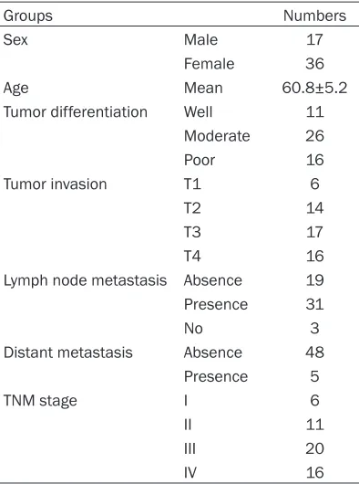
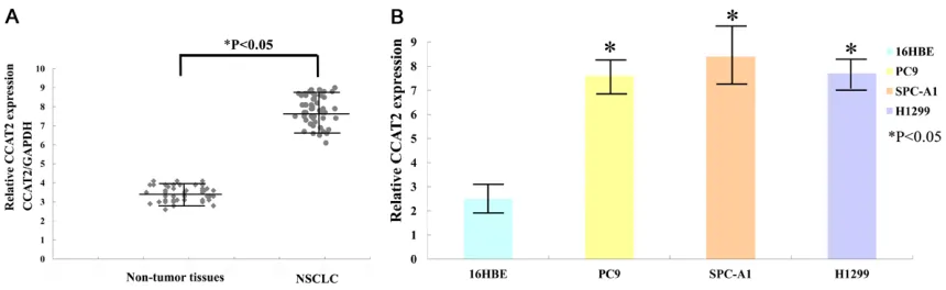
The qRT-PCR was performed to evaluate the expression of CCAT2 in 53 NSCLC and corre-sponding non-tumor tissues. The clinical and pathological information of the patients were shown in Table 2. The level of CCAT2 was increased significantly in NSCLC tissues com-pared with the non-tumor tissues (Figure 1A). The cell lines comparison results also showed highly expressed CCAT2 to the NSCLC cells (Figure 1B).
Knockdown of CCAT2 in NSCLC cell lines
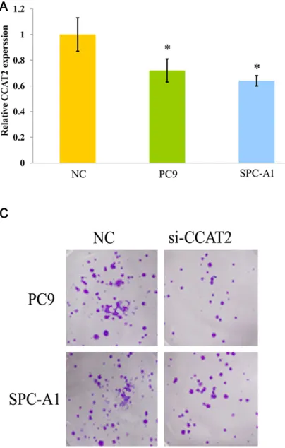
[image:3.612.90.520.69.201.2]The CCAT2 was knockdown in NSCLC cell lines, accessed the role of CCAT2 in NSCLC growth. Two cell lines were used, PC9 and SPC-A1. The decreased CCAT2 expression levels were selected for more than 85%. The CCAT2
expressions were statistically significant de- creased in all the cell lines (Figure 2A). Cell pro-liferation results detected by CCK-8 revealed that NSCLC cells growth were inhibited both in PC9 and SPC-A1 compared with negative con-trol (Figure 2B), and the colony forming experi-ment also showed reduced phenomenon by CCAT2 silenced in PC9 and SPC-A1 cell lines (Figure 2C).
The cell cycle progression and apoptosis of the NSCLC cell lines
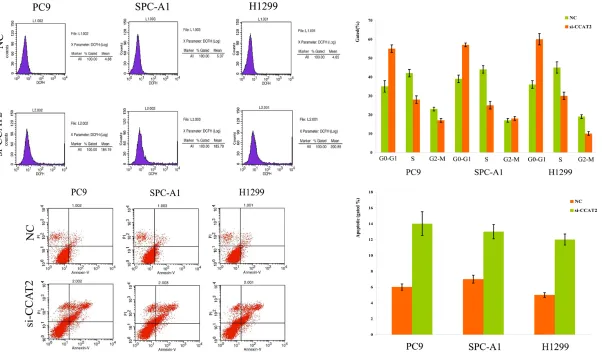
We performed the cell-cycle arrest and apopto-sis test for knockdown CCAT2 in NSCLC cell lines. The results showed that si-CCAT2 of PC9, SPC-A1 and H1299 had an obvious cell-cycle arrest at the G1-G0 phase and had a decreased at G2-S phase (Figure 3A). The flow-cytometry and TUNEL staining analysis was also
per-formed, and the results showed that all the NSCLC cell lines transfected with si-CCAT2 increased the apoptosis proportion compared with the control groups (Figure 3B). These data suggested that si-CCAT2 could promote the proliferation of NSCLC cell lines.
Si-CCAT2 on the signal regulated Wnt/β-catenin pathway
[image:4.612.92.294.73.390.2]CCAT2 regulated NSCLC cells’ proliferation and metastasis mechanisms were determined by western blot, and the results showed that the β-catenin, CCND1, and c-myc were significantly reduced for knockdown of CCAT2 compared with the control (Figure 4). These data suggest-ed that Wnt/β-catenin pathway might involve in the CCAT2 mediated proliferation and metasta-sis of NSCLC cell lines.
Discussion
NSCLC is any type of epithelial lung cancer other than small cell lung carcinoma (SCLC) [17, 18]. NSCLC accounted for about 85% of all lung cancers. As a class, NSCLCs were relatively insensitive to chemotherapy, compared to small cell carcinoma. When possible, they were primarily treated by surgical resection with curative intent, although chemotherapy was increasingly being used both pre-operatively and post-operatively [19-22]. Therefore, the most important strategy for NSCLC was to find an effective marker of early diagnosis and prog-nosis of cancer cells. With the rapid develop-ment of high-throughput gene transcription methods, a large number of evidences showed that at least 90% of the mammalian genome was active transcription, but only less than 2% of the genome sequence was capable of encod-ing proteins. Genome transcription contained a lot of non-coding RNAs, including small RNAs (miRNA) and long chain non-coding RNAs (lncRNA) [9, 23]. It was generally believed that lncRNA played an important role in cancer development, such as tumor formation, inva-sion and metastasis. LncRNA is a RNA mole-cule that is more than 200 nt in length and is not encoded protein. At present, lncRNA has become a hot research topic, which shows that the occurrence and development of a variety of tumors are closely related with lncRNA, and plays an important role for regulation effects, such as the homeobox gene antisense gene RNA (HOTAIR) [24], growth arrest specific tran-script 5 (GAS5) [25], lung cancer metastasis related transcript 1 (MALAT1) [26].
ma-specific lncRNA and promoted invasion of non-small cell lung cancer, and CCAT2 could be used as a potential biomarker for lymph node metastasis. Wang et al [28] analyzed that CCAT2 correlated with smoking in esophageal squamous cell carcinoma, and that CCAT2 showed higher diagnostic performance than conventional serum biomarkers. In addition, Wang et al [29] reported that upregulation of CCAT2 was correlated with gastric cancer pro-gression, and CCAT2 might be a potential molecular biomarker for predicting the progno-sis of patients. These studies suggested that CCAT2 might exert its biological function as an oncogene in several kinds of cancers; however, the exact function of CCAT2 in NSCLC was still uncertain.
In this study, we performed a study in terms of the expression, biological functions and mech-anism of CCAT2 in NSCLC and found that CCAT2 was significantly up-regulated in NSCLC tis-sues. Knockout of CCAT2 in NSCLC cell lines significantly inhibited the proliferation and metastasis of the NSCLC cells. Most important-ly, Wnt/β-catenin pathway was found to be inactivated in the NSCLC cell lines after CCAT2 knockout experiment. These results indicated that CCAT2 maybe serve as an oncogenic lncRNA that promoted proliferation and metas-tasis of NSCLC and activated the Wnt/β-catenin pathway.
Acknowledgements
This work was supported by the Liaoning Province Natural Science Fund from the Liaoning Province Natural Science Foundation of China (2014020103).
Disclosure of conflict of interest
[image:6.612.89.289.73.206.2]None.
Address correspondence to: Yi Ren, Department of Thoracic Surgery, Cancer Hospital of China Medical University, Liaoning Cancer Hospital & Institute, Shenyang 110042, China. Tel: 86-24-31916273; Fax: 86-24-24315679; E-mail: [email protected]
References
[1] Shim J, Brindle L, Simon M and George S. A systematic review of symptomatic diagnosis of lung cancer. Fam Pract 2014; 31: 137-148. [2] Pendharkar D, Ausekar BV and Gupta S.
Mo-lecular biology of lung cancer-a review. Indian J Surg Oncol 2013; 4: 120-124.
[3] Cavalheri V, Tahirah F, Nonoyama M, Jenkins S and Hill K. Exercise training for people follow-ing lung resection for non-small cell lung can-cer-a Cochrane systematic review. Cancer Treat Rev 2014; 40: 585-594.
[4] Yu Y and He J. Molecular classification of non-small-cell lung cancer: diagnosis, individual-ized treatment, and prognosis. Front Med 2013; 7: 157-171.
[5] Ohtaki Y, Hishida T, Yoshida J, Ishii G, Kawase A, Aokage K, Nishimura M and Nagai K. The clinical outcome of non-small cell lung cancer patients with adjacent lobe invasion: the opti-mal classification according to the status of the interlobar pleura at the invasion point. Eur J Cardiothorac Surg 2013; 43: 302-309. [6] Sukumaran R, Mathews A, Somanathan T and
Kattoor J. Renal cell carcinoma metastasizing to adenocarcinoma of esophagogastric junc-tion: a rare case of tumor-to-tumor metastasis. Indian J Pathol Microbiol 2012; 55: 525-527. [7] Cizmarevic B, Lanisnik B and Dinevski D.
Cervi-cal lymph node metastasis of squamous cell carcinoma from unknown primary tumor. Coll Antropol 2012; 36 Suppl 2: 27-32.
[8] Kivela T and Kujala E. Prognostication in eye cancer: the latest tumor, node, metastasis classification and beyond. Eye (Lond) 2013; 27: 243-252.
[9] Guo L, Zhao Y, Yang S, Zhang H, Wu Q and Chen F. An integrated evolutionary analysis of miRNA-lncRNA in mammals. Mol Biol Rep 2014; 41: 201-207.
[10] Xue Y, Wang M, Kang M, Wang Q, Wu B, Chu H, Zhong D, Qin C, Yin C, Zhang Z and Wu D. As-sociation between lncrna PCGEM1 polymor-phisms and prostate cancer risk. Prostate Can-cer Prostatic Dis 2013; 16: 139-144, S131. [11] Jiang YJ and Bikle DD. LncRNA profiling reveals
new mechanism for VDR protection against skin cancer formation. J Steroid Biochem Mol Biol 2014; 144: 87-90.
[12] Jalali S, Bhartiya D, Lalwani MK, Sivasubbu S and Scaria V. Systematic transcriptome wide
analysis of lncRNA-miRNA interactions. PLoS One 2013; 8: e53823.
[13] Ling H, Spizzo R, Atlasi Y, Nicoloso M, Shimizu M, Redis RS, Nishida N, Gafa R, Song J, Guo Z, Ivan C, Barbarotto E, De Vries I, Zhang X, Fer-racin M, Churchman M, van Galen JF, Beverloo BH, Shariati M, Haderk F, Estecio MR, Garcia-Manero G, Patijn GA, Gotley DC, Bhardwaj V, Shureiqi I, Sen S, Multani AS, Welsh J, Yama-moto K, Taniguchi I, Song MA, Gallinger S, Casey G, Thibodeau SN, Le Marchand L, Tiiri-kainen M, Mani SA, Zhang W, Davuluri RV, Mi-mori K, Mori M, Sieuwerts AM, Martens JW, Tomlinson I, Negrini M, Berindan-Neagoe I, Foekens JA, Hamilton SR, Lanza G, Kopetz S, Fodde R and Calin GA. CCAT2, a novel noncod-ing RNA mappnoncod-ing to 8q24, underlies metastat-ic progression and chromosomal instability in colon cancer. Genome Res 2013; 23: 1446-1461.
[14] Wang YJ, Liu JZ, Lv P, Dang Y, Gao JY and Wang Y. Long non-coding RNA CCAT2 promotes gas-tric cancer proliferation and invasion by regu-lating the E-cadherin and LATS2. Am J Cancer Res 2016; 6: 2651-2660.
[15] Chen S, Wu H, Lv N, Wang H, Wang Y, Tang Q, Shao H and Sun C. LncRNA CCAT2 predicts poor prognosis and regulates growth and me-tastasis in small cell lung cancer. Biomed Pharmacother 2016; 82: 583-588.
[16] Redis RS, Sieuwerts AM, Look MP, Tudoran O, Ivan C, Spizzo R, Zhang X, de Weerd V, Shimizu M, Ling H, Buiga R, Pop V, Irimie A, Fodde R, Bedrosian I, Martens JW, Foekens JA, Berin-dan-Neagoe I and Calin GA. CCAT2, a novel long non-coding RNA in breast cancer: expres-sion study and clinical correlations. Oncotarget 2013; 4: 1748-1762.
[17] Ulivi P, Zoli W, Capelli L, Chiadini E, Calistri D and Amadori D. Target therapy in NSCLC pa-tients: relevant clinical agents and tumour mo-lecular characterisation. Mol Clin Oncol 2013; 1: 575-581.
[18] Bar J, Damianovich M, Hout Siloni G, Dar E, Co-hen Y, Perelman M, Ben Nun A, Simansky D, Yellin A, Urban D and Onn A. Genetic mutation screen in early non--small-cell lung cancer (NSCLC) specimens. Clin Lung Cancer 2014; 15: 159-165.
[19] Errico A. Radiotherapy: a double-edged sword for NSCLC? Nat Rev Clin Oncol 2014; 11: 66. [20] Larici AR, Calandriello L, Amato M, Silvestri R,
teractions. Sci China Life Sci 2013; 56: 876-885.
[24] Wang B, Su Y, Yang Q, Lv D, Zhang W, Tang K, Wang H, Zhang R and Liu Y. Overexpression of long non-coding RNA HOTAIR promotes tumor growth and metastasis in human osteosarco-ma. Mol Cells 2015; 38: 432-440.
[25] Ma C, Shi X, Zhu Q, Li Q, Liu Y, Yao Y and Song Y. The growth arrest-specific transcript 5 (GAS5): a pivotal tumor suppressor long non-coding RNA in human cancers. Tumour Biol 2016; 37: 1437-1444.
R and Xu L. Long noncoding RNA CCAT2 corre-lates with smoking in esophageal squamous cell carcinoma. Tumour Biol 2015; 36: 5523-5528.