Original Article
Co-expression of CXCL16 and CXCR6 is a risk factor
for poor prognosis of patients with diffuse
large B Cell lymphoma
Bo Qiu1, Fang Liu2, Hongping Tang3, Jing Liu2, Lian Liu2, Jing Wen1, Tong Zhao4
1Department of Pathology, School of Basic Medical Science, Southern Medical University, Guangzhou,
Guang-dong, P. R. China; 2Department of Basic Medical Sciences, Medical School, Foshan University, Foshan,
Guang-dong, P. R. China; 3Department of Pathology, Shenzhen Maternity & Child Healthcare Hospital, Shenzhen,
Guangdong, P. R. China; 4Department of Pathology, The Third Affiliated Hospital of Southern Medical University,
Guangzhou, Guangdong, P. R. China
Received January 18, 2016; Accepted March 27, 2016; Epub May 1, 2016; Published May 15, 2016
Abstract: This study examined the role and clinical significance of CXC chemokine ligand 16 (CXCL16) and its che-mokine (C-X-C motif) (CXCR6) receptor 6 expression in human diffuse large B cell lymphoma (DLBCL) tissues and cell lines. The expression of CXCL16 and CXCR6 was investigated by immunohistochemistry, quantitative real time PCR (qRT-pCR), Western blotting and immunofluorescence. In comparison with that in the control reactive lymphoid hyperplasia tissues, significantly higher positive rate of CXCL16 expression, but lower CXCR6 expression were de-tected in DLBCL tissues. Varying levels of CXCR6 were expressed in the membrane-associated cytoplasm of three DLBCL cell lines. Clinically, co-expression of CXCL16/CXCR6 was an independent risk factor for poor survival in patients with DLBCL. Together, our findings suggest that CXCL16 and CXCR6 co-expression may be valuable for evaluating the survival of patients with DLBCL.
Keywords: CXCL16, CXCR6, DLBCL, prognosis
Introduction
Non Hodgkin’s lymphoma (NHL) is a group of tumors originating from lymphoid tissues, and accounts for 4% of the new cancer cases and 3% of the tumor associated deaths [1]. Diffuse large B cell lymphoma (DLBCL) is the most common type of aggressive NHL [2]. Previous studies have suggested that inflammation is associated with the development and progres-sion of malignant tumors [3, 4]. The interaction between chemokines and their receptors is crucial for inflammatory cell infiltration in tumors. During the formation of DLBCL, there are different types of non-tumor immunocom-petents, such as T lymphocytes, macrophages and mast cells, and stromal cells creating a unique microenvironment that regulates the development and progression of DLBCL [5]. However, little is known about the role of che-mokines and their receptors in the develop-ment of DLBCL.
on whether the CXCR6 is also expressed by DLBCL and on how the clinical significance of CXCL16 and CXCR6 expression in DLBCL has not been reported.
In this study, the CXCL16 and CXCR6 expres-sion in 46 fresh DLBCL tissues and 76 DLBCL tissue microarray samples as well as three human DLBCL cell lines were examined and the prognostic significance of CXCL16 and/or CXCR6 expression in DLBCL were explored. Materials and methods
Ethics statement
Written informed consent was obtained from individual subjects. The experimental protocols were approved by the Clinical Research Ethics Committee of Nanfang Hospital, Southern Medical University.
DLBCL tissue sample collection
A total of 46 patients with newly diagnosed DLBCL were recruited at Nanfang Hospital, Southern Medical University, Guangzhou, Ch- ina, between 2009 and 2012. Patients with DLBCL were diagnosed and classified accord-ing to the criteria of the World Health Organization (WHO). Individual patients with DLBCL were excluded if she/he had received chemotherapy or radiation therapy. Further- more, 27 non-tumor patients with reactive lym-phoid hyperplasia tissues were recruited and served the controls. In addition, 76 DLBCL tis-sue microarray samples were obtained from US Biomax (Rockville, USA). All clinical samples were immediately fixed in 10% formalin over-night and paraffin-embedded.
Cell lines and culture
Human DLBCL OCI-Ly3, OCI-Ly8, OCI-Ly10 cells were obtained by the Guangdong Provincial Key Laboratory of Molecular Oncopathology, Gu- angzhou, China. The cells were maintained in RPMI1640 medium supplemented with 10% fetal bovine serum, 100 U/ml penicillin and 100 U/ml streptomycin (complete medium) at 37°C in a humidified atmosphere of 5% CO2.
Immunhistochemical staining
The levels of CXCL6 and CXCR6 expression in DLBCL tissues and DLBCL cells were
character-tochemistry using the ChemMate™ EnVision™ Detection kit (Dako, Carpinteria, USA) [19]. Briefly, the paraffin-embedded tissue sections (3 µm) were dewaxed, rehydrated, blocked with 3% BSA in staining buffer. The sections were incubated with rabbit anti-CXCR6 (1:200) and anti-CXCL16 (1:100, Abcam, Cambridge, UK). The bound antibodies were detected with HRP-conjugated goat anti-rabbit IgG and visualized with reagents provided in the kit. The stained sections were evaluated by two pathologists (T.Z. and HP.T.) in a blinded manner. The per-centages of positive cells were graded semi-quantitatively, according to a scoring system: negative (-); 0%-5%, weakly positive (+); 6%- 25%, moderately positive (++); or 26%-50%, strongly positive (+++). A prostate cancer sec-tion was used as a positive control because of its CXCL16/CXCR6 expression. Similarly, DLBCL cells were cultured in glass-slides and the CXCR6 expression in DLBCL cells was charac-terized by immunocytochemistry.
Quantitative real time-PCR (qRT-PCR) analysis
Total RNA was extracted from individual types of DLBCL cells using the RNeasy kit according to the manufacturers’ protocol (Qiagen, Valen- cia, USA) and reversely transcribed into cDNA using the SurperScript II reverse transcriptase (Fisher, Pittsburgh, USA). The relative levels of CXCR6 mRNA transcripts in individual samples were determined by qRT-PCR using the fluores-cent TaqMan kit in the 7500 Real-Time PCR system (Applied Bio-systems) [19]. The sequ- ences of specific primers were forward 5’-GACTATGGGTTCAGCAGTTTCA-3’ and reverse 5’-GGCTCTGCAACTTATGGTAGAAG-3’ for CXCR6 (90 bp); forward 5’-AC-AGTCAGCCGCATCTTC- TT-3’ and reverse 5’-GACAAGCTTCCCGTTCTC- AG-3’ for glyceraldehydes-3-phosphate dehy-drogenase (GAPDH, 256 bp). A portion of untranscribed RNA from individual samples served the negative controls. The data were analyzed by the formula 2-ΔΔCt.
Immunofluorescence
medi-deposited in cover-slits, fixed and permeabi-lized, the cells were incubated with rabbit anti-human CXCR6 (1:100) and stained with PE-conjugated donkey anti-rabbit IgG (1:150; Proteintech, Chicago, USA), followed by coun-terstained with DAPI. Rabbit IgG from healthy animals served the negative control. The cells were observed under a florescence microscope (Nikon, Tokyo, Japan).
Western blot assay
[image:3.612.89.525.74.506.2]DLBCL cells were cultured in complete medium for 24 h. The cells were harvested and lyzed. The relative levels of CXCR6 to the control GAPDH were determined by Western blot assay using anti-CXCR6 and anti-GAPDH. The results were analyzed by densimetric scanning and ImageJ software.
Statistical analysis
Data are expressed as the case numbers. The difference among groups was statistically ana-lyzed by Pearson’s Chi-square x2 test using the SPSS19.0. Univariate survival analysis was per-formed using Kaplan-Meier, and the difference in the survival curves was analyzed by the log-rank test. Multivariate survival analysis was evaluated by Cox proportional hazards model with the log likelihood ratio significance test. A two-tailed P-value of <0.05 was considered sta-tistically significant.
Results
CXC16 and CXCR6 are widely expressed in DLBCL samples
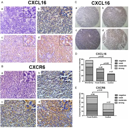
To explore the importance of CXCL16 or CXCR6 expression in DLBCL development, 76 DLBCL tissue microarray samples and 46 fresh DLBCL tissue samples as well as 27 non-tumor lymph
node tissues were collected. Among the 46 DLBCL patients, there were 30 male and 16 female, with an average age of 47.61±15.19. There was no significant difference in the distri-bution of gender and age between patients and non-tumor subjects in this population (data not shown). The levels of CXCL16 and CXCR5 expression in individual samples were charac-terized by immunohistochemistry and semi-quantitatively analyzed (Figure 1). While there was no clear anti-CXCL16 and anti-CXCR stain-ing in the interstitial cells in the DLBCL tissues there was obvious anti-CXCL16 and CXCR6 staining in the membrane-associated cyto-plasm and cytolemma of tumor cells. Semi-quantitative analysis indicated that the positive rate of anti-CXCL16 staining in DLBCL was high-er than that in the control (tissue microarray 69.74% vs. the control 7.41%, P<0.05; fresh DLBCL samples 76.09% vs. the control 7.41%,
[image:4.612.91.526.83.269.2]P<0.05). In contrast, the positive rate of anti-CXCR6 staining was significantly lower than Table 1. Relation between CXCR6/CXCL16 expression and clinical features of DLBCL
Feature CXCR6 expression CXCL16 expression
n - + ++ +++ PR P n - + ++ +++ PR P
Age (y)*
<60 36 11 10 13 2 69.44 0.190 85 21 36 17 11 75.29 0.238
≥60 10 1 5 2 2 90.00 37 13 14 7 3 64.86
Sex*
Male 30 9 10 9 2 70.00 0.408 77 21 33 13 10 72.73 0.848
Female 16 3 5 6 2 81.25 45 13 17 11 4 71.11
LDH 0.017 0.172
Low 29 11 9 6 3 62.07 29 9 13 5 2 68.97
High 17 1 6 9 1 92.12 17 2 8 6 1 88.24
Extranodal organs involvement
No 18 6 6 5 1 66.67 0.370 18 5 8 4 1 77.22 0.622
Yes 28 6 9 10 3 78.57 28 6 13 7 2 78.57
*Including tissue microarray specimens. Abbreviation: PR, positive rate. P, P values.
[image:4.612.96.523.302.404.2]that in the controls (fresh DLBCL samples 73.71% vs. the control 96.30%, P<0.05). The positive rate of both anti CXCL16 and anti-CXCR6 staining in the fresh DLBCL tissues was significantly higher than that in the control (65.22% vs. 7.41%, P<0.05). Clearly, both CXCL16 and CXCR6 are widely expressed in human DLBCL tissues.
CXCR6 and CXCL16 co-expression is an independent risk factor for poor prognosis of patients with DLBCL
Stratification analyses indicated that the posi-tive rate of anti-CXCR6 staining in patients with DLBCL was significantly associated with higher levels of serum LDH, but not with age >60, gen-der, extranodal organs involved (Table 1). Similarly, there was no significantly association of the positive rate of anti-CXCL16 staining with any measure tested in this population.
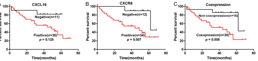
Further analysis revealed that the mean cumu-lative survival of patients with CXCR6+ DLBCL was significantly shorter than those with nega-tive DLBCL (24 moths in CXCR6+ vs. 60 months in CXCR6-, P<0.05). The CXCR6 expression was significantly associated with poor cumulative survival in patients with DLBCL (P=0.007, Figure 2). Although the mean cumulative sur-vival of patients with CXCL16+ DLBCL was shorter than that in those with CXCL16- DLBCL the CXCL16 expression in DLBCL was not sig-nificantly associated with cumulative survival of patients with DLBCL in this population (P=0.125). Interestingly, CXCL16 and CXCR6 co-expression in DLBCL was significantly asso-ciated with poor cumulative survival of patients with DLBCL in this population (P=0.008). Mu- ltivariate analysis revealed that gender, age >60 years, R-chop, levels of LDH, extranodal organs involved, single positive anti-CXCL16 or
anti-CXCR6 staining was not a significant risk factor for poor prognosis of patients with DLBCL (Table 2). However, the CXCL16 and CXCR6 co-expression was an independent risk factor for poor prognosis of patients with DLBCL in this population P=0.013).
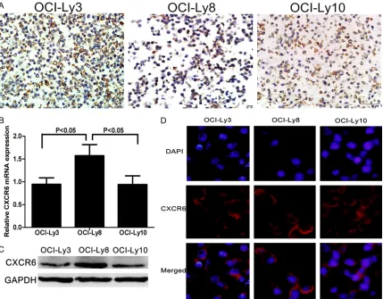
CXCR6 expression in DLBCL cell lines
Our previous study has shown that CXCL16 is highly expressed in human DLBCL OCI-Ly3, OCI-Ly8, and OCI-Ly10 cells [18]. We further investigated the CXCR6 expression in these cells by immunohistochemistry, qRT-PCR, Western blot and immunofluorescent assays. The results showed that many cells in each cell lines had anti-CXCR6 staining, particularly in their membrane-associated cytoplasm and cytolemma (Figure 3A). Furthermore, while sim-ilar levels of CXCR6 mRNA transcripts were detected in both OCI-Ly3 and OCI-Ly10 signifi-cantly higher levels of CXCR6 mRNA transcripts were observed in OCI-Ly8 cells (P<0.05, Figure 3B). A similar pattern of CXCR6 expression was detected among those cell lines (Figure 3C). Finally, immunofluorescence revealed that CXCR6 was expressed in the membrane-asso-ciated cytoplasm of those cells lines (Figure 3D). Together, varying levels of CXCR6 were expressed by DLBCL cell lines tested.
Discussion
[image:5.612.91.523.83.204.2]The CXCL16/CXCR6 axis is crucial for lympho-cyte chemotaxis [20, 21]. Previous studies have shown that CXCL16 is expressed in HL and NHL tissues and cell lines and is associat-ed with tumorigenesis of gastric mucosa asso-ciated lymphoma (MALT) [22-24]. Our previous study shows that varying levels of CXCL16 are expressed by HL and NHL cell lines [18]. In this study, we found that the frequency of DLBCL Table 2. Multivariate analysis of prognostic risk factors in DLBCL
Clinical parameters B Wald Relative risk (95% CI) P Value
Sex (male/female) -1.091 3.264 0.336 (0.103-1.097) 0.071
Age (>60 y/≤60 y) 0.019 0.964 1.019 (0.982-1.057) 0.326
R-chop (yes/no) 0.576 0.966 0.779 (0.564-1.187) 0.437
LDH (high/low) 0.589 1.026 1.779 (0.564-5.613) 0.326
Extranodal organs involved (yes/no) 0.027 0.002 1.028 (0.038-3.435) 0.965
CXCL16 (positive/negative) 0.323 0.911 1.381 (0.712-2.678) 0.340
CXCR6 (positive/negative) 0.401 1.495 1.493 (0.785-2.841) 0.221
tissues with positive CXCL16 expression was similar to that in prostatic cancer tissues [23] and was significantly higher than that in the non-tumor control tissues. However, the posi-tive rate of anti-CXCR6 staining in DLBCL tis-sues was significantly lower than that in the controls in this population. Stratification analy-sis indicated that the positive rate of CXCR6 expression in DLBCL tissues was significantly associated higher levels of serum LDH, Further analyses suggested that CXCR6 or both CXCL16 and CXCR6 expression in DLBCL tissues was significantly associated with a shorter survival of patients with DLBCL. Multivariate analysis revealed that co-expression of CXCR6 and CXCL16 in DLBCL tissues was an independent risk factor for poor prognosis of patients with DLBCL. To the best of our knowledge, this was the first report on the value of CXCL16 and
CXCR6 co-expression in prognosis of DLBCL. The co-expression of CXCL16 and CXCR6 in DLBCL tissues may be valuable for evaluating the prognosis of patients with DLBCL.
[image:6.612.94.523.73.408.2]and metastasis of gastric cancer [6]. In addi-tion, CXCL16 expression in renal cancer is associated with a better survival of patients with renal cancer [31] while high levels of CXCL16 in stromal cells and positive CXCR6 expression in NSCLC cells promote the survival of patients with NSCLC [32]. Thus, the CXCL16 and CXCR6 axis regulates the progression of different types of tumors, depending on the organ origins of the tumors.
The CXCL16/CXCR6 axis is associated with inflammation in the tumor environment. Given that CXCL16 can be resident as the membrane-associated molecule or proteolytically cleaved by the disintegrin-like metalloproteinase ADA- M10 to secrete [33, 34]. It is possible that CXCL16 can regulate CXCR6+ tumor cells in an autocrine or paracrine manner to create an positive feedback loop for the progression and metastasis of tumor cells [16, 35, 36]. The membrane-associated CXCL16 can enhance the adhesion of CXCR6+ cells [37, 38]. The secreted CXCL16 can also recruit CXCR6+ in- flammatory cells, such as dendritic cells, mac-rophages, and T cells and promote the prolifer-ation and migrprolifer-ation of CXCR6+ tumor cells [39, 40]. Furthermore, the CXCL16/CXCR6 axis can enhance the Akt/mTOR signaling, interleukin 8 (IL-8) and endothelial growth factor (VEGF) expression to promote the formation of mic- rovessels in prostate cancer cells [41]. In addi-tion, Recombinant soluble CXCL16 promoted the epithelial-mesenchymal transition (EMT) process and migration of CRC cells [42] and blockade of CXCR6 signaling suppresses the growth and invasion of hepatocellular carcino-ma cells by inhibiting the VEGF expression [43]. In contrast, the CXCR6 signaling can inhibit the AKT activation and MMP-2, and MMP-9 as well as the migration and invasion of gastric cancer cells [44]. Given that co-expression of CXCL16 and CXCR6 was associated with a poor progno-sis of patients with DLBCL it is possible that the CXCL16/CXCR6 axis may also promote the AKT/mTOR signaling and the EMT process to inhibit the proliferation and migration of DLBCL cells.
Moreover, we detected varying levels of CXCR6 expression in three DLBCL cell lines, extending our previous findings [18]. These data indicated that CXCL16 and CXCR6 were co-expressed by different DLBCL cell lines. Interestingly, the lev-els of CXCR6 and CXCL16 expression in GC sub-type OCI-Ly8 were significantly higher than that
in the non-GC subtype OCI-Ly3 and OCI-Ly10 cells. A previous study has shown that patients with GC-subtype DLBCL usually have a better survival rate than those with non-GC subtype DLBCL [45]. The high levels of CXCL16 and CXCR6 expression in GC subtype of DLBCL cells and poor prognosis of patients with CXCL16/ CXCR6 expressing DLBCL may reflect the differ-ence between in vivo DLBCL and in vitro main-tained DLBCL cells. Therefore, the clinical sig-nificance of CXCL16 and CXCR6 co-expression in different subtypes of DLBCL remains further determined.
We recognized that our study had limitations, including small sample size, the lack of molecu-lar mechanistic studies and the precise signal-ing in DLBCL cells. Thus, further studies in a bigger population are warranted to validate our findings and to investigate the molecular mech-anisms underlying the action of the CXCL16/ CXCR6 axis in regulating the development and progression of DLBCL.
In this study, we found that CXCR6/CXCL16 was wildly expressed in in the cytolemma and cyto-plasm of DLBCL. CXCR6 or CXCR6 and CXCL16 co-expression was significantly associated with shorter survival of patients with DLBCL. Clini- cally, co-expression of CXCL16/CXCR6 was an independent risk factor for t poor prognosis of patients with DLBCL in this population. Th- erefore, the expression of CXCR6 or CXCL16/ CXCR6 in DLBCL may be valuable for evaluating the prognosis of patients with DLBCL.
Acknowledgements
This study was supported by grants from the National Natural Science Foundation of China (no. 81101537 and 81570202), high technolo-gy development special foundation of Gu- angdong, China (2012A030400066) and the National Science Foundation of Guangdong Province (2015A030310177).
Disclosure of conflict of interest
None.
Hebing Road, Chancheng, Foshan 528000, Guangdong, P. R. China. Tel: +86-18038864836; E-mail: [email protected]
References
[1] Siegel R, Naishadham D, Jemal A, Cancer sta-tistics. CA Cancer Clin 2013; 63: 11-30. [2] vander Poel MW, Mulder WJ, Ossenkoppele GJ,
Maartense E, Hoogendoorn M, Wijermans P, Schouten HC. Factors that influence treatment decision-making in elderly DLBCL patients: a case vignette study. Ann Hematol 2015; 94: 1373-9.
[3] Pikarsky E, Porat RM, Stein I, Abramovitch R, Amit S, Kasem S, Gutkovich-Pyest E, Urieli-Shoval S, Galun E, Ben-Neriah Y. NF-kappaB functions as a tumour promoter in inflamma-tion-associated cancer. Nature 2004; 23: 461-6.
[4] Greten FR, Eckmann L, Greten TF, Park JM, Li ZW, Egan LJ, Kagnoff MF, Karin M. IKK-beta links inflammation and tumorigenesis in a mouse model of colitis-associated cancer. Cell 2004; 118: 285-96.
[5] Scott DW, Gascoyne RD. The tumour microen-vironment in B cell lymphomas. Nat Rev Can-cer 2014; 14: 517-34.
[6] Xing YN, Xu XY, Nie XC, Yang X, Yu M, Xu HM, Liu YP, Takano Y, Zheng HC. Role and clinico-pathologic significance of CXC chemokine li-gand 16 and chemokine (C-X-C motif) receptor 6 expression in gastric carcinomas. Hum Pathol 2012; 43: 2299-307.
[7] Deng L, Chen N, Li Y, Zheng H, Lei Q. CXCR6/ CXCL16 functions as a regulator in metastasis and progression of cancer. Biochem Biophys Acta 2010; 1806: 42-9.
[8] Zhang L, Ran L, Garcia GE, Wang XH, Han S, Du J, Mitch WE. Chemokine CXCL16 regulates neutrophil and macrophage infiltration into in-jured muscle, promoting muscle regeneration. Am Pathol 2009; 175: 2518-27.
[9] Diegelmann J, Seiderer J, Niess JH, Haller D, Göke B, Reinecker HC, Brand S. Expression and regulation of the chemokine CXCL16 in Crohns disease and models of intestinal in-flammation. Inflamm Bowel Dis 2010; 16: 1871-81.
[10] van Lieshout AW, Popa C, Meyer-Wentrup F, Lemmers HL, Stalenhoef AF, Adema GJ, van Riel PL, van Tits LJ, Radstake TR. Circulating CXCL16 is not related to circulating oxLDL in patients with rheumatoid arthritis. Biochem Biophys Res Commun 2007; 355: 392-7. [11] Hattermann K, Ludwig A, Gieselmann V,
Held-Feindt J, Mentlein R. The chemokine CXCL16 induces migration and invasion of glialprecur-sor cells via its receptor CXCR6. Mol Cell Neu-rosci 2008; 39: 133-41.
[12] Garcia GE, Truong LD, Li P, Zhang P, Johnson RJ, Wilson CB, Feng L. Inhibition of CXCL16 at-tenuates inflammatory and progressive phas-es of anti-glomerular basement membrane antibody-associated glomerulonephritis. Am Pathol 2007; 170: 1485-96.
[13] Morgan AJ, Guillen C, Symon FA, Huynh TT, Berry MA, Entwisle JJ, Briskin M, Pavord ID, Wardlaw AJ. Expression of CXCR6 and its li-gand CXCL16 in the lung in health and dis-ease. Clin Exp Allergy 2005; 35: 1572-80. [14] Smith C, Halvorsen B, Otterdal K, Waehre T,
Yn-destad A, Fevang B, Sandberg WJ, Breland UM, Frøland SS, Oie E, Gullestad L, Damås JK, Auk-rust P. High levels and inflammatory effects of soluble CXC ligand 16 (CXCL16) in coronary artery disease: down-regulatory effects of statins. Cardiovasc Res 2008; 79: 195-203. [15] Ruth JH, Haas CS, Park CC, Amin MA, Martinez
RJ, Haines GK 3rd, Shahrara S, Campbell PL, Koch AE. CXCL16-mediated cell recruitment to rheumatoid arthritis synovial tissue and mu-rine lymph nodes is dependent upon the MAPK pathway. Arthritis Rheum 2006; 54: 765-78. [16] Meijer J, Ogink J, Kreike B, Nuyten D, de Visser
KE, Roos E. The chemokine receptor CXCR6 and its ligand CXCL16 are expressed in carci-nomas and inhibit proliferation. Cancer Res 2008; 68: 4701-8.
[17] Richardsen E, Ness N, Melbø-Jørgensen C, Jo-hannesen C, Grindstad T, Nordbakken C, Al-Saad S, Andersen S, Dønnem T, Nordby Y, Bremnes RM, Busund LT. The Prognostic Sig-nificance of CXCL16 and Its Receptor C-X-C Chemokine Receptor 6 in Prostate. Cancer Am Pathol 2015; 185: 2722-30.
[18] Liu F, Zhang Y, Tang H, Zhou X, Wu Z, Tang D, Zhao T. CXC chemokine ligand 16, inversely correlated with CD99 expression in Hodgkin Reed-Sternberg cells, is widely expressed in diverse types of lymphomas. Oncol Rep 2013; 30: 783-92.
[19] Huang X, Zhou X, Wang Z, Li F, Liu F, Zhong L, Li X, Han X, Wu Z, Chen S, Zhao T. CD99 triggers upregulation of miR-9-modulated PRDM1/ BLIMP1 in Hodgkin/Reed-Sternberg cells and induces redifferentiation. Int Cancer 2012; 131: E382-94.
[20] Wehr A, Baeck C, Heymann F, Niemietz PM, Hammerich L, Martin C, Zimmermann HW, Pack O, Gassler N, Hittatiya K, Ludwig A, Lu-edde T, Trautwein C, Tacke F. Chemokine re-ceptor CXCR6-dependent hepatic NK T Cell ac-cumulation promotes inflammation and liver fibrosis. Immunol 2013; 190: 5226-36. [21] Isozaki T, Arbab AS, Haas CS, Amin MA, Arendt
chemo-taxis: studies in mice with K/BxN serum-in-duced arthritis. Arthritis Rheum 2013; 65: 1736-46.
[22] Hanamoto H, Nakayama T, Miyazato H, Takeg-awa S, Hieshima K, Tatsumi Y, Kanamaru A, Yoshie O. Expression of CCL28 by Reed-Stern-berg cells defines a major subtype of classical Hodgkin’s disease with frequent infiltration of eosinophils and/or plasma cells. Am Pathol 2004; 164: 997-1006.
[23] Darash-Yahana M, Gillespie JW, Hewitt SM, Chen YY, Maeda S, Stein I, Singh SP, Bedolla RB, Peled A, Troyer DA, Pikarsky E, Karin M, Farber JM. The chemokine CXCL16 and its re-ceptor, CXCR6, as markers and promoters of inflammation-associated cancers. PLoS One 2009; 4: e6695.
[24] Deutsch AJ, Steinbauer E, Hofmann NA, Strunk D, Gerlza T, Beham-Schmid C, Schaider H, Neumeister P. Neumeister. Chemokine recep-tors in gastric MALT lymphoma: loss of CXCR4 and upregulation of CXCR7 is associated with progression to diffuse large B-cell lymphoma. Mod Pathol 2013; 26: 182-94.
[25] Huang Y, Zhang J, Cui ZM, Zhao J, Zheng Y. Ex-pression of the CXCL12/CXCR4 and CXCL16/ CXCR6 axes in cervical intraepithelial neopla-sia and cervical cancer. Chin J Cancer 2013; 32: 289-96.
[26] Lee JT, Lee SD, Lee JZ, Chung MK, Ha HK. Ex-pression analysis and clinical significance of 26CXCL16/CXCR6 in patients with bladder cancer. Oncol Lett 2013; 5: 229-235.
[27] Guo L, Cui ZM, Zhang J, Huang Y. Chemokine axes CXCL12/CXCR4 and CXCL16/CXCR6 cor-relate with lymph node metastasis in epithelial ovarian carcinoma. Chin Cancer 2011; 30: 336-43.
[28] Airoldi I, Cocco C, Morandi F, Prigione I, Pistoia V. CXCR5 may be involved in the attraction of human metastatic neuroblastoma cells to the bone marrow. Cancer Immunol Immunother 2008; 57: 541-8.
[29] Seidl H, Richtig E, Tilz H, Stefan M, Schmidbau-er U, AsslabSchmidbau-er M, Zatloukal K, HSchmidbau-erlyn M, Schaider H. Profiles of chemokine receptors in melanocytic lesions: de novo expression of CXCR6 in melanoma. Hum Pathol 2007; 38: 768-80.
[30] Hattermann K, Held-Feindt J, Ludwig A, Men-tlein R. The CXCL16-CXCR6 chemokine axis in glial tumors. Neuroimmunol 2013; 260: 47-54.
[31] Gutwein P, Schramme A, Sinke N, Abdel-Bakky MS, Voss B, Obermüller N, Doberstein K, Kozi-olek M, Fritzsche F, Johannsen M, Jung K, Schaider H, Altevogt P, Ludwig A, Pfeilschifter J, Kristiansen G. Tumoural CXCL16 expression is a novel prognostic marker of longer survival
times in renal cell cancer patients. Eur Cancer 2009; 45: 478-89.
[32] Hald SM, Kiselev Y, Al-Saad S, Richardsen E, Johannessen C, Eilertsen M, Kilvaer TK, Al-Shibli K, Andersen S, Busund LT, Bremnes RM, Donnem T. Prognostic impact of CXCL16 and CXCR6 in non-small cell lung cancer: combined high CXCL16 expression in tumor stroma and cancer cells yields improved survival. BMC Cancer 2015; 15: 441.
[33] Shimaoka T, Kume N, Minami M, Hayashida K, Kataoka H, Kita T, Yonehara S. Molecular clon-ing of a novel scavenger receptor for oxidized low density lipoprotein, SR-PSOX, on macro-phages. Biol Chem 2000; 275: 40663-6. [34] Wilbanks A, Zondlo SC, Murphy K, Mak S, Soler
D, Langdon P, Andrew DP, Wu L, Briskin M. Ex-pression cloning of the STRL33/BONZO/TYM-STRligand reveals elements of CC, CXC, and CX3C chemokines. Immunol 2001; 166: 5145-54.
[35] Hu W, Liu Y, Zhou W, Si L, Ren L. CXCL16 and CXCR6 are coexpressed in human lung cancer in vivo and mediate the invasion of lung cancer cell lines in vitro. PLoS One 2014; 9: e99056. [36] Gough PJ, Garton KJ, Wille PT, Rychlewski M,
Dempsey PJ, Raines EW. A disintegrin and me-talloproteinase 10-mediated cleavage and shedding regulates the cell surface expression of cxc chemokine ligand 16. Immunol 2004; 172: 3678-85.
[37] Raman D, Baugher PJ, Thu YM, Richmond A. Role of chemokines in tumor growth. Cancer Lett 2007; 256: 137-65.
[38] De Marzo AM, Nakai Y, Nelson WG. Inflamma-tion, atrophy, and prostate carcinogenesis. Urol Oncol 2007; 25: 398-400.
[39] Shimaoka T, Nakayama T, Fukumoto N, Kume N, Takahashi S, Yamaguchi J, Minami M, Hayashida K, Kita T, Ohsumi J, Yoshie O, Yone-hara S. Cell surface-anchored SR-PSOX/CXC chemokine ligand 16 mediates firmadhesion of CXC chemokine receptor 6-expressing cells. Leukoc Biol 2004; 75: 267-74.
[40] Chandrasekar B, Bysani S, Mummidi S. CXCL16 signals via giphosphatidylinositol 3-ki-nase, Akt, IκB ki3-ki-nase, and nuclear factor-κb and induces cell-cell adhesion and aortic smooth muscle cell proliferation. Biol Chem 2004; 279: 3188-96.
[41] Wang J, Lu Y, Wang J, Koch AE, Zhang J, Taich-man RS. CXCR6 induces prostate cancer pro-gression by the AKT/mammalian target of ra-pamycin signaling pathway. Cancer Res 2008; 68: 10367-76.
metastases in colorectal cancer patients. Ann Surg Oncol 2012; Suppl 3: S518-27.
[43] Li Y, Fu LX, Zhu WL, Shi H, Chen LJ, Ye B. Block-ade of CXCR6 reduces invasive potential of gastric cancer cells through inhibition of AKT signaling. Int Immunopathol Pharmacol 2015; 28: 194-200.
[44] Xu JM, Weng MZ, Song FB, Chen JY, Zhang JY, Wu JY, Qin J, Jin T, Wang XL. Blockade of the CXCR6 signaling inhibits growth and invasion of hepatocellular carcinoma cells through inhi-bition of the VEGF expression. Int Immuno-pathol Pharmacol 2014; 27: 553-61.