Original Article
Expression of Fas/FasL and c-myc in bladder cancer
and their correlation with tumor immune function
Jian Li1, Yibin Zhou2, Qingwen Li1, Jiajun Zhang1, Wenyan Sun1, Changyuan Dai1, Yuxi Shan2
1Department of Urology, The First Affiliated Hospital of Bengbu Medical College, Bengbu, Anhui, China;
2Department of Urology, The Second Hospital Affiliated to Soochow University, Suzhou, Jiangsu, China
Received January 6, 2016; Accepted April 25, 2016; Epub June 1, 2016; Published June 15, 2016
Abstract: Fas/FasL can induce cell apoptosis and maintain body homeostasis. As one transmembrane glycoprotein, it exerts important biological functions. C-myc is one important oncogene involving in progression of multiple tumors and is closely correlated with tumor proliferation, differentiation and apoptosis. This study thus investigated the ex-pression level of Fas/FasL and c-myc in bladder cancer, to analyze its correlation with tumor immunity. A total of 40 bladder cancer patients were recruited. Enzyme linked immunosorbent assay (ELISA) and immunohistochemistry (IHC) were employed to detect the expression of Fas, FasL and c-myc in both serum and bladder cancer tissues.
In situ end labeling approach was used to determine the apoptotic rate of cancer and adjacent tissues along with
infiltrated lymphocytes (TIL) in bladder mucus. The correlation between Fas/FasL and c-myc expression and TIL
apoptotic rate were analyzed. Serum Fas in experimental group was lower than control group, with elevated FasL and c-myc (P<0.05). The expression of Fas in tumor tissues was lower than adjacent or control tissues, while FasL and c-myc levels were elevated (P<0.05). TIL apoptotic rate in bladder cancer tissues was higher than adjacent tis-sues (P<0.05). The expression of FasL and c-myc along with TIC apoptotic rate was higher in higher grade, multiple lesion, recurrent patients (P<0.05). FasL and TIL were positively correlated with TIL apoptotic rates, while Fas was negatively correlated (P<0.05). FasL was over-expressed while Fas was down-regulated in bladder cancer, facilitat-ing apoptosis of lymphocytes. C-myc expression can up-regulate the immune escape latency of tumor cells.
Keywords: Bladder cancer, Fas/FasL, C-myc, lymphocytes, apoptosis
Introduction
Previous study regarding immune surveillance indicated the body’s potency to recognize tumor cells via body’s immune system and exerts timely eradication via immune cells [1]. Both basic and clinical studies in immunology revealed that, although T cells could recognize tumor-specific antigens at the early stage, those recognized tumor cells are impossible for elimination, thus can proliferate and differenti-ate for tumor infiltration and migration [2]. Fas/ FasL is one important signal pathway for clear-ing aged cells, as it can induce apoptosis at all body sites and help the normal cellular metabo-lism, thus accelerating immune process for clearing abnormal cells to inhibit unfavorable consequences [3, 4]. As one important mem-ber of apoptotic factors, Fas/FasL plays a cri- tical role in immune escape mechanism of tumors. Fas is one transmembrane
Materials and methods
General information
A total of 40 bladder cancer patients in our hos-pital from January 2014 to January 2015 in the First Affiliated Hospital of Bengbu Medical College were recruited in this study. The con-firmed diagnosis was made by lab indexes, imaging and post-operative pathological meth-ods. There were 20 males and 20 females in patient group, with aging between 25 and 75 years (average = 46.1±9.4 years old). The aver-age period of disease onset was 22.5±9.1 months. All patients had hematuria and pollaki-uria, precipitant urea and urodynia, Under UIC standard, there were 14 cases of T0-T1 phase for superficial bladder cancer, 26 cases with infiltrated bladder cancer (stage T2-T4). In WHO criteria, there were 10, 19 and 11 patients for G1, G2 and G3, respectively. In all patients, there were 21 with primary diagnosis and 19 recurrent cases. Single lesion occupied 23 cases while 17 cases belong to multiple lesions. All patients had not received chemo-/radio-/ immune therapy before endoscopy. Another 20 individuals from out hospital were recruited as the control group (12 males and 8 fema- les, aging between 20 and 70 years, avera- ge age = 45.1±3.4 years. No significant dif-ference regarding sex or age existed be- tween two groups (P>0.05), which were thus comparable.
The experimental protocol has been pre-approved by the ethical committee of the First Affiliated Hospital of Bengbu Medical College and written consents have been obtained from all patients and healthy volunteers.
Inclusive criteria: All patients have been diag-nosed by pathology. There was no connective tissue disease, immune disorder, or severe major heart failure including heart, liver and kidney. Patients had not received any anti-tumor treatment such as radio-, chemo- or immune therapy before surgery.
Resin. Ultra-clean workstation (Formal, US); Centrifuge (Feige, China); Inverted microscope (Olympus, Japan); Microplate reader (TECNA, UK); Tissue embedding workstation (SAKURA, Japan); Tissue processor (TIYODA, Japan); Ultra-think microtome (Leica, Germany); Temperature-controlled vibrator (Jinghong, China); Image analyzing system (HP, US).
ELISA
Fasted venous blood samples were collected from all patients. After centrifugation, superna-tants were saved for further use. ELISA was applied to quantify serum level of Fas, FasL and c-myc. EDTA-Na anticoagulant; ELISA kits for Fas, FasL and c-myc; PBS buffer; Blocking solu-tion plus primary antibody for Fas, FasL and c-myc were purchased from Abcam, USA. In brief, the test kit was incubated at room tem-perature for 30 min. Standard samples were diluted along with test samples (N = 5 each). Samples were loaded into the plate for wash-ing, adding reaction buffer, development and quenching. A microplate reader was used to measure absorbance value at 450 nm by Microplate reader (TECNA, UK). Linear regres-sion formula was employed to calculate sample concentration.
Expression of Fas, FasL and c-myc
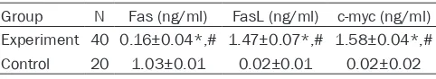
[image:2.612.92.333.83.127.2]Tissues were fixed in formalin, dehydrated, immersed in paraffin and embedded with tis -sue embedding workstation (SAKURA, Japan) and ultra-think microtome (Leica, Germany). Tissues sections were de-waxed, re-hydrated and were heated for antigen retrieval with and tissue processor (TIYODA, Japan). Hydrogen peroxide (Sigma, USA) was used to quench slides, which were then blocked in normal goat serum. Primary antibody (Abcam, USA) was then added for 1 h incubation at room tempera-ture, followed by secondary antibody conjugat-ed with horseradish peroxidase (Rabbit anti-mouse IHC secondary antibody, Abcam, USA) for 10 min incubation. Streptomycin-peroxidase Table 1. Serum Fas, FasL and c-myc contents
Group N Fas (ng/ml) FasL (ng/ml) c-myc (ng/ml) Experiment 40 0.16±0.04*,# 1.47±0.07*,# 1.58±0.04*,# Control 20 1.03±0.01 0.02±0.01 0.02±0.02
Note: *, P<0.05 compared to control group.
Reagents and instruments
complex (Abnova, USA) was then added for 10 min incubation. DAB (Sigma, USA) was used to develop slides. After stopping, hematoxylin (Sigma, USA) was used to counter-stain slides, which were differentiated by HCl-ethanol. Sli- des were dehydrated and mounted. Imaging system was used to capture 5 randomly select-ed fields for recording by invertselect-ed microscope (Olympus, Japan) and image analyzing system (HP, US).
IHC results judgment
Using positive controlled slides and negative (PBS) controlled slides, IHC staining results were analyzed with HE staining as the re- ference.
The positive staining for Fas, FasL and c-myc was identified as brown or dark yellow granules in cytoplasm. Negative (-): ≤10% positive cells; Weak positive (+): 11%~25% positive cells; Positive (++): 26%~50% positive cells; Strong positive (+++): >50% positive cells. 5 fields were randomly selected from each slide for image capture and recording in softwares.
TUNEL assay
Tissue samples were prepared for paraffin-based slides, which were incubated in 2% hydrogen peroxide in PBS. After rinsing, TdT enzyme buffer and reaction buffer were added for reaction (Thermofisher, USA). Anti-DIG anti -body conjugated with peroxidase (Abcam, USA) was then added to develop slides in DAB sub-strates. Positive signal was identified as brown granules in nucleus. 5 randomly selected fields were selected to calculate cell apoptotic rate (AR), which was equal to positive cell number/ total cell number ×100%.
Statistical analysis
SPSS 17.0 software (IBM, USA) was used to process all collected data, of which
enumera-Results
Serum Fas, FasL and c-myc contents
ELISA approach was used to determine serum levels of Fas, FasL and c-myc from peripheral venous blood samples. Results showed that Fas level in experimental group were (0.16± 0.04) ng/ml, which was lower than control group. FasL and c-myc levels were (1.47±0.07) ng/ml and (1.58±0.04) ng/ml, respectively, which were significantly higher than control group (P<0.05, Table 1).
Fas and FasL in bladder tissues
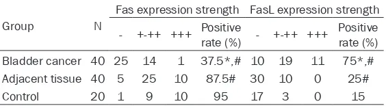
IHC staining was performed to test the expres-sion level of Fas and FasL in bladder tissues. Results found strong positive expression of Fas (1 case) and 14 cases of positive expression, making the positive rate at only 37.5%, which was significantly lower than adjacent and con -trol tissues (P<0.05). There were 11 strong positive and 19 positive cases of FasL in blad-der tissues, comprising 75% positive rate, which was elevated than adjacent and control tissues (P<0.05, Table 2; Figure 1).
Expression of c-myc in patient bladder tissues by IHC staining
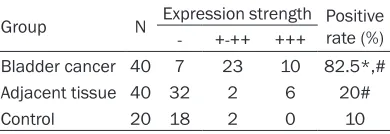
The expression of c-myc was tested in bladder tissues in all patients. Result showed 10 cases of strong positive and 23 positive cases, com-prising positive rate as high as 82.5%, which was significantly higher than adjacent tissues or control ones. See Table 3 and Figure 2 for details.
TUNEL assay of lymphocytes
Lymphocytes were distributed in the lamina propria of bladder mucous and has infiltrated into tumor mesenchymal and tumor solid. Positive signals were shown as yellow-brown granules in nucleus of lymphocytes. The
[image:3.612.91.373.85.164.2]apop-tion data were processed by chi-square analysis, while mea-surement data were compared by analysis of variance (ANOVA) and were presented as mean ± standard deviation. Logistic regression model was used in multi-factorial analysis. A sta-tistical analysis was defined when P<0.05.
Table 2. Fas and FasL expression in bladder tissues
Group N
Fas expression strength FasL expression strength - +-++ +++ Positive rate (%) - +-++ +++ Positive rate (%) Bladder cancer 40 25 14 1 37.5*,# 10 19 11 75*,# Adjacent tissue 40 5 25 10 87.5# 30 10 0 25#
Control 20 1 9 10 95 17 3 0 15
totic rates of lymphocytes infiltrated in bladder cancer tissue, adjacent tissue and normal blad-der mucous were (31.47±17.99)%, (6.59±7.46)% and (3.37±1.50)%, respectively, with statistical significance (P<0.05). See Figure 3 for details.
Correlation between Fas, FasL, c-myc expres-sion and TIL apoptotic rate
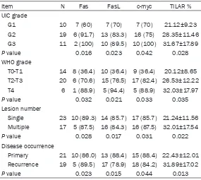
The analysis between expression of Fas, FasL and c-myc in bladder tissues showed elevated expression of FasL and c-myc in bladder cancer tissues compared to adjacent or control groups, while Fas expression was lower than adjacent or control tissues (P<0.05). In bladder cancer tissues, the expression of FasL and c-myc, along with TILAR were elevated while Fas was
down-regulated in tumors with higher UIC or G1-G3 grade (P<0.05). In tumors with advanced WHO grade (T0-T4), FasL, c-myc expression and TILAR were also elevated while Fas expression was decreased (P<0.05). Regarding number of lesions, FasL, c-myc and TILAR were all elevat-ed in multiple lesions while Fas was down-regu-lated (P<0.05). Compared to primary cases, recurrent patients had higher expression of FasL and c-myc, along with higher TILAR but lower Fas expression (P<0.05). See Table 4 for details.
In analyzing the correlation between all param-eters, we found a positive correlation between FasL and TILAR (r = 0.84, P<0.05), between c-myc and TILAR (r = 0.51, P<0.05) and a nega-tive correlation between Fas and TILAR (r = -0.65, P<0.05). C-myc was positively correlated with FasL (r = 0.38, P<0.05), and was negative- ly correlated with Fas (r = -0.39, P<0.05). FasL was negatively correlated with Fas (r = -0.27, P<0.05).
Discussion
[image:4.612.82.525.72.170.2] [image:4.612.92.287.257.324.2]Fas/FasL belongs to the tumor necrosis factor/ neural growth factor receptor family. Fas/FasL had interaction for body signal transduction and can participate in the induction of cell apoptosis to maintain the homeostasis of inter-nal environment, thus exerting important bio-logical functions [9]. Study has indicated that FasL could induce apoptosis of infiltrated T lym -phocytes, thus facilitating immune escape in eyes or testicles [10]. Fas is expressed in vari-ous tumor tissues. FasL is mainly existed in the surface of immune activated spleen cells or thyroid cells, including activated T cells and natural killer cells [11]. In multiple tumors, Fas and FasL were co-expressed. The positive Figure 1. The expressions of Fas and FasL in bladder tissues detected by Immunohistochemistry (×200). A. Fas expression in tumor adjacent tissues; B. Fas expression in bladder cancer tissues; C. FasL expression in adjacent tissues; D. FasL expression in bladder cancer tissues.
Table 3. C-myc expression in bladder tissues by IHC staining
Group N Expression strength Positive rate (%) - +-++ +++
Bladder cancer 40 7 23 10 82.5*,# Adjacent tissue 40 32 2 6 20#
Control 20 18 2 0 10
Note: *, P<0.05 compared to adjacent tissues; #, P<0.05 compared to control group.
[image:4.612.88.290.366.455.2]expression of FasL had anti-attack potency to resist against death signal, endowing tumor cells the ability of immune escape, thus facili-tating tumor proliferation and differentiation [12, 13]. C-myc is one transcriptional regulatory protein, and exerts its function via facilitating/ inhibiting target gene transcription. The abnor-mal expression of c-myc can induce the devel-opment of tumors. When c-myc was inactivat-ed, cell growth suppressor genes p15, p21 and
[image:5.612.93.376.72.256.2]tissues found higher FasL and c-myc expres-sion along with TICAR and lower Fas expresexpres-sion in those tumors with higher G1-G3 grade or WHO T0-T4 grade, multiple lesions or recur -rence. These results suggested that bladder cancer with higher grade and lower differentia-tion grade may manifest with elevated FasL expression, which may be correlated with the differentiation grade of tumors. For those tumor tissues with good differentiation, FasL is Figure 3. TUNEL assay for apoptosis of lymphocytes (TILAR %). *, P<0.05
compared to adjacent tissue; #, P<0.05 compared to control tissue.
Table 4. Fas, FasL, c-myc expression and apoptotic rates
Item N Fas FasL c-myc TILAR %
UIC grade
G1 10 7 (60) 7 (70) 7 (70) 21.12±9.23 G2 19 6 (91.7) 13 (83.3) 16 (75) 28.35±11.46 G3 11 2 (100) 10 (89.5) 10 (100) 31.67±17.89
P value 0.016 0.023 0.042 0.028
WHO grade
T0-T1 14 8 (36.4) 10 (36.4) 9 (36.4) 20.12±8.65 T2-T3 20 6 (70.6) 15 (76.5) 17 (82.4) 26.53±12.22 T4 6 1 (88.9) 5 (94.4) 5 (88.9) 32.03±17.97
P value 0.032 0.021 0.033 0.035
Lesion number
Single 23 10 (89.3) 14 (85.7) 17 (85.7) 21.24±11.56 Multiple 17 5 (87.5) 16 (84.3) 16 (87.5) 32.01±17.54
P value 0.028 0.017 0.031 0.022
Disease occurrence
Primary 21 10 (86.0) 13 (88.4) 15 (88.4) 22.43±12.01 Recurrence 19 5 (89.5) 17 (78.9) 18 (84.2) 31.89±17.02
P value 0.023 0.015 0.044 0.013
p27 were all up-regulated. In cells with over-expression of c-myc, these genes were do- wn-regulated.
In this study, bladder cancer patients were recruited to col-lect peripheral blood sam-ples, in which serum Fas, FasL and c-myc contents were determined by ELISA. Results showed lower Fas and higher FasL or c-myc in experimental groups compared to control group. Further IHC staining found elevated FasL and c-myc along with lower Fas expressions in bladder cancer tissues. These results sug-gested depressed Fas expres-sion level and elevated FasL/ c-myc level in bladder cancer patients. Previous study has shown the down-regulation of Fas in various malignant tumor cells. Under such cir-cumstances, tumor cell can escape from body immune killing, thus avoiding cell apo- ptosis. Meanwhile, in tumor cells with co-expression with those two factors, the local increase of FasL can facilitate the killing of body immune cells by tumors, further aggra-vating local environment for immune escape, helping tu- mor cells to survive under immune surveillance for con-tinuous infiltration and migra -tion [14-16].
[image:5.612.91.376.320.575.2]highly expressed in peripheral tumor cells around the lesion, thus benefiting tumor cell infiltration and migration. C-myc protein expres -sion is also related with the stage and differen-tiation grade of bladder cancer patients, thus may working as one important factor evaluating disease condition and prognosis of bladder cancer patients. C-myc was found to be posi-tively correlated with FasL while negaposi-tively cor-related with Fas, which was negatively cor-related with Fas. The combined assay of those three factors in disease diagnosis and prognosis pre-diction may better analyze the biological activi-ty of bladder cancer, thus having clinical impli-cations [17].
This study assayed TIL by TUNEL method and found more infiltration of TIL in mesenchymal and mucosal layer of bladder cancer than solid. TILAR in cancer infiltrated tissues was also higher than adjacent tissue or normal bladder mucosal tissues. These results suggested the facilitating of TILAR by bladder cancer cells. Further analysis of related indexes found the negative correlation between Fas and TILAR, and positive correlation among FasL, c-myc and TILAR. Inside body TIL can received antigen stimulus for rapid activation. The abundantly positive expression of Fas increased FasL sen-sitivity. For TIL which kill tumor cells, they were susceptible for strong attack for FasL-positive tumor cells, leading to abundant apoptosis. Those apoptotic TIL exist in cancer lesion or the peripheral region [18]. Basic study has found 4-times decrease of TIL in FasL-positive tumor cells. For those tumor cells with FasL-negative, TIL was increased by almost 2 folds [19]. These data suggested the close correlation between TIL loss and FasL expression by tumor cells. Inside the body, activation of TIL rapidly down-regulate Fas-resistant TIL to highly express FasL by tumor cells, which strongly attack TIL to induce their apoptosis, thus escaping immune surveillance and toxicity, making tumor growth and progression [20].
In summary, in bladder cancer, tumor cells highly expressed FasL while expressed Fas at low levels. They can help to clear locally specific lymphocytes for facilitating lymphocyte apopto-sis, thus making the focal growth, proliferation, infiltration and migration of malignant tumor cells. Moreover, c-myc was up-regulated in bladder cancer and facilitated the interaction between Fas and FasL, thus potentiating im-
mune escape ability of tumors. There might be one coordinated function between Fas/FasL and c-myc to modulate the occurrence and pro-gression of lymphocyte apoptosis. The detailed mechanism of Fas/FasL and c-myc still requi- re substantiation. Further exploration can be made via inhibiting its transduction pathway and may help in treating bladder cancer.
Disclosure of conflict of interest
None.
Address correspondence to: Dr. Yuxi Shan, De-
partment of Urology, The Second Hospital Affiliated
to Soochow University, 1055, San Xiang Road, Suzhou 215004, Jiangsu, China. Tel: +86-512-677- 84135; Fax: +86-512-67784135; E-mail: [email protected]
References
[1] Brown MA. Genetics of ankylosing spondylitis.
Curr Opin Rheumatol 2010; 22: 126-32.
[2] Iero M, Valenti R, Huber V, Filipazzi P, Parmiani
G, Fais S, Rivoltini L. Tumour-released exo-somes and their implications in cancer immu-nity. Cell Death Differ 2008; 15: 80-8.
[3] Punsawad C, Viriyavejakul P, Setthapramote
C3, Palipoch S. Enhanced expression of Fas and FasL modulates apoptosis in the lungs of severe P. falciparum malaria patients with pul-monary edema. Int J Clin Exp Pathol 2015; 8: 10002-13.
[4] Wu GZ, Pan CX, Jiang D, Zhang Q, Li Y, Zheng
SY. Clinicopathological significance of Fas and
Fas ligand expressions in esophageal cancer. Am J Cancer Res 2015; 5: 2865-2871. [5] Matsumoto H, Murakami Y, Kataoka K, Notomi
S, Mantopoulos D, Trichonas G, Miller JW, Gregory MS, Ksander BR, Marshak-Rothstein
A, Vavvas DG. Membrane-bound and soluble
Fas ligands have opposite functions in photo-receptor cell death following separation from the retinal pigment epithelium. Cell Death Dis 2015; 6: e1986.
[6] Gmeiner WH, Jennings-Gee J, Stuart CH, Pardee TS. Thymineless death in F10-treated AML cells occurs via lipid raft depletion and Fas/FasL co-localization in the plasma mem-brane with activation of the extrinsic apoptotic pathway. Leuk Res 2015; 39: 229-35. [7] Calmon-Hamaty F, Audo R, Combe B, Morel J,
Hahne M. Targeting the Fas/FasL system in Rheumatoid Arthritis therapy: Promising or risky? Cytokine 2015; 75: 228-33.
Pitiakoudis M, Vaos G, Simopoulos C. The ef -fects of apigenin on the expression of Fas/ FasL apoptotic pathway in warm liver ischemia-reperfusion injury in rats. Biomed Res Int 2014; 2014: 157216.
[9] Han W, Zhou Y, Zhong R, Wu C, Song R, Liu L, Zou L, Qiao Y, Zhai K, Chang J, Huang L, Liu L, Lu X, Lou J, Yu D, Tan W, Zhang J, Wang H, Miao X. Functional polymorphisms in FAS/ FASL system increase the risk of
neuroblasto-ma in Chinese population. PLoS One 2013; 8:
e71656.
[10] Abebe M, Doherty TM, Wassie L, Aseffa A, Bobosha K, Demissie A, Zewdie M, Engers H, Andersen P, Kim L, Huggett J, Rook G, Yamuah
LK, Zumla A; VACSEL study group. Expression
of apoptosis-related genes in an Ethiopian co-hort study correlates with tuberculosis clinical status. Eur J Immunol 2010; 40: 291-301. [11] Leon-Bollotte L, Subramaniam S, Cauvard O,
Plenchette-Colas S, Paul C, Godard C, Martinez-Ruiz A, Legembre P, Jeannin JF, Bettaieb A. S-nitrosylation of the death receptor fas pro-motes fas ligand-mediated apoptosis in cancer cells. Gastroenterology 2011; 140: 2009-18, 2018.e1-4.
[12] Li CG, Li ML, Shu XH, Liu YJ, Wu WS. Antitumor effects of recombinant human interleukin-6 on mouse bladder carcinoma through Fas-mediated apoptosis. Cancer Chemother Phar- macol 2010; 66: 981-6.
[13] Svatek RS, Herman MP, Lotan Y, Casella R, Hsieh JT, Sagalowsky AI, Shariat SF. Soluble Fas--a promising novel urinary marker for the
detection of recurrent superficial bladder can -cer. Cancer 2006; 106: 1701-7.
[14] Zhang J and Xu G. Suppression of FasL expres-sion in tumor cells and preventing tumor ne-crosis factor-induced apoptosis by adenovirus 14.7K is an effective escape mechanism for immune cells. Cancer Genet Cytogenet 2007; 179: 112-7.
[15] Ryan AE, Shanahan F, O’Connell J, Houston
AM. Fas ligand promotes tumor immune eva-sion of colon cancer in vivo. Cell Cycle 2006; 5: 246-9.
[16] Ayari C, Bergeron A, LaRue H, Ménard C, Fradet Y. Toll-like receptors in normal and malignant human bladders. J Urol 2011; 185: 1915-21. [17] Mitra AP and Cote RJ. Molecular pathogenesis
and diagnostics of bladder cancer. Annu Rev Pathol 2009; 4: 251-85.
[18] Jiang G, Wang Y, Yun J, Hajrasouliha AR, Zhao Y, Sun D, Kaplan HJ, Shao H. HMGB1 release triggered by the interaction of live retinal cells and uveitogenic T cells is Fas/FasL
activation-dependent. J Neuroinflammation 2015; 12:
179.
[19] Miller MR, Mandell JB, Beatty KM, Harvey SA, Rizzo MJ, Previte DM, Thorne SH, McKenna KC. Splenectomy promotes indirect elimina-tion of intraocular tumors by CD8+ T cells that is associated with IFNgamma- and Fas/FasL-dependent activation of intratumoral macro-phages. Cancer Immunol Res 2014; 2: 1175-85.