Original Article
Overexpression of PER2 inhibits the progression of
atherosclerosis via the Akt-eNOS signaling in
apolipoprotein e-null mice
Gang Su1*, Guangli Sun2*, Hai Liu1, Liliang Shu1, Jingchao Zhang1, Longhui Guo1, Chen Huang1, Jing Xu1
1Department of Cardiovascular Surgery, The First Affiliated Hospital of Zhengzhou University, Zhengzhou, Henan,
P. R. China; 2Department of Ophthalmology, The First Affiliated Hospital of Zhengzhou University, Zhengzhou,
Henan, P. R. China. *Equal contributors.
Received January 15, 2016; Accepted March 26, 2016; Epub June 1, 2016; Published June 15, 2016
Abstract: Atherosclerosis is an inflammatory disease. The circadian clock gene has a connection with inflammation, including vascular inflammation. However, whether circadian clock gene has a regulatory effect on atherosclerosis is rarely studied. Our aim is to determine whether Period 2 (Per2), a main clock gene, exerts a therapeutical effect on atherosclerosis in Apolipoprotein E-null (ApoE-/-) mice. Atherosclerotic mice were divided into three groups, includ-ing the Control group (mice received an intravenous injection of phosphate buffered saline at 13 weeks old), the Ad-GFP group (mice received an intravenous infection of adenovirus vector carrying GFP at 13 weeks old) and the Ad-PER2 group (mice received an intravenous infection of adenovirus vector carrying PER2 at 13 weeks old). PER2 overexpression decreased lesion areas of aorta and aortic root, levels of inflammatory factors and adhesion mol-ecules, and the serum levels of total cholesterol (TC) and low density lipoprotein cholesterol (LDL-C). Moreover, PER2 exerted an inhibitory effect on atherosclerosis by activating the protein kinase B-endothelial nitric oxide synthase (Akt-eNOS) signaling pathway in ApoE-/- mice. Taken together, our findings suggest that PER2 has an inhibitory effect on atherosclerosis in ApoE-/- mice via activation of the Akt-eNOS pathway.
Keywords: Atherosclerosis, PER2, Akt-eNOS pathway, inflammation, adhesion molecules
Introduction
Atherosclerosis is considered as a complex
inflammatory disease which could cause seri
-ous cardiovascular diseases to human health
[1]. The accumulation of lipid fibrous tissue and
inflammatory cells in arteries is the main char
-acteristic of atherosclerosis [2]. Monocytes in the endothelial dysfunction adhere to the artery
wall and move to the tunica intima where they
differentiate to macrophages and then engulf modified LDL to form early atherosclerosis lesions [3]. Inflammatory cytokines including tumor necrosis factor-α (TNF-α), interleukin-1β (IL-1β) and interleukin-6 (IL-6) contribute to the inflammatory response of multiple chronic
inflammatory disorders [4]. Inflammatory cyto
-kines expressed on vascular and extravascular tissues elicit the development of coronary
lesion [5]. Cellular adhesion molecules such as cellular adhesion molecules such as vascular
cell adhesion molecule 1 (VCAM-1), intercellular cell adhesion molecule-1 (ICAM-1) and
selec-tins (P, E and L) mediate early phase of athero
-sclerosis [6]. For instance, VCAM-1, originated
from cytokine-stimulated endothelial cells, products accumulation of inflammatory cells by binding monocytes and T lymphocytes in the initiation of atherosclerotic plaque [7, 8].
Circadian genes such as CLOCK, BMAL1, NPAS2, Cryptochrome genes (Cry1/2), and
Period genes (Per1/2/3) are the main
media-tors of circadian clock to achieve biological syn
-chronization [9]. Circadian disruption is
respon-sible for inflammatory immune responses and
vascular disease [10, 11]. Per2, a clock gene,
plays an important role in maintenance of nor
-mal cardiovascular functions, mutating which
could lead to premature vascular senescence
that correlates with impaired endothelial func
oxide (NO) release and vascular relaxation [13]. Additionally, Per2 plays a crucial role in control
of cardiac metabolism and myocardial isch
-emia [14, 15]. Although recent evidence has implicated Per2 gene in cardiovascular
dys-functions and cardiovascular events physiologi
-cally and pathologi-cally, no investigation
direct-ly implicate the association of Per2 gene and
atherosclerosis.
Akt, a serine-threonine kinase, has been
thought as a key factor in protection of cardio
-myocytes against apoptosis through eNOS
phosphorylation induced by activation of Akt [16, 17]. eNOS the main productor of NO, has
various originations such as endothelium, the heart and blood vessels, cardiac myocytes and
blood platelets. The phosphorylation of eNOS is stimulated by its upstream activator Akt at site
Ser1177, allowed by endothelial NO generation,
which leads to cell proliferation, migration, and
angiogenesis [18]. Endothelial NO plays a cru
-cial role in vascular protection, because it pre-vents vasoconstriction, thrombus and
athero-ma forathero-mation via enhancement of vascular relaxation and inhibition of platelet aggrega -tion, monocytes adhesion and vascular smooth
muscle cell (VSMC) proliferation [19, 20].
Recently, several studies have reported that
the Akt/eNOS pathway could mediate the ame
-lioration of atherosclerosis [21].
In the present study, the effect of PER2 overex
-pression on the prevention of atherosclerosis
was studied in ApoE-/- mice. In addition, the
underlying mechanism responsible for the inhi
-bition of atherosclerosis was investigated.
Materials and methods
Animals and experimental design
Male apolipoprotein E-deficient mice (ApoE-/-, 5
weeks old, weighing 20-25 g) were purchased from Beijing Biocytogen Co., Ltd (Beijing, China)
and raised as the standard protocols. All the
mice were housed under normal specific patho
-gen-free (SPF) conditions. Room temperature of the housing facility was maintained at 25 ± 1°C with a relative humidity of 60 ± 10% and a
12-h light/dark cycle. To establish atheroscle
-rotic mouse models, mice were fed on high-cholesterol diet (20% fat, 20% sugar and 1.25% cholesterol) with free access to water in the
whole experimental process. Wild-type mice (5
weeks old, weighing 20-25 g) also purchased from Biocytogen were fed normal rodent chow
with free access to water (defined as the nor
-mal group). All ani-mal experiments were
per-formed under a protocol approved by the Institutional Animal Care and Use Committee of the First People’s Hospital of Shangqiu.
At 13 weeks old, atherosclerotic mice were ran
-domly divided into three groups (8 in each group). Mice received an injection of phosphate buffered saline were defined as the control
group. Mice were injected with adenovirus
vec-tor carrying GFP were defined as the Ad-GFP
group. Mice were injected with adenovirus
vec-tor carrying PER2 were defined as the Ad-PER2 group. Mice were injected with PBS,
PER2-expressing adenovirus vector or GFP-adeno- virus vector (1 × 10⁸ pfu in 100 μl of PBS; Biowit, Shenzhen, China) by tail vein injection for 4 weeks. All the mice were euthanized with sodium pentobarbital at 17 weeks old. The mRNA and protein levels of PER2 in aorta and inflammatory cytokines as well as adhesion
molecules in aortic endothelial cells were
deter-mined by using qRT-PCR and western blot anal
-ysis, respectively.
To confirm the regulatory effect of the Akt/
eNOS signaling on atherosclerosis, ApoE-/- mice
were divided into three groups (8 in each group). At 9 weeks old, L-NAME, an eNOS inhibitor, was
dissolved in water and added directly to the
drinking water of the animals for 8 weeks. The
L-NAME concentration in each group is 0, 0.1 and 0.2 g/L. All the mice were euthanized with sodium pentobarbital to collect aortas and
aor-tic roots. The protein levels of Akt, 3-phos
-phoinositide-dependent kinase-1 (PDK1), and
eNOS in aorta were detected by using western blot analysis.
Quantitative real-time polymerase chain reac-tion (qRT-PCR)
After the mice were humanely sacrificed, total RNA was isolated from the mouse aorta and aortic endothelial cells using TRIzol reagent (Invitrogen) according to the manufacturer’s protocol. Total RNA of 5 µg was reverse-tran
-scribed into cDNA using M-MLV reverse tran
Supermix (Bio-Rad), 1 µl of cDNA and 2 µl of each of the forward and reverse primers (1 µM).
The PCR amplification was performed with ini
-tial denaturation at 94°C for 5 min; 35 cycles of denaturation at 94°C for 20 s, annealing at 55°C for 30 s and extension at 72°C for 20 s, and melt curve from 65 to 95°C. β-actin was used as internal reference gene for normalizing
gene expression. The data obtained were cal
-culated by 2-ΔΔCT.
Tissue preparation and analysis
The en-face preparation was prepared as fol
-lows: mice were euthanized and the perfusion of aortic tree was performed using PBS through the left ventricle. Subsequently, the aorta was
separated from the aortic arch to approximate
-ly the iliac bifurcation. After overnight fixation of the isolated aortas in 4% paraformaldehyde,
the adventitia was removed from the surround
-ing material and the aortas were cut longitudi-nally and secured on a silicone-coated dish
with black paraffin under a dissecting
microscope.
The aortic section was soak in 60% isopropanol (Aladdin, shanghai, China) for 25 s and then
stained with oil red (Sigma, St. Louis, MO, USA )
for 8 min. Next, soaking the slide in 60% isopro
-panol for 10 s again to remove redundant dye. To determine the plaque size in the aortic root, The cryosections of the aortic root were soaked in 60% isopropanol for 30 s and then in oil red O for 20 min. Sections were rinsed 3 times and
counterstained with hematoxylin (Amresco,
Shanghai, China). The images were captured
and analyzed using Image Pro-Plus 6.0 soft
-ware (Media Cybernetics, Inc., Bethesda, MD,
USA).
Western blot analysis
After mice tissues of the aorta were homoge
-nized and aortic endothelial cells were lysed, the protein concentration were determined
using the BCA protein quantification kit (Pierce
Chemical Co., Rockford, IL, USA). Fifty μg pro
-teins was fractionated by 12% SDS-PAGE elec
-trophoresis and transferred to a nitrocellulose
membrane (Amersham, Little Chalfont, UK) fol
-lowed by incubation with TBS containing 0.1% Tween-20 and 5% skimmed milk in Tris-buffered saline for 1 h at room temperature to block non-specific binding. After blocking, the filters
were incubated with primary antibody at 4°C
overnight. The primary antibodies are as fol
-lowed: anti-TNF-α (Abcam, Cambridge, UK), IL-1β (Abcam), IL-6 (Abcam),
anti-VCAM-1 (Abcam), anti-ICAM-1 (Abcam),
anti-E-selectin (Abcam), anti-Akt (Cell Signaling Tech-nology, Danvers, MA, USA), anti-Akt (phospho
Ser473, Cell Signaling Technology), anti-3-Phos
-phoinositide-dependent Kinase-1 (PDK1)
(Ab-cam), anti-eNOS (phospho Ser1177, Abcam)
and anti-GAPDH (Abcam). After washing and
incubated with horseradish
peroxidase-conju-gated goat anti-rabbit IgG (Abcam) for 1 h,
membranes were visualized with Odyssey
imag-ing system (LI-COR Biosciences, Lincoln, NE,
USA).
Serum sample analysis
Under anesthesia, blood samples were ob-
tained from mouse orbital venous plexus using
capillary glass tubes followed by the centrifuga
-tion at 3000 g for 5 min. Levels of total choles
-terol (TC), triglyceride (TG), free fatty acids (FFA), low-density lipoprotein cholesterol
(LDL-C), and high-density lipoprotein cholesterol
(HDL-C) were measured using ELISA kit (Dirui,
Changchun, China), according to the manufac
-turer’s instructions.
Statistical analysis
Data were expressed as the mean ± SD. Comparisons among different groups were
undertaken using the using the ANOVA. The cri
-teria of statistical significance were a P value
less than 0.05. All statistical analyses were
done with SPSS software version 23.0 (SPSS
Inc., Chicago, IL, USA).
Results
PER2 was down-regulated in atherosclerotic mice
Previous studies showed that Clock protein
mutant increases atherosclerosis in ApoE
-/-mice and main clock genes, including Per2,
were significantly attenuated in human
plaque-derived vascular smooth muscle cells (VSMCs) compared with normal human carotid VSMCs
[22, 23]. To understand the expression of PER2
in atherosclerotic mice, mRNA and protein
Ad-GFP groups were significantly decreased
compared to the normal group (Figure 1A and
1B). Thus, we hypothesized that overexpres
-sion of PER2 might contribute to prevention of
atherosclerosis.
Overexpression of PER2 attenuated athero-sclerosis
We examined PER2 mRNA and protein levels in
aorta after mice were injected with PER2-expressing adenovirus vector. The results showed that PER2 mRNA was markedly
increased in the Ad-PER2 group compared with
the control or Ad-GFP group (Data not shown).
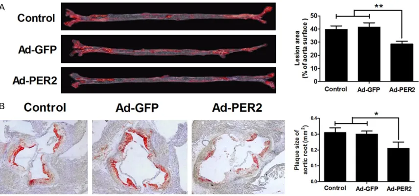
To exam the role of PER2 on atherosclerosis,
lesion areas of aorta and aortic root were ana
-lyzed. The results showed that the Ad-PER2
group had a remarkable smaller areas com
-pared with the Ad-GFP or control group (Figure 2A and 2B), which suggested that
overexpres-sion of PER2 significantly attenuated
athero-sclerosis.
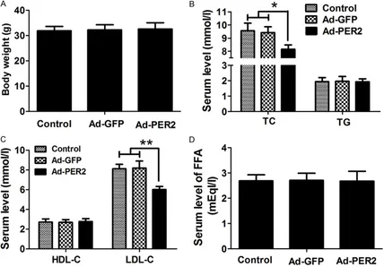
Overexpression of PER2 altered lipid profiles
To further explore the effect of PER2 overex -pression on atherosclerosis, body weight and
serum levels of LDL-C, HDL-C, total cholesterol,
[image:4.612.92.522.70.232.2]triglycerides, and FFA were measured. As Figure 1. Expression of PER2 in atherosclerotic mice. The mRNA level (A) and protein level (B) of PER2 in aorta of mice were determined using qRT-PCR and western blot, respectively. **P < 0.01. n = 8.
[image:4.612.93.521.283.484.2]shown in Figure 3A-D, overexpression of PER2 significantly decreased the serum levels of total cholesterol and LDL-C, compared with the
Ad-GFP or control group. Nevertheless, body
weight and the serum levels of HDL-C, triglycer
-ides, and FFA were almost unchanged in the Ad-PER2 group compared with those in the Ad-GFP or control group.
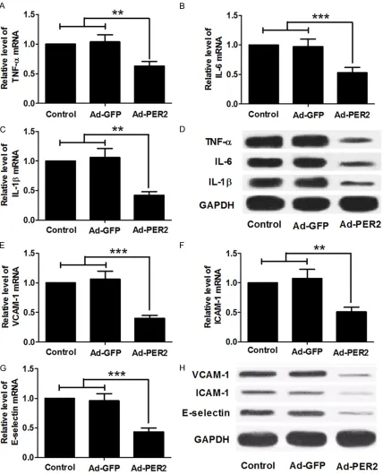
Overexpression of PER2 decreased the levels
of inflammatory factors and adhesion mol -ecules in ApoE-/- mice
To examine whether the overexpression of PER2 has a negative effect on inflammatory cytokines expression, mRNA levels of TNF-α, IL-6 and IL-1β in the aortic endothelial cells
were determined by qRT-PCR (Figure 4A-C),
and protein levels were determined by western blot (Figure 4D). Overexpression of PER2 mark
-edly decreased levels of TNF-α, IL-6 and IL-1β in
the Ad-PER2 group compared with the Ad-GFP
or control group. To evaluate the effect of PER2
overexpression on the adhesion molecules in
aortic tissues, qRT-PCR and western blot analy
-sis were performed. The mRNA levels of these adhesion molecules were markedly decreased
in the Ad-PER2 groups, compared with the Ad-GFP or control group (Figure 4E-G). Western
blot analysis showed that the levels of VCAM-1,
ICAM-1 and E-selectin proteins had a similar trend in these groups (Figure 4H).
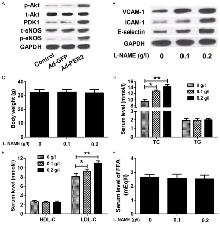
The Akt-eNOS signaling was activated by over-expression of PER2
It was reported that the Akt/eNOS signaling
pathway are involved in atherosclerotic inflam
-matory reaction [21] and circadian clock
enhances NO production by regulating
phos-phorylation of eNOS to protect endothelial function from injury [24]. To examine whether
the Akt-eNOS signaling is activated by overex
-pression of PER2, total (t)-Akt, phosphorylated
(p)-Akt, PDK1, t-eNOS and p-eNOS were mea
-sured by using western blot analysis in aortic
tissues. The results showed that the expres
[image:5.612.92.520.74.372.2]-sion levels of p-Akt, PDK1 and p-eNOS in the
Ad-PER2 group were significantly increased
compared with the Ad-GFP or control group.
The levels of t-Akt and t-eNOS were unchanged
in these groups (Figure 5A). These findings indi
-cated that overexpression of PER2 activated
[image:6.612.93.521.71.601.2]the Akt/eNOS signaling pathway in ApoE-/- mice.
To further identify the effect of the Akt/eNOS
signaling on atherosclerosis, we detected the
levels of VCAM-1, ICAM-1 and E-selectin in
endothelial cells in ApoE-/- mice treated with L-NAME, an eNOS inhibitor. As shown in Figure
5B, L-NAME significant increased the levels of
VCAM-1, ICAM-1 and E-selectin in a concentra-tion-dependent manner. Additionally, we
evalu-ated the body weight and lipid profiles in
ApoE-/- mice treated with L-NAME to further
understand the effect of the Akt/eNOS signal
-ing. We found that serum levels of total choles
-terol and LDL-C were increased in mice treated
L-NAME in a concentration-dependent manner,
while body weight and the serum levels of
HDL-C, triglycerides, and FFA were unchanged among these groups (Figure 5C-F).
Discussion
[image:7.612.89.523.71.518.2]In the current study, the findings suggest that PER2 is beneficial to control atherosclerosis as
a regulator in the inflammatory process.
Moreover, by over-expressing PER2 in ApoE -/-mice, we provide evidence that PER2 may
con-tribute to relieve atherosclerosis via the Akt-eNOS signaling pathway. Our findings might
shed lights on development of effective treat
-ments for cardiovascular disease therapy. Dysregulated lipid metabolism is of special
interest in atherosclerosis. For example, high
serum concentration of TC and LDL-C are always involved in atherosclerosis formation. In
this study, as expected, atherosclerotic
pro-cess elicited aberrant lipid profiles with high
serum levels of TC and LDA-C, while those met
-abolic parameters were alleviated markedly by
overexpression of PER2. These findings sug
-gested that PER2 is capable to attenuated ath-erosclerosis by interacting with the lipid.
Atherosclerosis is known as an inflammatory
disease to cause cardiovascular disorder,
therefore, in the development of atherosclero
-sis, multiple of pro-inflammatory factors and
cell adhesion molecules are involved to facili
-ties this progression [25]. For example, inflam
-matory mediators including TNF-α, IL-6 and
IL-1β are measured as the characteristic of car
-diovascular disease [5, 26]. In this study, the
levels of these inflammatory mediators reduced markedly by overexpression of PER2, which
indicates that PER2 possesses anti-inflamma
-tory effects and could impede atherosclerosis formation.
The formation of atherosclerosis involves migration of blood leukocytes and medial
smooth muscle cells into the arterial intima to
form lesions [3]. Adhesion molecules such as
VCAM-1, ICAM-1 and E-selectin expressed on
vascular endothelium and circulating leuko
-cytes may be one of the contributions to lesion
initiation in the earliest stages of atherogene
-sis, because they could recruit blood mono-cytes and lymphomono-cytes to the intima in which these cells mature into macrophages and then
turn into foam cells to develop and progress
into the atherosclerosis progressively [6, 26]. In the present study, the data demonstrated that
overexpression of PER2 has significantly inhibi
-tory effect to the expression of these genes.
Cardiovascular system owns typical circadian rhythmicity, because pressure and heart rates have distinct circadian pattern physiologically and pathophysiologically [27]. As an important
Period gene, Period 2 gene possesses diverse
biological functions such as the regulation of bone formation [28], tumor suppression [29],
and periodic pattern maintain [30] and
regula-tion of vascular endothelial funcregula-tion [31]. In line with these findings, our results indicated that
PER2 suppressed atherosclerosis and
stabi-lized plaques in ApoE-/- mice.
Through regulation of vascular tone and relax
-ation and maintenance of vascular integrity, endothelium-derived relaxing factor nitric
oxide, a gaseous transmitter, plays a vital role
in endothelium events, dysfunction of which
involved in the pathogenesis of various vascu
-lar disease including atherosclerosis, hyperten-sion and aging [32, 33]. eNOS, the mainly
gen-erator of NO, catalyzes conversion of L-arginine
to L-citrulline and NO [34]. Akt, a serine-threo
-nine kinase, regulates several biological pro
-cesses in protection of cardiomyocytes against
apoptosis through eNOS phosphorylation [35]. Various studies have demonstrated that the
cir-cadian clock regulates phosphorylation of
eNOS which enhance NO production to protect
endothelial function from injury and maintain
circadian rhythm [24]. In parallel with these studies, a similar trend occurred in this study,
the p-Akt, PDK1 and p-eNOS increased signifi
-cantly by overexpression of PER2, which indi
-cated that the Akt-eNOS signaling was activat
-ed by PER2, even though the complicat-ed
mechanism of this finding is still being explored.
In addition, the eNOS inhibitor L-NAME
increased VCAM-1, ICAM-1, Selectins-E, TC and LDL-C. These results suggest that the ApoE
-/-mice require the Akt/eNOS signal pathway to
eliminate the atherosclerosis.
In conclusion, PER2 has a significantly inhibito
-ry effect on atherosclerosis via the Akt-eNOS
pathway. Our data also demonstrated that
manipulation of PER2 attenuates the inflamma
-tory progression by inhibiting the inflamma-tory factors and adhesion molecules. Furthermore, overexpression of PER2 suppressed lesion
area significantly and attenuated atherosclero
-sis by altering aberrant lipid profiles. These
results indicate that PER2 is a promising target
for atherosclerosis treatment.
Acknowledgements
Education Department of Henan Science and Technology (162102310550).
Disclosure of conflict of interest
None.
Address correspondence to: Dr. Guangli Sun, De- partment of Ophthalmology, The First Affiliated Hospital of Zhengzhou University, 1 Jianshe East Road, Zhengzhou 450052, Henan, P. R. China. Tel: +86-371-66862211; E-mail: sunguangli126@163. com
References
[1] Libby P, Ridker PM, Maseri A. Inflammation and atherosclerosis. Circulation 2002; 105: 1135-43.
[2] Hansson GK, Robertson AK, Söderberg-Nauclér C. Inflammation and atherosclerosis. Annu Rev Pathol 2006; 1: 297-329.
[3] Epstein FH, Ross R. Atherosclerosis-an inflam-matory disease. N Engl J Med 1999; 340: 115-26.
[4] Hougee S, Sanders A, Faber J, Graus YM, van den Berg WB, Garssen J, Smit HF and Hoijer MA. Decreased inflammatory cytokine pro-duction by LPS-stimulated PBMC upon in vitro incubation with the flavonoids apigenin, luteo-lin or chrysin, due to selective elimination of monocytes/macrophages. Biochem Pharma- col 2005; 69: 241-8.
[5] Mazurek T, Zhang L, Zalewski A, Mannion JD, Diehl JT, Arafat H, Sarov-Blat L, O’Brien S, Keiper EA and Johnson AG. Human epicardial adipose tissue is a source of inflammatory me-diators. Circulation 2003; 108: 2460-6. [6] Blankenberg S, Barbaux S, Tiret L. Adhesion
molecules and atherosclerosis. Atherosclerosis 2003; 170: 191-203.
[7] Cybulsky MI, Gimbrone M. Endothelial expres-sion of a mononuclear leukocyte adheexpres-sion molecule during atherogenesis. Science 1991; 251: 788-91.
[8] Li H, Cybulsky MI, Gimbrone M, Libby P. An ath-erogenic diet rapidly induces VCAM-1, a cyto-kine-regulatable mononuclear leukocyte adhe-sion molecule, in rabbit aortic endothelium. Arterioscler Thromb Vasc Biol 1993; 13: 197-204.
[9] Reppert SM, Weaver DR. Coordination of circa-dian timing in mammals. Nature 2002; 418: 935-41.
[10] Anea CB, Zhang M, Stepp DW, Simkins GB, Reed G, Fulton DJ and Rudic RD. Vascular dis-ease in mice with a dysfunctional circadian clock. Circulation 2009; 119: 1510-7.
[11] Castanon-Cervantes O, Wu M, Ehlen JC, Paul K, Gamble KL, Johnson RL, Besing RC, Menaker M, Gewirtz AT and Davidson AJ. Dysregulation of inflammatory responses by chronic circadian disruption. J Immunol 2010; 185: 5796-805.
[12] Wang CY, Wen MS, Wang HW, Hsieh IC, Li Y, Liu PY, Lin FC and Liao JK. Increased vascular se-nescence and impaired endothelial progenitor cell function mediated by mutation of circadi-an gene Per2. Circulation 2008; 118: 2166-73.
[13] Viswambharan H, Carvas JM, Antic V, Marecic A, Jud C, Zaugg CE, Ming XF, Montani JP, Albrecht U and Yang Z. Mutation of the circa-dian clock gene Per2 alters vascular endothe-lial function. Circulation 2007; 115: 2188-95. [14] Bonney S, Kominsky D, Brodsky K, Eltzschig H,
Walker L and Eckle T. Cardiac Per2 functions as novel link between fatty acid metabolism and myocardial inflammation during ischemia and reperfusion injury of the heart. PLoS One 2013; 8: e71493.
[15] Eckle T, Hartmann K, Bonney S, Reithel S, Mittelbronn M, Walker LA, Lowes BD, Han J, Borchers CH and Buttrick PM. Adora2b-elicited Per2 stabilization promotes a HIF-dependent metabolic switch crucial for myocardial adap-tation to ischemia. Nature Med 2012; 18: 774-82.
[16] Matsui T, Tao J, del Monte F, Lee KH, Li L, Picard M, Force TL, Franke TF, Hajjar RJ and Rosenzweig A. Akt activation preserves cardiac function and prevents injury after transient cardiac ischemia in vivo. Circulation 2001; 104: 330-5.
[17] Dimmeler S, Fleming I, Fisslthaler B, Hermann C, Busse R, Zeiher AM. Activation of nitric oxide synthase in endothelial cells by Akt-dependent phosphorylation. Nature 1999; 399: 601-5. [18] Zheng H, Fu G, Dai T, Huang H. Migration of
endothelial progenitor cells mediated by stro-mal cell-derived factor-1α/CXCR4 via PI3K/ Akt/eNOS signal transduction pathway. J Cardiovasc Pharmacol 2007; 50: 274-80. [19] Lloyd-Jones M, Donald M, Bloch M, Kenneth D.
The vascular biology of nitric oxide and its role in atherogenesis. Ann Rev Med 1996; 47: 365-75.
[20] Förstermann U, Münzel T. Endothelial nitric ox-ide synthase in vascular disease from marvel to menace. Circulation 2006; 113: 1708-14. [21] Hwang HJ, Jung TW, Hong HC, Choi HY, Seo JA,
Kim SG, Kim NH, Choi KM, Choi DS and Baik SH. Progranulin protects vascular endothelium against atherosclerotic inflammatory reaction via Akt/eNOS and nuclear factor-kappaB path-ways. PLoS One 2013; 8: e76679.
atherosclero-sis in clock mutant mice. Circulation 2013; 128: 1758-69.
[23] Lin C, Tang X, Zhu Z, Liao X, Zhao R, Fu W, Chen B, Jiang J, Qian R and Guo D. The rhythmic ex-pression of clock genes attenuated in human plaque-derived vascular smooth muscle cells. Lipids Health Dis 2014; 13: 14.
[24] Ko ML, Jian K, Shi L, Ko GY. Phosphatidylinositol 3 kinase-Akt signaling serves as a circadian output in the retina. J Neurochem 2009; 108: 1607-20.
[25] Seno T, Hamaguchi M, Ashihara E, Kohno M, Ishino H, Yamamoto A, Kadoya M, Nakamura K, Murakami K and Matoba S. 15-deoxy-δ 12, 14 prostaglandin J 2 reduces the formation of atherosclerotic lesions in apolipoprotein e knockout mice. PLoS One 2011; 6: e25541. [26] Liu J, Ren Y, Kang L, Zhang L. Overexpression
of CCN3 inhibits inflammation and progression of atherosclerosis in apolipoprotein E-deficient mice. PLoS One 2014; 9: e94912.
[27] Hickie IB, Naismith SL, Robillard R, Scott EM and Hermens DF. Manipulating the sleep-wake cycle and circadian rhythms to improve clinical management of major depression. BMC Med 2013; 11: 79.
[28] Fu L, Patel MS, Bradley A, Wagner EF and Karsenty G. The molecular clock mediates leptin-regulated bone formation. Cell 2005; 122: 803-15.
[29] Fu L, Pelicano H, Liu J, Huang P and Lee C. The circadian gene Period2 plays an important role in tumor suppression and DNA damage re-sponse in vivo. Cell 2002; 111: 41-50.
[30] Schmalen I, Reischl S, Wallach T, Klemz R, Grudziecki A, Prabu JR, Benda C, Kramer A and Wolf E. Interaction of circadian clock pro-teins CRY1 and PER2 is modulated by zinc binding and disulfide bond formation. Cell 2014; 157: 1203-15.
[31] Bhatwadekar AD, Yan Y, Qi X, Thinschmidt JS, Neu MB, Calzi SL, Shaw LC, Dominiguez JM, Busik JV and Lee C. Per2 mutation recapitu-lates the vascular phenotype of diabetes in the retina and bone marrow. Diabetes 2013; 62: 273-82.
[32] Cecchi E, Giglioli C, Valente S, Lazzeri C, Gensini GF, Abbate R and Mannini L. Role of hemodynamic shear stress in cardiovascular disease. Atherosclerosis 2011; 214: 249-56. [33] Sun D, Huang A, Yan EH, Wu Z, Yan C, Kaminski
PM, Oury TD, Wolin MS and Kaley G. Reduced release of nitric oxide to shear stress in mes-enteric arteries of aged rats. Am J Physiol Heart Circul Physiol 2004; 286: H2249-H56. [34] Arnal JF, Dinh-Xuan AT, Pueyo M, Darblade B,
Rami J. Endothelium-derived nitric oxide and vascular physiology and pathology. Cell Mol Life Sci 1999; 55: 1078-87.