Original Article
Identification of microRNA expression in a rat model of
post-infarction heart failure
Yan Li1, Jing Dong2, Yan-Hua Xuan2, Shuang-Shuang Liu2, Jia-Ying Luo2, Yang-Jie Xiao3, Zhi-Peng Tian4, Zhi-Jun Sun2
Departments of 1Geriatrics, 2Cardiology, 3Ultrasound, Shengjing Hospital of China Medical University, Shenyang, Liaoning, P. R. China; 4Department of Cardiology, Central Hospital Affiliated to Shenyang Medical College, Shenyang, Liaoning, P. R. China
Received November 29, 2016; Accepted January 16, 2017; Epub April 1, 2017; Published April 15, 2017
Abstract: Although the mechanisms for development and progression of heart failure (HF) have been extensively studied, the molecular mechanisms underlying the post transcriptional regulation of HF have not been fully eluci-dated. Functional miRNA studies reported that a variety of miRNAs play a role in pathogenic mechanisms leading to heart failure, such as remodelling, hypertrophy, apoptosis, and hypoxia. In the present study, six rats were as-signed into two different treatment groups: rats with chronic heart failure (CHF group; n=3), and sham-operated rats (SO group; n=3). The miRCURYTM LNA Array (v.18.0, Exiqon) was utilized to detect the miRNA transcriptome
in SO and CHF rats. And bioinformatics were utilized to identify putative miRNA target genes and their associated biological functions. Forty-one miRNAs were found to be differentially expressed between SO and CHF rats. The five most upregulated and downregulated miRNAs were selected to obtain more representative biological meanings for the putative genes. And the biological analysis further showed that the differentially expressed miRNAs are mainly involved in Wnt signaling pathway and TGF-beta signaling pathway. In conclusion, the data from the present study indicated changes in the miRNA expression levels in the rat myocardium under heart failure post myocardium infarc-tion. When compared with that of the control group, heart failure exerted differential effects on miRNA expression. Bioinformatic analysis of the differentially expressed miRNAs showed that major affected pathways are known to be involved in cardiac remodeling and fibrosis.
Keywords: Heart failure, microRNA, rat, microarray, infarction
Introduction
Heart failure (HF) is a complex syndrome de- fined by a cardiac output inadequate to meet the metabolic demands of body tissues [1]. It results from a wide range of congenital and acquired cardiovascular or metabolic diseases leading to structural and functional impairment of the heart [2]. Although there have been dra-matic innovations in medical and device treat-ments for heart failure in recent decades, the incidence of heart failure is increasing [3]. The heart failure syndrome affects an estimated more than 23 million people worldwide [4, 5]. Although the mechanisms for development and progression of HF have been extensively stud-ied, the molecular mechanisms underlying the post transcriptional regulation of HF have not been fully elucidated [6-8].
MicroRNAs (miRNAs) are a kind of non-coding small RNAs that can post-transcriptionally re- gulate the expression of hundreds of their tar-get genes. Functional miRNA studies reported that a variety of miRNAs play a role in patho-genic mechanisms leading to heart failure, such as remodelling, hypertrophy, apoptosis, and hypoxia [9-11]. Evidence from experimental platforms points to potential applications of miRNAs for diagnostic and therapeutic purpos-es [12, 13].
Identification of microRNA expression in heart failure rat
4730 Int J Clin Exp Pathol 2017;10(4):4729-4738
CHF rats. The five most upregulated and down-regulated miRNAs were selected to obtain more representative biological meanings for the putative genes. The biological analysis further showed that the differentially expressed miR-NAs are mainly involved in adipocytokine sig-naling pathway, vascular smooth muscle con-traction signaling pathway, Wnt signaling pathway and TGF-beta signaling pathway.
Materials and methods
Animals
Male Wistar rats (7-8 weeks old; weighing 250-300 g) were obtained from the Animal Center of the Chinese Academy of Medical Sciences (Beijing, China) for use as a model of CHF in the present study. The animals were housed in stainless steel wire-mesh cages (5 rats per cage) under standard laboratory conditions (25°C, relative humidity 60%, 12 hours dark/ light periods) and were allowed free access to water and food. The investigations conformed to the Guide for the Care and Use of Laboratory Animals, published by the National Institutes of Health (NIH Publication no. 85-23; revised 1985). The present study was approved by the Animal Research Ethics Committee of Shengjing hospital of China Medical University (Shenyang, China). The rats were assigned into two different treatment groups: Rats with chronic heart failure, (CHF group; n=3), and sham-operated rats, (SO group; n=3).
Rat model of CHF
Acute myocardial infarction was induced in the rats by left anterior descending artery ligation, as previously described [12]. Briefly, the rats were anesthetized via intraperitoneal injection of 10% chloral hydrate (3 ml⁄kg). The rats were then tracheotomied, intubated and mechani-cally ventilated, with arterial pH, PO2 and PCO2 maintained within the physiological range by supplying O2 and altering the respiratory rate using an Ahx-300S Animal Respirator (Chengdu Tme Technology Co, Ltd., Chengdu, China). A thoracotomy was performed through the fourth intercostal space, the heart was exposed and an electrocardiogram was monitored on a BL-420S Data Acquisition & Analysis System (Chengdu Tme Technology Co, Ltd., China). A prolene suture (Ethicon, Inc., Somerville, NJ,
USA) was placed around the left coronary artery, close to its origin, and the ends were tied firmly in the CHF group and loosely in the SO group. Acute myocardial infarction was deemed successful on the basis of regional cyanosis of the myocardial surface distal to the suture, accompanied by elevation of the ST segment on the electrocardiogram. At 3 weeks-post myocardial infarction, echocardiography was performed on the surviving rats. The rats, which exhibited left ventricular wall infarction, with an infarcted area >40% of the left ventricu-lar wall area were enrolled for the subsequent investigations. The infarcted rats were allocat-ed into the CHF group (n=3). Heart rate, mean blood pressure, weight, and echocardiography were recorded, and blood samples were obtained in the rats of the two groups. The present study was approved by the ethics com-mittee of Shengjing Hospital of China Medical University (Shenyang, China).
Echocardiographic and hemodynamic mea-surements
Echocardiography was performed 3 weeks after surgery. A transthoracic 2D M-mode echo-cardiogram was obtained using a Philips iE33 (Philips Electronics, Amsterdam, The Nether- lands), equipped with a transducer S12-4. The rats were anesthetized with 3 ml/kg 10% chlo-ral hydrate, and echocardiography was per-formed. The chest each mouse was shaved and the rats were placed in a supine position. Images were captured by placing the transduc-er against the chest. Images wtransduc-ere obtained by placing the transducer against the chest. Two-dimensional echocardiography measureme- nts of left ventricular end-diastolic diameter (LVEDD), left ventricular end-systolic diameter (LVESD) and left ventricular wall thickness were recorded. Left ventricular fractional shortening (LVFS) was calculated according to the formula: LVFS = (LVEDD-LVESD)/LVEDD×100%. Left ve- ntricle ejection fraction (LVEF) was measured using the Biplane Modified Simpson’s method.
Blood sampling
the experiments. For each rat, the entire heart was rapidly excised and washed with cold phos-phate buffer (Experimental Center of Shengjing Hospital of China Medical University, Shenyang, China, containing 137 mmol/l NaCl, 2.7 mmol/l KCl, 10 mmol/l Na2HPO4, 2 mmol/l KH2PO4 (pH 7.4). The heart was then frozen at -80°C, prior to its use in the subsequent miRNA microarray analyses.
Detection of brain natriuretic peptide (BNP)
The blood samples were collected into serum separator tubes, and serum was obtained by centrifugation at 4,500 g for 15 min. The serum BNP was measured using a commercially
avail-able human/mouse/rat BNP enzyme immuno-assay kit (RayBiotech, Norcross, GA, USA), according to the manufacturer’s instructions. All samples and standards were measured in triplicate.
Methods of miRNA microarray
[image:3.612.92.521.73.424.2]Identification of microRNA expression in heart failure rat
4732 Int J Clin Exp Pathol 2017;10(4):4729-4738
lated using TRIzol (Invitrogen) and miRNeasy mini kit (QIAGEN) according to manufacturer’s instructions, which efficiently recovered all RNA species, including miRNAs. RNA quality and quantity was measured by using nanodrop spectrophotometer (ND-1000, Nanodrop Te- chnologies) and RNA Integrity was determined by gel electrophoresis. RNA labeling. After RNA isolation from the samples, the miRCURY™ Hy3™/Hy5™ Power labeling kit (Exiqon, Ved- baek, Denmark) was used according to the manufacturer’s guideline for miRNA labeling. One microgram of each sample was 3’-end-labeled with Hy3TM fluorescent label, using T4 RNA ligase by the following procedure: RNA in 2.0 μL of water was combined with 1.0 μL of CIP buffer and CIP (Exiqon). The mixture was incubated for 30 min at 37°C, and was termi-nated by incubation for 5 min at 95°C. Then 3.0 μL of labeling buffer, 1.5 μL of fluorescent label (Hy3TM), 2.0 μL of DMSO, 2.0 μL of labe- ling enzyme were added into the mixture. The labeling reaction was incubated for 1 h at 16°C, and terminated by incubation for 15 min at 65°C. Array hybridization. After stopping the labeling procedure, the Hy3TM-labeled samples were hybridized on the miRCURYTM LNA Array (v.18.0) (Exiqon) according to array manual. The total 25 μL mixture from Hy3TM-labeled sam-ples with 25 μL hybridization buffer were first denatured for 2 min at 95°C, incubated on ice for 2 min and then hybridized to the microarray for 16-20 h at 56°C in a 12-Bay Hybridiza- tion Systems (Hybridization System-Nimblegen Systems, Inc., Madison, WI, USA), which pro-vides an active mixing action and constant incubation temperature to improve hybridiza-tion uniformity and enhance signal. Following hybridization, the slides were achieved, washed several times using Wash buffer kit (Exiqon), and finally dried by centrifugation for 5 min at 400 rpm. Then the slides were scanned using the Agilent DNA Microarray Scanner (part num-ber G2505C). Data analysis. Scanned images were then imported into GenePix Pro 6.0 soft-ware (Axon) for grid alignment and data extrac-tion. Replicated miRNAs were averaged and miRNAs that intensities ≥30 in all samples were chosen for calculating normalization fac-tor. Expressed data were normalized using the Median normalization. After normalization, dif-ferentially expressed miRNAs with statistical significance were identified through Volcano Plot filtering.
Statistical analysis
All statistical analyses were performed using SPSS 18.0 software (SPSS, Inc., Chicago, IL, USA). Data are expressed as the mean ± stan-dard deviation. Statistical analyses were per-formed using Student’s t-test for comparison between two groups). P<0.05 was considered to indicate a statistically significant difference.
Results
Characteristics of the rat models
The rats of CHF group exhibited left ventricular wall infarction. Figure 1A shows the represen-tative M-mode image of CHF group and SO group. As shown in Figure 1B, 3 weeks post myocardial infarction, the LVEF and the LVFS value decreased significantly in the CHF group, compared with the SO group (LVEF, 45.97±2.28, vs. 71.30±1.93%, P=0.000 and LVFS, 19.93± 1.27, vs. 59.67±8.08%, P=0.001). In addition, myocardial infarction increased the LVEDV and the LVESV in the CHF rats, compared with the SO group (LVEDV, 1.31±0.26, vs. 0.82±0.09 cm3, P=0.038 and LVESV, 0.71±0.17, vs. 0.24±0.02 cm3, P=0.008). Figure 1C shows serum levels of BNP increased significantly in the CHF rats compared with the SO rats, (11.85±1.53, vs. 3.30±0.90 ng/ml, P=0.001), 3 weeks post myocardial infarction.
miRNA expression is altered after heart failure
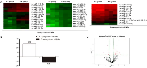
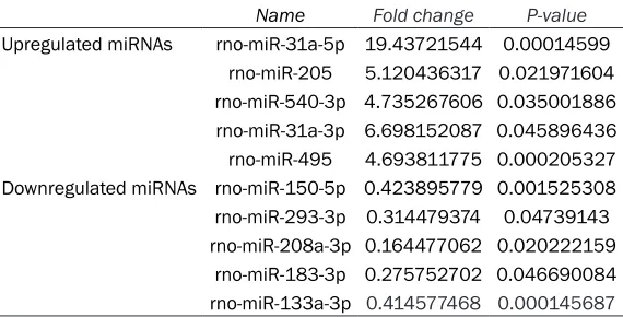
To analyze heart failure changes in miRNA expression in heart, miRNA microarray analysis was performed using arrays containing 3100 capture probes, covering all human, mouse and rat microRNAs annotated in miRBase 18.0, as well as all viral microRNAs related to these spe-cies. Total RNA was extracted from left ventric-ular of normal and CHF rats. miRNAs from CHF rats that were upregulated (n=29) and down-regulated (n=12) at least 1.5-fold compared with those of normal rats were identified (Figure 2), and lists of the top five most differentially upregulated or downregulated miRNAs are pre-sented in Table 1.
Prediction of target genes and functional an-notation
Identification of microRNA expression in heart failure rat
4734 Int J Clin Exp Pathol 2017;10(4):4729-4738
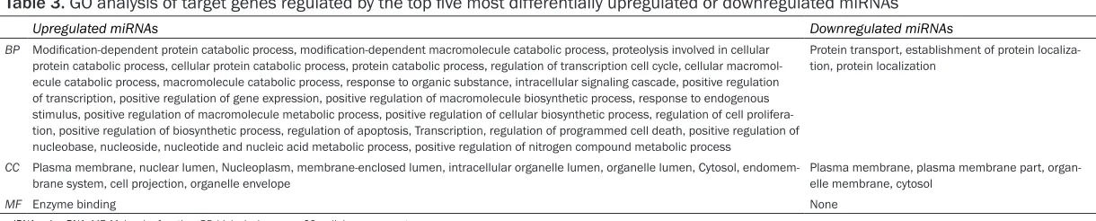
altered >2.0-fold (Table 1). So, the five most upregulated and downregulated miRNAs were selected to obtain more representative biologi-cal meanings for the putative genes. We used the Intersection of four miRNA target prediction database to identify putative target genes. TargetScan, miRanda, miRDB, miRWalk, four miRNA target prediction database, were used to identify 258 putative target genes for upreg-ulated and downregupreg-ulated miRNAs (Table 2). The analysis revealed 189 putative targets for the top 5 upregulated miRNAs and 69 putative targets for the top 5 downregulated miRNAs. GO analysis was performed to determine the functions of the target genes regulated by these differentially expressed miRNAs. Each putative targets gene was subjected to GO analysis to reveal its cell function. GO was ana-lyzed using three significant categories as fol-lows: Molecular function, biological process and cellular component. The analysis revealed a wide distribution of cellular functions, which are presented in Table 3.
KEGG pathway annotations of the candidate target genes regulated by differentially expre- ssed miRNAs were also performed. The result indicated that the significant pathways of the candidate target genes are mainly involved in Adipocytokine signaling pathway, vascular smooth muscle contraction signaling pathway, Wnt signaling pathway and TGF-beta signaling pathway, which are presented in Table 4.
Discussion
The present study demonstrated that heart failure post myocardium infarction induced
and categorized their reported biological functions.
[image:6.612.92.377.97.242.2]A number of miRNAs determined to be differen-tially expressed in the present study are known to be involved in function in the rat myocardi-um. Firstly, some of microRNAs we found in the present study have been reported to have close relationship with heart failure. For example, recently miR-150 has been described as a reg-ulator in cardiac hypertrophy. In a mouse model, a study identified the crucial role of miR-150 in protecting against cardiac hypertrophy and fibrosis in response to pressure overload through the downregulation of serum response factor [13]. Prior study have showed a change in several circulating miRNA levels after left ventricular assist device implantation, with decreased levels of the myomirs miR-208a after 3 months [14]. Another study validated miR-208 as a potent therapeutic target for the modulation of cardiac function and remodeling during heart disease progression [15]. Rooij et al found mutant mice overexpressing miR-208 to not develop cardiomyocyte fibrosis despite being exposed to an increased after load [16, 17]. miR-133a, a muscle specific miRNA, is functionally cooperative in promoting meso-derm differentiation in embryonic stem (ES) cells whilst repressing ectodermal and endo-dermal differentiation [1, 18, 19]. Loss-of-function miR-133a mutants exhibit increased proliferation of cardiomyocytes and up-regula-tion of smooth muscle cell-specific genes. miR-133 regulates cardiomyocyte proliferation by repressing cyclin D2 and serum response fac-tor [20]. In both animal and human models, miR-133 was identified as regulator of cardiac
Table 1. The top five most differentially upregulated or
downregu-lated miRNAs
Name Fold change P-value
Upregulated miRNAs rno-miR-31a-5p 19.43721544 0.00014599 rno-miR-205 5.120436317 0.021971604 rno-miR-540-3p 4.735267606 0.035001886 rno-miR-31a-3p 6.698152087 0.045896436 rno-miR-495 4.693811775 0.000205327 Downregulated miRNAs rno-miR-150-5p 0.423895779 0.001525308 rno-miR-293-3p 0.314479374 0.04739143 rno-miR-208a-3p 0.164477062 0.020222159
rno-miR-183-3p 0.275752702 0.046690084 rno-miR-133a-3p 0.414577468 0.000145687
miRNA, microRNA.
Table 2. Predicted target of the top five most differentially upregulated or downregulated miRNAs
Target genes
Upregulated miRNAs Serpinb2, Trim39, Tmem50b, Dhrs8, Mgrn1, Siah1a, Klhl12, Vil2, RGD708449, MGC124555, Podxl, Rnd3, Acsl1, RGD1359713, Nars, Fkbp9, Trak2, RAMP4, Sel1h, MGC109455, Ssbp3, Esrrg, SPP24_RAT, Pja2, RGD1305453, RGD1305440, Mmd, Errfi1, St6galnac3, Bet1l, RGD1305246, Cep55, RGD1305314, Prdx3, Ap3m1, Paqr4, Pcdha2_predicted, Pcdha2, Pcdhac2, Pir, Drb1, Dctn2, Tmbim1, Cnbp1, Slc1a4, Shank1, C1qtnf6, Mfng, Arf3, Errfi1, Clp1, Slc25a19, Cep55, Abca2, Slc5a7, Xrcc4, Lypla1, Fbxo11, Dncli2, Vnn1, Flt1, Sgk, Tmed9, LOC305166, Nuak2, Bat1a, Cpox, Gmfb, Siah1a, SEPT7, RGD1309892, Sdc1, Rg9mtd1, Btbd15, Pcdha2_predicted, Pcdha2, Pcdhac2, Khdrbs2, Tcfcp2l2, Ssr1, Zfp297b, Yt521, Hspa5, Akr1b4, Acsl4, Dusp1, Tgfbr1, Ptn, Smad1, Tagln3, Dnajb9, Ppp6c, Cbfb, Ccng1, Strn3, Stx6, Slc12a2, LOC303059, Akap6, Ube4a, Serpine1, Pafah1b2, Nupl1, Cirh1a, Nip7, Pscdbp, TSEN34, Tmpo, Mgat2, Hnrph1, 7-Mar, Tnfsf4, Grm6, Asah1, RGD1306658, LOC683711, Dusp6, Rab18, RGD1359509, Prei3, Gpr64, Mtr, Smad7, Ccna2, Psip1, Gad1, Fcgr3a, RGD1306635_predicted, Afap, Cand1, Pigm, Klhl24, MGC124653, Klf5, Ddit4, Arl5b, Kcnk10, Ppp1cc, Ankrd46, Ube2v2, B3galt3, Ssbp3, Cnbp1, Cugbp1, Gucy1b3, MGC108974, Cmpk, Cd164, Irs1, Dclk2_predicted, Ube1c, Tsc22d3, Git2, Cd44, Lrrn1, Chfr, Ptk2, Gatad2b, Actc1, Them4, Snx27, Zfp422, Tgif, F101B_RAT, Pvalb, Cacna1c, Tnfaip1, Myod1, Sycp1, Tob2, Slc16a1, Slc17a6, Apba2, Prom2, Slc38a2, Bcat1, Ube2z, Crkrs, MGC94113, Lphn2, Stat3, Psme3, Adm, Kpna2, Clcf1, Cep55, Scd2, Nolc1
Downregulated miRNAs Dhrs8, Sgk, Syt2, Rab8a, Timm17a, Ptbp1, Clta, Gpm6a, Glra2, Ppp2ca, Ppp2cb, Zfp672, Agt, Whsc2, Sec61a1, Cmpk, Vps54, Ndrg1, Slc6a1, Sv2a, MGC94600, RGD1305778, Cybasc3, Ncoa4_predicted, Drd1a, Freq, Plp2, RGD1305117, RGD708449, Hspa5, Acsl4, Tmem24, Rad23b, Ppp2r1a, St3gal2, Cd276, Gdi2, Egr2, Mtfmt, Abcb9, Pvrl2, Mcf2l, Rnf34, Hspb8, Gpi, Ptk9, Bcat1, Commd7, Ap3m1, RGD1305356, Gpr85, Vil2, Commd10, Ppp2r1a, Tmpo, Ppp2ca, Bnip3l, Prkch, Ppp2cb, Snx1, Ssrp1, Amd1, Irs1, Vtcn1, Wbp11, Scpep1, Plcb4, Sfrs2
[image:7.792.90.700.265.388.2]miRNA, microRNA.
Table 3. GO analysis of target genes regulated by the top five most differentially upregulated or downregulated miRNAs
Upregulated miRNAs Downregulated miRNAs
BP Modification-dependent protein catabolic process, modification-dependent macromolecule catabolic process, proteolysis involved in cellular protein catabolic process, cellular protein catabolic process, protein catabolic process, regulation of transcription cell cycle, cellular macromol-ecule catabolic process, macromolmacromol-ecule catabolic process, response to organic substance, intracellular signaling cascade, positive regulation of transcription, positive regulation of gene expression, positive regulation of macromolecule biosynthetic process, response to endogenous stimulus, positive regulation of macromolecule metabolic process, positive regulation of cellular biosynthetic process, regulation of cell prolifera-tion, positive regulation of biosynthetic process, regulation of apoptosis, Transcripprolifera-tion, regulation of programmed cell death, positive regulation of nucleobase, nucleoside, nucleotide and nucleic acid metabolic process, positive regulation of nitrogen compound metabolic process
Protein transport, establishment of protein localiza-tion, protein localization
CC Plasma membrane, nuclear lumen, Nucleoplasm, membrane-enclosed lumen, intracellular organelle lumen, organelle lumen, Cytosol,
endomem-brane system, cell projection, organelle envelope Plasma membrane, plasma membrane part, organ-elle membrane, cytosol
MF Enzyme binding None
miRNA, microRNA; MF, Molecular function; BP, biological process; CC, cellular component.
Table 4. Significant pathway of target genes regulated by the top five most differentially upregulated or downregulated miRNAs
Upregulated miRNAs Downregulated miRNAs
KEGG_PATHWAY Adipocytokine signaling pathway, Vascular smooth muscle contraction, Lysosome Long-term depression, Tight junction, Wnt signaling pathway, TGF-beta signaling pathway Oocyte meiosis Lysosome
Identification of microRNA expression in heart failure rat
4736 Int J Clin Exp Pathol 2017;10(4):4729-4738
hypertrophy, with lower levels in heart failure and cardiac hypertrophy compared with con-trols [9, 21-23] miR-133a also plays an impor-tant role in β-adrenergic signaling pathway and Akt pathway [9].
Some of microRNAs we found in this study is new to heart failure. miRNA-31 can manifest antifibrotic effect via downregulating Islet-1, a key factor during the TGF-β-induced epithelial-mesenchymal transition [24]. These findings identify atrial-specific up-regulation of miR-31 in human AF as a key mechanism causing atrial dystrophin and nNOS depletion, which in turn contributes to the atrial phenotype begetting this arrhythmia. A study described miR-495 mediated the expression of endothelial or angiogenic genes by directly targeting vascular endothelial zinc finger 1. After transplantation in immunodeficient myocardial infarction mice, the derived endothelial cells significantly inc- reased neovascularization in the infarcted heart, prevented functional worsening, and attenuated expansion of infarct size. The func-tional integration of the implanted ECs into cor-onary networks was also enhanced by inhibiting miR-495 [25].
In addition, the present study identified puta-tive target genes of the up- and downregulated miRNAs and categorized their reported biologi-cal functions by GO into molecular function (MF), biological process (BP), cellular compo-nent (CC). Among the Significant pathway of tar-get genes regulated by the top five most differ-entially upregulated or downregulated miRNAs, Wnt signaling pathway and TGF-beta signaling pathway were assigned to the highest number of target genes. Wingless related integration site (Wnt) signaling has proven to be a funda-mental mechanism in cardiovascular develop-ment as well as disease [26]. There is increas-ing evidence that Wnt signalincreas-ing is involved in adverse cardiac remodeling and the progres-sion to heart failure [27]. TGF-β is a powerful profibrotic cytokine with diverse and often con-tradictory functions [28]. TGF-β has both profi-brotic and hypertrophic actions and has been linked to fibrosis in heart disease [28, 29]. In animal models of ischemic HF, positive benefits from inhibition of TGF-β signaling is controver-sial as inhibition of TGF-β before or immediately following MI has been shown to increase mor-tality and exacerbate dysfunction, yet inhibition
started 24 hours post-MI attenuated remodel-ing and improved function [28, 30-33].
In conclusion, the data from the present study indicated changes in the miRNA expression lev-els in the rat myocardium under heart failure post myocardium infarction. When compared with that of the control group, heart failure exerted differential effects on miRNA expres-sion. Bioinformatic analysis of the differentially expressed miRNAs showed that major affected pathways are known to be involved in cardiac remodeling and fibrosis.
The present study has some limitations. It has a small number of samples. Secondly, further study of using quantitative real time-PCR tech-nique to verify microarray results is needed. Furthermore, the target genes and pathways of CHF associated miRNAs remain elusive and deserve further investigations.
Acknowledgements
This study was supported by Liaoning Provincial Science and Technology Department (grant. no. 2011404013-7).
Disclosure of conflict of interest
None.
Address correspondence to: Dr. Zhi-Jun Sun, De- partment of Cardiology, Shengjing Hospital of China Medical University, 36 Sanhao Street, Heping District, Shenyang 110004, P. R. China. Tel: +86 24 2599 8745; Fax: +86 24 2599 8745; E-mail: sunzj_ [email protected]
References
[1] Wong LL, Wang J, Liew OW, Richards AM and Chen YT. MicroRNA and heart failure. Int J Mol Sci 2016; 17: 502.
Heart Association Task Force on practice guidelines. Circulation 2013; 128: e240-327. [3] Chaudhry SP and Stewart GC. Advanced heart
failure: prevalence, natural history, and prog-nosis. Heart Fail Clin 2016; 12: 323-333. [4] Bui AL, Horwich TB and Fonarow GC.
Epidemi-ology and risk profile of heart failure. Nat Rev Cardiol 2011; 8: 30-41.
[5] Go AS, Mozaffarian D, Roger VL, Benjamin EJ, Berry JD, Borden WB, Bravata DM, Dai S, Ford ES, Fox CS, Franco S, Fullerton HJ, Gillespie C, Hailpern SM, Heit JA, Howard VJ, Huffman MD, Kissela BM, Kittner SJ, Lackland DT, Lichtman JH, Lisabeth LD, Magid D, Marcus GM, Marelli A, Matchar DB, McGuire DK, Mohler ER, Moy CS, Mussolino ME, Nichol G, Paynter NP, Sch-reiner PJ, Sorlie PD, Stein J, Turan TN, Virani SS, Wong ND, Woo D and Turner MB. Heart disease and stroke statistics--2013 update: a report from the American Heart Association. Circulation 2013; 127: e6-e245.
[6] Liu X, Meng H, Jiang C, Yang S, Cui F and Yang P. Differential microRNA expression and regu-lation in the rat model of post-infarction heart failure. PLoS One 2016; 11: e0160920. [7] Zhu X, Wang H, Liu F, Chen L, Luo W, Su P, Li W,
Yu L, Yang X and Cai J. Identification of micro-RNA networks in end-stage heart failure be-cause of dilated cardiomyopathy. J Cell Mol Med 2013; 17: 1173-1187.
[8] Naga Prasad SV, Duan ZH, Gupta MK, Suram-pudi VS, Volinia S, Calin GA, Liu CG, Kotwal A, Moravec CS, Starling RC, Perez DM, Sen S, Wu Q, Plow EF, Croce CM and Karnik S. Unique mi-croRNA profile in end-stage heart failure indi-cates alterations in specific cardiovascular sig-naling networks. J Biol Chem 2009; 284: 27487-27499.
[9] Vegter EL, van der Meer P, de Windt LJ, Pinto YM and Voors AA. MicroRNAs in heart failure: from biomarker to target for therapy. Eur J Heart Fail 2016; 18: 457-468.
[10] Melman YF, Shah R and Das S. MicroRNAs in heart failure: is the picture becoming less miRky? Circ Heart Fail 2014; 7: 203-214. [11] Tijsen AJ, Pinto YM and Creemers EE.
Non-car-diomyocyte microRNAs in heart failure. Cardio-vasc Res 2012; 93: 573-582.
[12] Li Y, Xuan YH, Liu SS, Dong J, Luo JY and Sun ZJ. Shortterm vagal nerve stimulation im-proves left ventricular function following chron-ic heart failure in rats. Mol Med Rep 2015; 12: 1709-1716.
[13] Liu W, Liu Y, Zhang Y, Zhu X, Zhang R, Guan L, Tang Q, Jiang H, Huang C and Huang H. Mi-croRNA-150 protects against pressure over-load-induced cardiac hypertrophy. J Cell Bio-chem 2015; 116: 2166-2176.
[14] Akat KM, Moore-McGriff D, Morozov P, Brown M, Gogakos T, Correa Da Rosa J, Mihailovic A, Sauer M, Ji R, Ramarathnam A, Totary-Jain H, Williams Z, Tuschl T and Schulze PC. Compara-tive RNA-sequencing analysis of myocardial and circulating small RNAs in human heart fail-ure and their utility as biomarkers. Proc Natl Acad Sci U S A 2014; 111: 11151-11156. [15] Montgomery RL, Hullinger TG, Semus HM,
Dickinson BA, Seto AG, Lynch JM, Stack C, Lat-imer PA, Olson EN and van Rooij E. Therapeutic inhibition of miR-208a improves cardiac func-tion and survival during heart failure. Circula-tion 2011; 124: 1537-1547.
[16] Schulte C, Westermann D, Blankenberg S and Zeller T. Diagnostic and prognostic value of cir-culating microRNAs in heart failure with pre-served and reduced ejection fraction. World J Cardiol 2015; 7: 843-860.
[17] van Rooij E, Sutherland LB, Qi X, Richardson JA, Hill J and Olson EN. Control of stress-de-pendent cardiac growth and gene expression by a microRNA. Science 2007; 316: 575-579. [18] Wystub K, Besser J, Bachmann A, Boettger T
and Braun T. miR-1/133a clusters coopera-tively specify the cardiomyogenic lineage by adjustment of myocardin levels during embry-onic heart development. PLoS Genet 2013; 9: e1003793.
[19] Chen JF, Mandel EM, Thomson JM, Wu Q, Callis TE, Hammond SM, Conlon FL and Wang DZ. The role of microRNA-1 and microRNA-133 in skeletal muscle proliferation and differentia-tion. Nat Genet 2006; 38: 228-233.
[20] Liu N, Bezprozvannaya S, Williams AH, Qi X, Richardson JA, Bassel-Duby R and Olson EN. microRNA-133a regulates cardiomyocyte pro-liferation and suppresses smooth muscle gene expression in the heart. Genes Dev 2008; 22: 3242-3254.
[21] Care A, Catalucci D, Felicetti F, Bonci D, Ad-dario A, Gallo P, Bang ML, Segnalini P, Gu Y, Dalton ND, Elia L, Latronico MV, Hoydal M, Au-tore C, Russo MA, Dorn GW 2nd, Ellingsen O, Ruiz-Lozano P, Peterson KL, Croce CM, Peschle C and Condorelli G. MicroRNA-133 controls cardiac hypertrophy. Nat Med 2007; 13: 613-618.
[22] Danowski N, Manthey I, Jakob HG, Siffert W, Peters J and Frey UH. Decreased expression of miR-133a but not of miR-1 is associated with signs of heart failure in patients undergoing coronary bypass surgery. Cardiology 2013; 125: 125-130.
Identification of microRNA expression in heart failure rat
4738 Int J Clin Exp Pathol 2017;10(4):4729-4738
mechanism for progressive cardiac hypertro-phy. Hypertension 2010; 55: 946-952. [24] Bronnum H, Andersen DC, Schneider M,
Nos-sent AY, Nielsen SB and Sheikh SP. Islet-1 is a dual regulator of fibrogenic epithelial-to-mes-enchymal transition in epicardial mesothelial cells. Exp Cell Res 2013; 319: 424-435. [25] Liang J, Huang W, Cai W, Wang L, Guo L, Paul
C, Yu XY and Wang Y. Inhibition of microR-NA-495 enhances therapeutic angiogenesis of human induced pluripotent stem cells. Stem Cells 2017; 35: 337-350.
[26] Pahnke A, Conant G, Huyer LD, Zhao Y, Feric N and Radisic M. The role of Wnt regulation in heart development, cardiac repair and dis-ease: a tissue engineering perspective. Bio-chem Biophys Res Commun 2016; 473: 698-703.
[27] Hermans KC and Blankesteijn WM. Wnt signal-ing in cardiac disease. Compr Physiol 2015; 5: 1183-1209.
[28] Edgley AJ, Krum H and Kelly DJ. Targeting fibro-sis for the treatment of heart failure: a role for transforming growth factor-beta. Cardiovasc Ther 2012; 30: e30-40.
[29] Lim H and Zhu YZ. Role of transforming growth factor-beta in the progression of heart failure. Cell Mol Life Sci 2006; 63: 2584-2596.
[30] Frantz S, Hu K, Adamek A, Wolf J, Sallam A, Maier SK, Lonning S, Ling H, Ertl G and Bauer-sachs J. Transforming growth factor beta inhi-bition increases mortality and left ventricular dilatation after myocardial infarction. Basic Res Cardiol 2008; 103: 485-492.
[31] Ikeuchi M, Tsutsui H, Shiomi T, Matsusaka H, Matsushima S, Wen J, Kubota T and Takeshita A. Inhibition of TGF-beta signaling exacerbates early cardiac dysfunction but prevents late re-modeling after infarction. Cardiovasc Res 2004; 64: 526-535.
[32] Ellmers LJ, Scott NJ, Medicherla S, Pilbrow AP, Bridgman PG, Yandle TG, Richards AM, Protter AA and Cameron VA. Transforming growth fac-tor-beta blockade down-regulates the renin-angiotensin system and modifies cardiac re-modeling after myocardial infarction. Endocri- nology 2008; 149: 5828-5834.