Original Article
PTP4A3 expression is associated with the clinical
features and prognosis of cervical cancer
Li Tang1*, Hongjin Yu1,2*, Wenbing Zhong1, Fubing Yu1
1Department of Gynecology, Dongguan Maternal & Children Health Care Hospital, Dongguan, China; 2Department of Gynecology, Dongguan General Hospital, Dongguan, China. *Equal contributors.
Received November 9, 2016; Accepted January 6, 2017; Epub March 1, 2017; Published March 15, 2017
Abstract: Background:Protein tyrosine phosphatase 4A3 (PTP4A3) is overexpressed in different cancer types and
is involved in tumor progression. However, the clinical significance of PTP4A3 in cervical cancer is still unclear.
Methods: Real-time PCR was used to examine the mRNA expression levels of PTP4A3 in 8 pairs of cervical cancer tissues, and immunohistochemistry was used to investigate PTP4A3 protein expression pattern in 144 archived cervical cancer specimens. The correlation of PTP4A3 expression with clinicopathologic features was analyzed. Results: The expression of PTP4A3 was increased in all 8 cervical cancerous tissues compared with that in adjacent normal cervical tissues. PTP4A3 overexpression in patients was correlated with tumor stage (P = 0.004), tumor size (P = 0.024), and lymph node involvement (P = 0.025). Furthermore, patients with high PTP4A3 expression showed shorter overall and recurrence-free survival time after surgery (P = 0.012 and P = 0.005, respectively). Multivariate Cox regression analysis showed that PTP4A3 expression had a predictive value for overall survival of cervical cancer patients (P = 0.029). Conclusion:The expression of PTP4A3 was increased in cervical cancer and it plays an impor-tant oncogenic role in tumor progression of cervical cancer.
Keywords: PTP4A3, progression, cervical cancer, prognosis
Introduction
Cervical cancer is among the most preval- ent gynecological malignancies, with nearly 530,000 new cases and 275,000 deaths reported annually [1]. Despite the improvement of diagnostic and treatment strategies, most cervical cancer patients diagnosed at advanced stages are not eligible for potential curative therapies. The three-year survival and disease-free survival rate of patients with locoregional recurrent cervical cancer were 55.6% and 54.3% after concomitant chemo-radiation ther-apy [2]. Recently, more and more studies have been made in understanding the genetic altera-tions and biological processes in cervical can-cer preogression. However, the search for spe-cific molecular and genetic alterations in cervi-cal cancer that have clinicopathologic signifi-cance is limited.
Protein tyrosine phosphatase 4A3 (PTP4A3), also named as phosphatase of regenerating liver-3 (PRL-3), is a member of the protein
Although both PTP4A3 has been shown to be overexpressed in squamous cell carcinoma of the cervix [12], the prognostic role of PTP4A3 in cervical cancer patients has not been investi-gated. Therefore, we evaluated the expression levels of PTP4A3 in human cervical cancer tis-sues and analyzed the clinical significance of PTP4A3 expression in cervical cancer patients. Our data demonstrate that PTP4A3 expression is significantly correlated with cervical cancer progression and clinical prognosis. Thus, PTP- 4A3 may serve as a new prognostic marker and therapeutic target for cervical cancer therapy.
Materials and methods
Patients and tissue specimens
A total of 144 cervical cancer patients who underwent surgical resection from January 2004 to December 2009 at Dongguan General Hospital were analyzed. The records of patients were reviewed in the context of clinicopatho-logical and follow-up information. The tumor stage was classified according to the latest International Federation of Gynecology and Obstetrics criteria. The overall survival (OS) and recurrence-free survival (RFS) was calculated starting from the date of the initial surgery to the time of death and recurrence, respectively. After surgery, resected specimens were pro-cessed routinely for macroscopic pathological assessment. In addition, 8 pairs of snap-frozen cervical cancer and normal cervical samples were collected for Real-time PCR. Informed consent was obtained from each patient and this study was approved by the Research Ethics Committee of Dongguan General Hospital.
Quantitative real-time polymerase chain reac-tion (RT-PCR)
A total of 2 µg of tissue RNAwas reverse tran-scribed to cDNA with a high-capacity (Thermo,
Immunohistochemistry
The formalin-fixed, paraffin-embedded archival specimens were cut in four-μm sections, and were mounted on poly-L-lysine-coated slides. They were then deparaffinized in xylene and rehydrated through graded alcohol to distilled water. Endogenous peroxidase activity was then blocked by incubation in 3% hydrogen per-oxide-methanol for 10 min. After washing with phosphate-buffered saline, the slides were blocked with 1% BSA and then incubated over-night at 4°C with anti-PTP4A3 antibody (Abcam, Cambridge, USA; 1:200), followed by incubation with prediluted secondary antibody and strep-tavidin-horseradish peroxidase complex.
Semiquantitative immunohistochemical scor-ing
Evaluation of PTP4A3 immunoreactivity was carried out by two independent pathologists. The analysis was assessed according to both the proportion of positively stained tumor cells and the intensity of staining. The area of stain-ing was stratified into 5 scorstain-ing groups: 0, not detected; 1, <10% positive cells; 2, 11-50% positive cells; 3, 51-80% positive cells; and 4, >80% positive cells. Intensity of stained cells was graded semi-quantitatively into four levels: 0: no staining; 1: weak staining; 2: positive staining; and 3: strong staining. Using this method of assessment, the expression of PTP4A3 was evaluated by the staining index (scored as 0, 1, 2, 3, 4, 6, 8, 9 or 12). Staining index score ≥6 was identified as high expres-sion, while score <6 was low expression.
Statistical analysis
assess the association between PTP4A3 ex- pression and the clinicopathological character-istics. Overall survival (OS) and recurrence-free survival (RFS) curves were obtained by the Kaplan-Meier method and were compared with the log-rank test. Multivariate analysis was per-formed using the Cox regression model. A two tailed P-value of <0.05 was considered statisti-cally significant.
Results
PTP4A3 is upregulated in cervical cancer
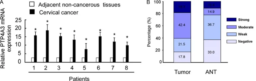
Analysis of the expression of PTP4A3 by real-time PCR determined that PTP4A3 was signifi-cantly upregulated in cervical cancer tissues compared with that in adjacent normal tissues (Figure 1A). Additionally, PTP4A3 staining inten-sity was scored as strong or moderate inteninten-sity in 60.7% of tumor tissues, whereas only 30.3% of corresponding adjacent normal cervical tis-sues were scored as strong or moderate inten-sity (Figure 1A, 1B). Our results showed that PTP4A3 was frequently overexpressed in human cervical cancer.
Association of PTP4A3 expression and clinico-pathological factors
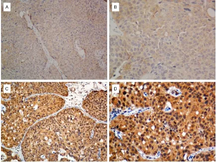
PTP4A3 expression in 144 primary cervical cancer specimens was determined by immuno-histochemistry. The representative immunos-taining of PTP4A3 in cervical cancer tissues was shown in Figure 2. PTP4A3 protein mainly localized at cell membrane and cytoplasm. Statistical analysis further showed positive associations of PTP4A3 expression with tumor
stage (P = 0.004), tumor size (P = 0.024), and lymph node involvement (P = 0.025) (Table 1). No significant difference between high and low levels of PTP4A3 was observed in terms of patient age, tumor differentiation, or histologi-cal type.
PTP4A3 expression predicted worse overcome in cervical cancer
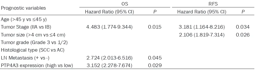
Statistical analysis also revealed a correlation between PTP4A3 expression and patients’ clini-cal prognosis. Kaplan-Meier analysis showed that higher PTP4A3 expression led to shorter overall survival and recurrence-free survival time, while lower PTP4A3 expression resulted in longer survival (P = 0.012 and P = 0.005, respectively; Figure 3). Multivariable Cox regr- ession analysis showed that PTP4A3 expres-sion might serve as an independent predictor of OS in patients with cervical cancer prognosis (P = 0.029; Table 2).
Discussion
[image:3.612.93.524.74.212.2]genic process and progression of cervical carcinoma.
[image:4.612.88.525.72.398.2]PTP4A3 belongs to a small class of PTPs, which are cell signaling molecules that contain a PTP domain and a characteristic C-terminal prenyl-ation motif. Together with tyrosine kinases, PTPs play regulatory roles in a variety of impor-tant signaling molecules and cellular processes [13]. The expression of PTPs is strictly tissue specific, and most cells express 30% to 60% of all the PTPs, while neuronal and hematopoietic cells express higher number of PTPs in com-pared with other cell types [14]. Studies of this class of PTPs demonstrated that dysregulation of PTP activity is involved in various diseases, including cancer, and that prenylation of PTPs is correlated with cancer development and metastasis [15]. As a prenylated PTP, PTP4A3 participates in many fundamental physiological processes and emerges as potential biomark-ers and therapeutic targets for various types of malignancy. Lian et al. demonstrated that PTP4A3 promotes tumor invasion, metastasis and cell adhesion by interacting with JAM2 in colon cancer [16]. Maacha et al. reported that
Table 1. Association between clinical param-eters with PTP4A3
Features Total PTP4A3 P
Low High
Age ≤45 y 90 35 55 0.978
>45 y 54 27 27
Tumor stage IB1+IB2 95 49 46 0.004 IIA1+IIA2 49 13 36
Tumor size ≤4 cm 87 44 43 0.024 >4 cm 57 18 39
Differentiation 1/2 108 43 65 0.174 3 36 19 17
Histological type SCC 89 36 53 0.422 AC 55 26 29
LN Metastasis No 110 53 57 0.025 Yes 34 9 25
144 62 82
[image:4.612.91.297.474.671.2]SCC: squamous cell cancer; AC: Adenocarcinoma.
PTP4A3 promotes human uveal melanoma aggressiveness through membrane accumula-tion of matrix metalloproteinase 14 (MMP14), while inhibition of MMP14 expression in uveal melanoma cells expressing PTP4A3 impairs their migration in vitro and invasiveness [17]. Liu et al. proposed that PTP4A3 plays a critical role in ovarian cancer tumorigenicity and main-taining the malignant phenotype of ovarian can-cer, and could be used as a promising thera-peutic target and potential early biomarker in ovarian cancer progression [18]. However, the roles of PTP4A3 in cervical cancer remain large-ly unknown. In our studies, PTP4A3 was highlarge-ly expressed in cervical cancer tissues in com-parison to that in adjacent normal cervix. We also found significantly positive correlations between PTP4A3 expression and tumor stage, tumor size, and lymph nodes status, indicating the specific involvement of PTP4A3 protein in cervical cancer development.
[image:5.612.90.523.72.256.2]Several recent studies have demonstrated the PTP4A3 positive expression has a significant worse overall survival compared with those do not express. Yeh et al. showed that PTP4A3 overexpression was significantly associated with higher tumor stage, lymph nodal metasta-sis, vascular invasion and unfavorable pro- gnosis in bladder cancer patients [19]. den Hollander et al. revealed that high expression of PTP4A3 could serve as an independent prog-nostic indicator for worse overall survival of patients with triple-negative breast cancer [20]. Xing et al. suggested that PTP4A3 may serve as a potential prognostic biomarker and an indica-tor of lymph node metastasis and vascular invasion in human gastric cancer [21]. Our results are consistent with some previous stud-ies, which found that patients with overexpres-sion of PTP4A3 have a significant worse overall and recurrent-free survival than that with low PTP4A3 expression. These facts may suggest Figure 3. Kaplan-Meier curves for overall survival (OS) and recurrence-free survival (RFS) based on PTP4A3 expres-sion in cervical cancer patients.
Table 2. Multivariate cox proportional hazards regression models for estimating OS and RFS
Prognostic variables OS RFS
Hazard Ratio (95% CI) P Hazard Ratio (95% CI) P
Age (>45 y vs ≤45 y)
Tumor Stage (IIA vs IB) 4.483 (1.774-9.344) 0.015 3.181 (1.164-8.216) 0.034
Tumor size (>4 cm vs ≤4 cm) 2.106 (1.819-7.314) 0.026
Tumor grade (Grade 3 vs 1/2) Histological type (SCC vs AC)
[image:5.612.91.525.321.441.2]and progression.
Disclosure of conflict of interest
None.
Address correspondence to: Fubing Yu, Department of Gynecology, Dongguan Maternal & Children Heal- th Care Hospital, Dongguan, China. Tel: +86 133-1668-0799; Fax: +86 769-2223-8375; E-mail: [email protected]
References
[1] Peiretti M, Zapardiel I, Zanagnolo V, Landoni F, Morrow CP and Maggioni A. Management of recurrent cervical cancer: a review of the litera-ture. Surg Oncol 2012; 21: e59-66.
[2] Cerrotta A, Gardan G, Cavina R, Raspagliesi F, Stefanon B, Garassino I, Musumeci R, Tana S and De Palo G. Concurrent radiotherapy and weekly paclitaxel for locally advanced or recur-rent squamous cell carcinoma of the uterine
cervix. A pilot study with intensification of dose.
Eur J Gynaecol Oncol 2002; 23: 115-119. [3] Guzinska-Ustymowicz K and Pryczynicz A.
PRL-3, an emerging marker of carcinogenesis, is strongly associated with poor prognosis. Anti-cancer Agents Med Chem 2011; 11: 99-108. [4] Walls CD, Iliuk A, Bai Y, Wang M, Tao WA and
Zhang ZY. Phosphatase of regenerating liver 3 (PRL3) provokes a tyrosine phosphoproteome to drive prometastatic signal transduction. Mol Cell Proteomics 2013; 12: 3759-3777. [5] Zimmerman MW, McQueeney KE, Isenberg JS,
Pitt BR, Wasserloos KA, Homanics GE and Lazo JS. Protein-tyrosine phosphatase 4A3 (PTP4A3) promotes vascular endothelial growth factor signaling and enables endotheli-al cell motility. J Biol Chem 2014; 289: 5904-5913.
[6] Peng L, Jin G, Wang L, Guo J, Meng L and Shou
C. Identification of integrin alpha1 as an inter -acting protein of protein tyrosine phosphatase PRL-3. Biochem Biophys Res Commun 2006; 342: 179-183.
erating liver 3 (PRL-3) promotes cell migration through Arf-activity-dependent stimulation of integrin alpha5 recycling. J Cell Sci 2012; 125: 3883-3892.
[10] Min G, Lee SK, Kim HN, Han YM, Lee RH, Jeong DG, Han DC and Kwon BM. Rhodanine-based PRL-3 inhibitors blocked the migration and in-vasion of metastatic cancer cells. Bioorg Med Chem Lett 2013; 23: 3769-3774.
[11] Cao Y, Tu Y, Mei J, Li Z, Jie Z, Xu S, Xu L, Wang S and Xiong Y. RNAimediated knockdown of PRL3 inhibits cell invasion and downregulates ERK1/2 expression in the human gastric can-cer cell line, SGC7901. Mol Med Rep 2013; 7: 1805-1811.
[12] Bardelli A, Saha S, Sager JA, Romans KE, Xin B, Markowitz SD, Lengauer C, Velculescu VE, Kinzler KW and Vogelstein B. PRL-3 expression in metastatic cancers. Clin Cancer Res 2003; 9: 5607-5615.
[13] Alonso A, Sasin J, Bottini N, Friedberg I, Fried-berg I, Osterman A, Godzik A, Hunter T, Dixon J and Mustelin T. Protein tyrosine phosphatases in the human genome. Cell 2004; 117: 699-711.
[14] Wang WQ, Sun JP and Zhang ZY. An overview of the protein tyrosine phosphatase superfamily. Curr Top Med Chem 2003; 3: 739-748. [15] Ostman A, Hellberg C and Bohmer FD.
Protein-tyrosine phosphatases and cancer. Nat Rev Cancer 2006; 6: 307-320.
[16] Lian S, Meng L, Xing X, Yang Y, Qu L and Shou C. PRL-3 promotes cell adhesion by interacting with JAM2 in colon cancer. Oncol Lett 2016; 12: 1661-1666.
[17] Maacha S, Anezo O, Foy M, Liot G, Mery L, Lau-rent C, Sastre-Garau X, Piperno-Neumann S, Cassoux N, Planque N and Saule S. Protein ty-rosine phosphatase 4A3 (PTP4A3) promotes human uveal melanoma aggressiveness through membrane accumulation of matrix metalloproteinase 14 (MMP14). Invest Oph-thalmol Vis Sci 2016; 57: 1982-1990.
[19] Yeh HC, Huang CN, Li CC, Chang LL, Lin HH, Ke HL, Huang AM, Liang PI, Li CF and Wu WJ. Overexpression of PTP4A3 is associated with metastasis and unfavorable prognosis in blad-der cancer. World J Urol 2016; 34: 835-846. [20] den Hollander P, Rawls K, Tsimelzon A,
Shep-herd J, Mazumdar A, Hill J, Fuqua SA, Chang JC, Osborne CK, Hilsenbeck SG, Mills GB and Brown PH. Phosphatase PTP4A3 promotes tri-ple-negative breast cancer growth and pre-dicts poor patient survival. Cancer Res 2016; 76: 1942-1953.