Original Article
MicroRNA-182-5p protects H9c2 cardiomyocytes from
hypoxia-induced apoptosis by down-regulation of PTEN
Lan Yao, Qingshan Zhou, Lu Wang, Guo Hou
Department of Critical Care Medicine, Renmin Hospital of Wuhan University, Wuhan, China
Received January 20, 2017; Accepted March 30, 2017; Epub May 1, 2017; Published May 15, 2017
Abstract: Recent studies suggest that miRNA may play an important role in the process of cell oxidative damage. In this study we made an attempt to explore the functional role of miR-182-5p in acute myocardial infarction. H9c2 cells were cultured and hypoxic H9c2 cells were incubated in hypoxic incubator to simulate hypoxia. Total RNA was
extracted and quantified for miR-182-5p. MiR-182-5p mimic, si-miR-182-5p and the NC controls were synthesized
and transfected into H9c2 cells. Transfected cells were then then assessed for viability by CCk8 assay, cell-apoptosis and cell migration. Result showed that hypoxia led to proportionate decrease in cell-viability, an increased
cell-apoptosis, and significant reduction in cell-migration at 24 hrs (P-value < 0.001). A significant increase in rela -tive expression of miR-182-5p was observed with time at 24 hrs (P-value < 0.001). Over-expression of miR-182-5p alleviated myocardial injury induced by hypoxia (P-value < 0.0001) while knockdown of miR-182-5p showed con-trary results. Cells transfected with miR-182-5p mimic down-regulated the expression of phosphatase and tensin homolog (PTEN) (P-value < 0.05) and PTEN expression was significantly up-regulated in silenced miR-182-5p cells.
The results also showed that over-expression of miR-182-5p inhibited PTEN expression and promoted Akt/GSK-3 beta signaling pathway, resulting in promotion of myocardial cell survival and reduction of cell injury. It was safely concluded that over-expression of miR-182-5p inhibited PTEN expression and enhanced Akt/GSK-3 beta signaling pathway which led to the promotion of myocardial cell survival and reduction of cell injury.
Keywords: Myocardial infarction, hypoxia, mir-182-5p, H9c2 cells, phosphatase and tensin homologue (PTEN), Akt/GSK-3 beta signaling pathway
Introduction
MicroRNAs (miRNAs) are produced endoge-nously and are short noncoding RNAs which regulate target messenger RNA translation by binding to the 3’ untranslated region of mRNA transcript, thereby leading to the degradation of RNA and/or inhibition of protein synthesis [1-3]. miRNAs are not only crucial for the devel-opment and maintenance of physiological homeostasis, but are also equally important in diverse pathological condition [4, 5]. A number of studies have proved that miRNAs play an indispensable role in gastrointestinal develop-ment and physiology [6-8]. Moreover, charac-teristic changes in miRNA expression are asso-ciated with cell apoptosis, oxidative stress and inflammation [7, 9, 10]. miR-182-5p is a family member of miR-182, which is highly expressed in prostate cancer cell. miR-182-5p can
pro-mote cell invasion and proliferation by down-regulating the expression of FOXF2, RECK and MTSS1 [11].
Recent studies suggest that miRNA may play an important role in the process of cell oxida-tive damage. In this study we made an attempt to explore the functional role of miR-182-5p in acute myocardial infarction (AMI).
Materials and methods Cell culture and treatment
H9c2 cells derived from rat embryonic ventricu-lar cardiomyocytes were cultured in DMEM con-tained with 10% (v/v) FBS, 100 U/ml penicillin and 100 lg/ml streptomycin at 37°C in an atmosphere of 95% air and 5% CO2. Hypoxic H9c2 cells were incubated in hypoxic incubator containing 94% N2, 5% CO2, and 1% O2 to simu-late hypoxia [11].
Quantitative-real-time PCR (QRT-PCR)
Total RNA was extracted from cells and tissues using Trizol reagent (Life Technologies Cor- poration, Carlsbad, CA, USA) according to the manufacturer’s instructions. The Taqman Micro- RNA Reverse Transcription Kit and Taqman Universal Master Mix II with the TaqMan MicroRNA Assay of miR-182-5p and U6 (Applied Biosystems, Foster City, CA, USA) were used for testing the expression levels of miR-182-5p in cells.
MiRNAs transfection
MiR-182-5p mimic, si-miR-182-5p and the NC controls were synthesized by GenePharma Co. (Shanghai, China). Cell transfections were con-ducted using Lipofectamine 3000 reagent (Invitrogen) following the manufacturer’s pro- tocol.
CCK-8 assay
Cells were seeded in 96-well plates with 5000 cells/well, cell proliferation was assessed by a Cell Counting Kit-8 (CCK-8, Dojindo Molecular Technologies, Gaithersburg, MD). Briefly, after stimulation, the CCK-8 solution was added to the culture medium, and the cultures were incu-bated for 1 hour at 37°C in humidified 95% air and 5% CO2. The absorbance was measured at 450 nm using a Microplate Reader (Bio-Rad, Hercules, CA).
Apoptosis assay
Flow cytometry analysis was performed to iden-tify and quaniden-tify the apoptotic cells by using
Annexin V-FITC/PI apoptosis detection kit (Beijing Biosea Biotechnology, Beijing, China). The H9c2 cells (100,000 cells/well) were seed-ed in 6 well-plates. Treatseed-ed cells were washseed-ed twice with cold PBS and resuspended in buffer. The adherent and floating cells were combined and treated according to the manufacturer’s instruction and measured with flow cytometer (Beckman Coulter, USA) to differentiate apop-totic cells (Annexin-V positive and PI-negative) from necrotic cells (Annexin-V and PI-positive). Migration assay
Cell migration was determined by using a modi-fied two-chamber migration assay with a pore size of 8 mm. For migration assay, cells sus-pended in 200 ml of serum-free medium were seeded on the upper compartment of 24-well Transwell culture chamber, and 600 ml of com-plete medium was added to the lower compart-ment. After incubation at 37°C, cells were fixed with methanol. Non-traversed cells were removed from the upper surface of the filter carefully with a cotton swab. Traversed cells on the lower side of the filter were stained with crystal violet and counted.
Western blot
0.05 was considered to indicate a statistically significant result.
Results
Hypoxia induced cardiomyocytes injury
Hypoxia led to a proportionate decrease in cell-viability and increase in cell-apoptosis with time compared to the percentage of normoxia. A statistically significant decrease in cell viabil-ity (P < 0.0001) and an increase in cell apopto-sis were observed at 24 hrs. A significant reduc-tion in cell-migrareduc-tion was observed at 24 hrs (P < 0.001, Figure 1). These results suggest that hypoxia induced injury in H9c2 cells.
Hypoxia induced high expression of miR-182-5p
A significant increase in relative expression of miR-182-5p was observed with time at 18 and 24 hrs (P-value < 0.001, Figure 2). These results suggest that hypoxia induced high expression of miR-182-5p in H9c2 cells.
Over-expression or silencing of miR-182-5p expression in cardiomyocytes
[image:3.612.95.378.73.485.2]MiR-182-5p mimic, si-miR-182-5p and the NC controls were synthesized and transfected into H9c2 cells. Over-expression or silencing of miR-182-5p expression was observed in cardiomyo-cytes when compared to the control cells (P < 0.0001 and P < 0.001, respectively, Figure 3). These findings suggested that miR-182-5p can Figure 1. Hypoxia inhibited the
[image:3.612.96.286.546.719.2]cell viability, promoted cell apop-tosis, and reduced the migration in cardiomyocytes. Data repre-sent the mean ± SD of three in-dependent experiments. **P < 0.01; ***P < 0.001.
Figure 2. MiR-182-5p was highly expressed by hypox-ia induction. Data represent the mean ± SD of three independent experiments. **P < 0.01.
Statistical analysis
All experiments were repeat-ed three times. The results of multiple experiments are presented as mean ± SD. Statistical analyses were performed using SPSS 19.0 statistical software. P-va- lues were calculated using one-way analysis of vari-ance (ANOVA). A P-value of < Figure 3. Over-expression or silenced the expression of miR-182-5p in cardiomyocytes. Data represent the mean ± SD of three independent ex-periments. **P < 0.01; ***P
be either overexpressed or silenced in cardiomyocytes.
the effects of miR0182-5p in hypoxia induced injury.
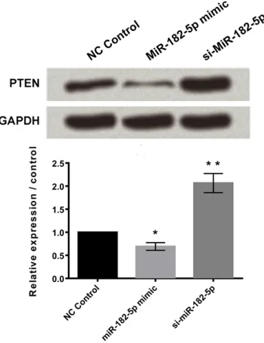
MiR-182-5p negatively regulated the expres-sion of PTEN
Western blotting results showed that cells transfected with miR-182-5p mimic down-regu-lated the expression of phosphatase and ten-sin homolog (PTEN) with P < 0.05. Similarly, significant up-regulation was observed in cells transfected with silenced miR-182-5p, both with P < 0.001 (Figure 5).
MiR-182-5p alleviated hypoxia-induced car-diomyocytes injury by regulation of PTEN ex-pression via activating Akt/GSK-3β signaling pathway
[image:4.612.91.377.70.322.2]PTEN is an inhibitory factor of AKT pa- thway. Hypoxia induced slight increase of AKT/GSK3β pathway. This was confirmed by the over-expression of miR-182-5p which inhibited the expression of PTEN, leading to the activation of AKT/GSK3β pathway, and then promoted cell survival and redu- ced apoptosis. The results were rever- sed by silencing of miR-182-5p (P < 0.05, Figure 6).
Figure 4. Over-expression of miR-182-5p alleviated hypoxia-induced prolifera-tion inhibiprolifera-tion, reduced cell apoptosis, and promoted cell migraprolifera-tion. Data rep-resent the mean ± SD of three independent experiments. *P < 0.05.
Figure 5. MiR-182-5p negatively regulated the ex-pression of PTEN. Data represent the mean ± SD of three independent experiments. *P < 0.05; **P < 0.01.
[image:4.612.93.285.389.639.2]Discussion
Myocardial hypoxia is known to trigger cell inju-ry and apoptosis, which play a key role in the pathogenesis of many cardiovascular diseases including myocardial infarction (MI), heart fail-ure, myocardial ischemia, and reperfusion inju-ry [14]. A number of scientific reports indicated that miRNAs are of vital importance in cardio-vascular diseases, thereby increasing the importance of demonstration of miRNAs in con-trolling apoptosis and identifying their direct and indirect targets [15-17].
A study by Zhang et al. showed that miR-182 is a tumor suppressor in lung cancer cell line [18], while a number of reports suggested miR-182-5p to be an oncogene in several types of can-cers [19-24].
In this paper, the effects and mechanisms of miR-182-5p on hypoxia-induced injury of car-diomyocytes were explored. Hypoxia induced upregulation of miR-182-5p. Over-expression of miR-182-5p led to alleviation of myocardial injury induced by hypoxia while knockdown of miR-182-5p showed contrary results. Further, the results showed that over-expression of miR-182-5p inhibited PTEN expression and promot-ed Akt/GSK-3 beta signaling pathway, resulting in promotion of myocardial cell survival and reduction of cell injury. PTEN when deleted on chromosome ten was found to antagonize the actions of phosphatidylinositol (PI)-3-kinase (PI3K) by de-phosphorylation of PI-3,4,5-trisphosphate (PIP3) to PI-4,5-bisphophate (PIP2) and Pi [25], which lead to enhanced cell
death and increased myocardial contractility in the heart [26-29]. Thus, pharmacological inhi-bition of PTEN was found to enhance PI3K activity and attenuate cardiac injury after isch-emia-reperfusion [30]. Moreover, inactivation of cardiac-specific PTEN protects the heart from functional failure and fibrosis as observed in a mouse model of pressure overload [31]. Further, constitutive PI3K (p110 alpha) and chronic Akt activation attenuates post-MI remodeling [32, 33]. Hence, the findings of our study revealed that the expression of PTEN inhibition led to the activation of Akt/GSK-3 beta signaling pathway which was subsequent-ly followed by promotion of myocardial cell sur-vival and reduction of cell injury was in line with observations in aforementioned reports. This paper thus lays a theoretical foundation for fur-ther study on the function of miR-182-5p, and also provides a new strategy for the clinical treatment of myocardial infarction.
Disclosure of conflict of interest
None.
Address correspondence to: Qingshan Zhou, De- partment of Critical Care Medicine, Renmin Hospital
of Wuhan University, 99, Zhangzhidong Street, Wuhan 430060, China. E-mail: zhouqingshan1109@
126.com
References
[image:5.612.98.519.76.222.2][1] Mendell JT and Olson EN. MicroRNAs in stress signaling and human disease. Cell 2012; 148: 1172-1187.
Figure 6. PTEN is a inhibitory factor of AKT pathway: Hypoxia induced weak increase of AKT/GSK3β pathway: Over-expression of miR-182-5p inhibited the Over-expression of PTEN, leading to the activation of AKT/GSK3β pathway, and
[2] Bartel DP. MicroRNAs: target recognition and regulatory functions. Cell 2009; 136: 215-233.
[3] Ambros V. The functions of animal microRNAs. Nature 2004; 431: 350-355.
[4] Schickel R, Boyerinas B, Park SM and Peter ME. MicroRNAs: key players in the immune system, differentiation, tumorigenesis and cell death. Oncogene 2008; 27: 5959-5974. [5] Ma L, Teruya-Feldstein J and Weinberg RA.
Tumour invasion and metastasis initiated by microRNA-10b in breast cancer. Nature 2007; 449: 682-688.
[6] McKenna LB, Schug J, Vourekas A, McKenna JB, Bramswig NC, Friedman JR and Kaestner KH. MicroRNAs control intestinal epithelial dif-ferentiation, architecture, and barrier function. Gastroenterology 2010; 139: 1654-1664, 1664, e1651.
[7] Miska EA. How microRNAs control cell division, differentiation and death. Curr Opin Genet Dev 2005; 15: 563-568.
[8] Zhang L, Cheng J and Fan XM. MicroRNAs: new therapeutic targets for intestinal barrier dys-function. World J Gastroenterol 2014; 20: 5818-5825.
[9] Nallamshetty S, Chan SY and Loscalzo J.
Hypoxia: a master regulator of microRNA bio-genesis and activity. Free Radic Biol Med 2013; 64: 20-30.
[10] Marques-Rocha JL, Samblas M, Milagro FI,
Bressan J, Martinez JA and Marti A. Noncoding RNAs, cytokines, and inflammation-related dis -eases. FASEB J 2015; 29: 3595-3611. [11] Hirata H, Ueno K, Shahryari V, Deng G, Tanaka
Y, Tabatabai ZL, Hinoda Y and Dahiya R. MicroRNA-182-5p promotes cell invasion and proliferation by down regulating FOXF2, RECK and MTSS1 genes in human prostate cancer. PLoS One 2013; 8: e55502.
[12] Santos CX, Anilkumar N, Zhang M, Brewer AC and Shah AM. Redox signaling in cardiac myo-cytes. Free Radic Biol Med 2011; 50: 777-793. [13] Cassavaugh J and Lounsbury KM. Hypoxia-mediated biological control. J Cell Biochem 2011; 112: 735-744.
[14] Liu B, Che W, Xue J, Zheng C, Tang K, Zhang J, Wen J and Xu Y. SIRT4 prevents hypoxia-in-duced apoptosis in H9c2 cardiomyoblast cells. Cell Physiol Biochem 2013; 32: 655-662. [15] Zhou M, Cai J, Tang Y and Zhao Q. MiR-17-92
cluster is a novel regulatory gene of cardiac ischemic/reperfusion injury. Med Hypotheses 2013; 81: 108-110.
[16] Li P. MicroRNAs in cardiac apoptosis. J Cardiovasc Transl Res 2010; 3: 219-224. [17] He S, Liu P, Jian Z, Li J, Zhu Y, Feng Z and Xiao
Y. miR-138 protects cardiomyocytes from hy-poxia-induced apoptosis via MLK3/JNK/c-jun pathway. Biochem Biophys Res Commun 2013; 441: 763-769.
[18] Zhang L, Liu T, Huang Y and Liu J. microR-NA-182 inhibits the proliferation and invasion of human lung adenocarcinoma cells through its effect on human cortical actin-associated protein. Int J Mol Med 2011; 28: 381-388. [19] Segura MF, Hanniford D, Menendez S, Reavie
L, Zou X, Alvarez-Diaz S, Zakrzewski J, Blochin
E, Rose A, Bogunovic D, Polsky D, Wei J, Lee P, Belitskaya-Levy I, Bhardwaj N, Osman I and Hernando E. Aberrant miR-182 expression pro-motes melanoma metastasis by repressing FOXO3 and microphthalmia-associated tran-scription factor. Proc Natl Acad Sci U S A 2009; 106: 1814-1819.
[20] Guttilla IK and White BA. Coordinate regulation of FOXO1 by miR-27a, miR-96, and miR-182 in breast cancer cells. J Biol Chem 2009; 284: 23204-23216.
[21] Jiang L, Mao P, Song L, Wu J, Huang J, Lin C, Yuan J, Qu L, Cheng SY and Li J. miR-182 as a prognostic marker for glioma progression and patient survival. Am J Pathol 2010; 177: 29-38.
[22] Liu Z, Liu J, Segura MF, Shao C, Lee P, Gong Y, Hernando E and Wei JJ. MiR-182 overexpres-sion in tumourigenesis of high-grade serous ovarian carcinoma. J Pathol 2012; 228: 204-215.
[23] Mihelich BL, Khramtsova EA, Arva N, Vaishnav A, Johnson DN, Giangreco AA,
Martens-Uzunova E, Bagasra O, Kajdacsy-Balla A and
Nonn L. miR-183-96-182 cluster is
overex-pressed in prostate tissue and regulates zinc
homeostasis in prostate cells. J Biol Chem 2011; 286: 44503-44511.
[24] Myatt SS, Wang J, Monteiro LJ, Christian M, Ho KK, Fusi L, Dina RE, Brosens JJ,
Ghaem-Maghami S and Lam EW. Definition of microR -NAs that repress expression of the tumor sup-pressor gene FOXO1 in endometrial cancer. Cancer Res 2010; 70: 367-377.
[25] Siddall HK, Warrell CE, Yellon DM and Mocanu MM. Ischemia-reperfusion injury and cardio-protection: investigating PTEN, the phospha-tase that negatively regulates PI3K, using a
congenital model of PTEN haploinsufficiency.
Basic Res Cardiol 2008; 103: 560-568. [26] Crackower MA, Oudit GY, Kozieradzki I, Sarao
R, Sun H, Sasaki T, Hirsch E, Suzuki A, Shioi T,
Irie-Sasaki J, Sah R, Cheng HY, Rybin VO, Lembo G, Fratta L, Oliveira-dos-Santos AJ,
Benovic JL, Kahn CR, Izumo S, Steinberg SF,
Wymann MP, Backx PH and Penninger JM. Regulation of myocardial contractility and cell
size by distinct PI3K-PTEN signaling pathways.
Cell 2002; 110: 737-749.
[28] Ruan H, Li J, Ren S, Gao J, Li G, Kim R, Wu H
and Wang Y. Inducible and cardiac specific
PTEN inactivation protects ischemia/reperfu-sion injury. J Mol Cell Cardiol 2009; 46: 193-200.
[29] Schwartzbauer G and Robbins J. The tumor
suppressor gene PTEN can regulate cardiac hypertrophy and survival. J Biol Chem 2001; 276: 35786-35793.
[30] Keyes KT, Xu J, Long B, Zhang C, Hu Z and Ye Y. Pharmacological inhibition of PTEN limits
myo-cardial infarct size and improves left ventricu -lar function postinfarction. Am J Physiol Heart Circ Physiol 2010; 298: H1198-1208.
[31] Oudit GY, Kassiri Z, Zhou J, Liu QC, Liu PP, Backx PH, Dawood F, Crackower MA, Scholey JW and Penninger JM. Loss of PTEN attenuates the development of pathological hypertrophy and heart failure in response to biomechanical stress. Cardiovasc Res 2008; 78: 505-514.
[32] Hua Y, Zhang Y, Ceylan-Isik AF, Wold LE, Nunn JM and Ren J. Chronic Akt activation accentu-ates aging-induced cardiac hypertrophy and myocardial contractile dysfunction: role of au-tophagy. Basic Res Cardiol 2011; 106: 1173-1191.
[33] Lin RC, Weeks KL, Gao XM, Williams RB,
Bernardo BC, Kiriazis H, Matthews VB,
Woodcock EA, Bouwman RD, Mollica JP, Speirs
HJ, Dawes IW, Daly RJ, Shioi T, Izumo S,
Febbraio MA, Du XJ and McMullen JR. PI3K(p110 alpha) protects against myocardial
infarction-induced heart failure: identification