Original Article
Protective effects of LM22A-4 on injured spinal cord
nerves
Guangzhe Yu1, Wenbo Wang2
Departments of 1Emergency Surgery, 2Orthopedic Surgery, The First Affiliated Hospital of Harbin Medical University, Harbin 150001, P.R. China
Received April 3, 2015; Accepted May 19, 2015; Epub June 1, 2015; Published June 15, 2015
Abstract: Objective: The goal of this study was to elucidate the protection by and potential mechanisms of LM22A-4,
a specific agonist of tyrosine kinase receptor B, against spinal cord injury (SCI). Methods: Spinal cord trauma was
induced by the application of vascular clips to the dura of mice via a four-level T7-T11 laminectomy. Thirty minutes
after the injury, an abdominal injection of LM22A-4 (at dosages of 10 mg/kg and 15 mg/kg) or an equal volume
of solvent was provided. Twenty-four hours after SCI, a Western blot was performed to examine the expression of
p-TrkB, p-Akt, p-ERK, cleaved-caspase-3, and Bcl-2; a TUNEL assay and Nissl staining were performed to study
apoptosis and the survival of neurons. In addition, another batch of mice was allowed to live for 14 days after the SCI treatment, during which the LM22A-4 was provided at the same time each day and the neurological function was assessed. Results: Spinal cord injury in mice resulted in severe trauma characterized by tissue damage and
apoptosis. Treatment of the mice with LM22A-4 (10 mg/kg) significantly reduced the degree of tissue injury (histo
-logical score) and apoptosis (TUNEL staining and caspase-3 and Bcl-2 expression) compared with vehicle treated
group (P < 0.05). In a separate set of experiments, chronic treatment with LM22A-4 also significantly ameliorated
the recovery of limb function (P < 0.05). Conclusion: This study provides an experimental evidence that LM22A-4
reduces the development of tissue injury associated with spinal cord trauma, and activation of the activity of TrkB
may represent a novel approach for the therapy of spinal cord trauma.
Keywords: SCI, LM22A-4
Introduction
Previous studies have indicated that brain-derived neurotrophic factor (BDNF) can acti-vate the TrkB signaling pathway to exert a pro-tective function against spinal cord trauma. LM22A-4 is a newly developed TrkB-specific agonist that has been demonstrated to play a neuroprotective role in brain injury. However, its role in SCI has not been reported. Hence, the LM22A-mediated protection and potential mechanisms against SCI are examined herein, which might shed light on strategies and meth-ods for clinically treating SCI patients.
Materials and methods
Materials
Experimental ICR mice were purchased from the Laboratory Animal Medical Center, X
University. A total protein extraction solution was purchased from Beyotime Technology. Phosphatase inhibitors were purchased from Sigma (USA). Antibodies for p-TrkB, p-Akt, p-ERK, p-CREB, cleaved-caspase-3, and Bcl-2 were purchased from Cell Signaling (USA). Vascular clips for aortic clamping in mice were purchased from Kent Scientific (USA). LM22A-4 was purchased from Sigma and was dissolved in DMSO.
Animal grouping and preparation of the mouse model of spinal cord injury
Protection of SCI by LM22A-4
mouse SCI model was based on a previous study [1, 2] and on a protocol employed by this group. Briefly, a mouse was anesthetized via the administration of chloral hydrate (4 mg/kg) before a 3-cm incision was introduced on its back. T7-T11 vertebrae were exposed under a surgical microscope before the laminae were removed with a vascular clip to fully expose the spinal cord. The spinal cord was clamped with the vascular clip for 1 minute with a force of 10 g. The animal was then subjected to complete staunching of the bleeding, and the incised dor-sal muscle and skin were sutured. Mice from the control group underwent laminectomy, full exposure of the spinal cord, and subsequent suturing, but without aortic clamping. After the surgery, the mice were placed on a warm blan-ket until fully awake and were then housed in cages accommodating a normal diet. After the SCI treatment, 14 mice were sacrificed to enable molecular and histological examina-tions; the other 14 mice were allowed to live for 20 days for neurological scoring and were sac-rificed on day 20 via cervical dislocation.
Western blot
Twenty-four hours after spinal cord trauma, one
allow opening of the thoracic cavity and expo-sure of the heart. An injection needle was inserted into the left apical position and fixed with a vascular clamp. Subsequently, the right atrial appendage was opened, and venous blood was drawn, followed by rapid infusion of 80 mL saline until clear fluid flowed out from the right atrial appendage. The original dorsal incision was reopened to expose the original dorsal incision to expose vertebrae T5-T9 and to remove the lamina. A 1.5-cm segment of spi-nal cord tissue flanking the clip mark was retrieved and stocked in liquid nitrogen. Consequently, 50 mg of spinal cord tissue was obtained from each mouse; it was mixed with tissue lysis buffer at a ratio of 1:1000 (W/V), fully ground, and centrifuged at 12000 r/min-ute to generate a supernatant. One portion of the supernatant was mixed with four parts of 5× loading buffer, which was boiled for 10 min-utes. Thirty-five micrograms total protein from each sample was loaded into each well, elec-trophoresed, and transferred to a membrane. The membrane was blocked with 5% skim milk at room temperature for 10 minutes and incu-bated with each primary antibody at 4°C. The membrane was then rinsed with TBST buffer, incubated with a secondary antibody at room temperature for 1 hour, and rinsed with TBST buffer. Bands were revealed by incubating the membrane with developer and were quantita-tively analyzed with Image J.
TUNEL assay and Nissl staining
A group of mice was injected with saline into the left apical position before 80 mL of 4% paraformaldehyde was injected around the spi-nal cord tissue, which was retrieved and paraf-fin-embedded. Consecutive sectioning at a thickness of 6 μm was then performed. A TUNEL assay was then conducted to examine neuronal apoptosis, following the manufactur-er’s instructions. Under 400× magnification, 10 fields were randomly chosen to enumerate pos-itively stained cells. The mean cell counts on each section slide were documented. In addi-tion, four section slides of spinal cord tissue from each mouse were subjected to Nissl stain-ing followstain-ing standard procedures.
Motor function scoring
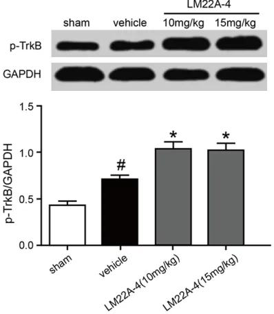
[image:2.612.90.289.74.302.2]Motor function was assessed via a new neuro-logical scoring system called Basso Mouse Figure 1. Influences of LM22A-4 treatment on p-TrkB
expression of injured spine cord tissue. In
compari-son with the solvent treatment, LM22A-4 significant
-ly upregulated the expression of p-TrkB. Two doses of 10 mg/kg and 15 mg/kg did not generate an ap
-preciable difference in p-TrkB expression. # P < 0.05
tent monitors the coordination of the forelimbs (FLs) and hindlimbs, consistent position of the paw during stepping, adequate toe clearance, maintenance of a stable trunk, and tail position of SCI-treated mice within 4 minutes. Our assessment included a major scoring system and an auxiliary scoring system. The first assessment was performed 24 hours after the SCI treatment, and the second assessment
was performed 13 days later at the same time point.
Statistical analysis
The experimental data were analyzed via the SPSS17.0 software. The data were expressed as mean ± standard deviation (x ± S). One-way ANOVA was performed to facilitate
compari-Figure 2. Influences of LM22A-4 on the expres
-sion of p-Akt, p-Erk, cleaved-caspase-3, and
Bcl-2 in SCI-treated mice. In comparison with the SCI treatment, LM22A-4 administration (10 mg/
kg) significantly improved the expression of p-Akt, p-Erk, and Bcl-2 and significantly inhibited
the production of cleaved-caspase-3 (P < 0.05).
# P < 0.05 compared with the sham-treatment
[image:3.612.85.521.66.589.2]Protection of SCI by LM22A-4
sons between groups. P < 0.05 was considered statistically significant.
Results
Influences of LM22A-4 on the expression of p-TrkB, p-Akt, and p-ERK
The dosage of LM22A-4 used in this study was based on a previous study [1]. Our results indi-cated that, in comparison with the solvent treatment, administration of LM22A-4 may sig-nificantly upregulate p-TrkB expression and that both doses (10 mg/kg and 15 mg/kg) dis-played no difference in this upregulation (Figure 1). Hence, 10 mg/kg was adopted as the dose in subsequent experiments. In comparison with the sham treatment, exposure to solvent appeared to cause increased expression of p-TrkB, p-Akt, and p-ERK, but the margins were not statistically significant (Figure 2, P > 0.05). By contrast, the administration of LM22A-4
sig-genes compared with those of the solvent-treated mice (P < 0.05).
LM22A-4 significantly inhibits the expression of the apoptosis-related protein cleaved-cas-pase-3 and augments the expression of the anti-apoptotic protein Bcl-2
In comparison with the sham-treated animals, SCI treatment caused apparently increased ex- pression of cleaved-caspase-3, which was sup-pressed by administration of LM22A-4 (10 mg/ kg, Figure 2, P < 0.05). In addition, as shown in Figure 2, SCI significantly reduced the expres-sion of the anti-apoptotic protein Bcl-2 (P < 0.05), which, however, was clearly increased by the LM22A-4 treatment (10 mg/kg, P < 0.05).
Morphological changes in the spinal cord
tis-sue
As shown in Figure 3, Nissl staining revealed that SCI-treated mice exhibited neuronal pyk-Figure 3. Nissl staining after LM22A-4 treatment. Nissl staining revealed that SCI-treated mice exhibited
neuro-nal pyknosis and significantly reduced neuroneuro-nal counts. LM22A-4 (10 mg/kg) treatment resulted in a significant
increase in neuron number. #P < 0.05 compared with the sham-treatment group; * P < 0.05 compared with the
[image:4.612.93.523.72.397.2]bers; whereas animals adminis-tered LM22A-4 (10 mg/kg) ex- hibited significantly increased neuron numbers. In addition, Figure 4 illustrates the positive nuclear staining of the apoptot-ic neurons. In comparison with the sham-treated animals, SCI treatment caused a significant elevation in the number of apoptotic neurons (P < 0.05), which was apparently de- creased by the LM22A-4 admin-istration (10 mg/Kg, P < 0.05).
LM22A-4 significantly amelio-rated SCI neurological scores
As shown in Figure 5, the neuro-logical scores of the sham-treat-ed animals did not exhibit Figure 4. Influence of LM22A-4 treatment on SCI-resulted apoptosis. The apoptosis-positive neurons are revealed by brown nuclear staining. LM22A-4 administration (10 mg/kg) significantly decreased the number of apoptotic neu -rons in the SCI-treated mice (P < 0.05). # P < 0.05 compared with the sham-treatment group; * P < 0.05 compared
with the vehicle-treatment group.
Figure 5. Influence of LM22A-4 administration on the neurological function
in SCI-treated mice. Animals in the sham treatment group displayed no ap-parent change during the observation period. In comparison with the
sol-vent treatment, LM22A-4 (10 mg/kg) administration significantly improved
Protection of SCI by LM22A-4
apparent changes during the observation peri-od. By contrast, LM22A-4 administration (10 mg/kg, P < 0.05) significantly improved the neurological scores compared with those of the solvent-treated animals (P < 0.05).
Discussion
Despite rapid economic development, spinal cord trauma still has a high incidence world-wide and yields considerable mortality and dis-ability rates. Moreover, the survivors often exhibit severe dysfunctions. Hence, a focus and challenge of global research remains how to improve the prognosis of SCI patients. Although many drugs have been experimentally indicat-ed to ameliorate the SCI prognosis, none has demonstrated reliable efficacy or been clinical-ly applied to treat SCI patients [4]. Hence, it is of vital importance to develop novel and effec-tive anti-SCI drugs that have few side effects. BDNF is a member of the family of endogenous neuroprotective factors. Previous studies have demonstrated that injection of BDNF can gen-erate neuroprotective activities and effectively improve the prognosis of spinal cord trauma. However, it is difficult for BDNF to penetrate the blood-brain barrier, which is compounded by the molecule’s short half-life [5]. These issues greatly restrict its clinical applications. BDNF mainly activates the TrkB signaling pathway to exert neuroprotection; therefore, the develop-ment of drugs that target TrkB, a BDNF ligand, is currently a research focus. LM22A-4 is a newly designed TrkB-specific agonist that has been demonstrated to play neuroprotective roles in multiple nervous system diseases. In agreement with previous results, our data revealed that LM22A-4 can significantly upreg-ulate the expression of p-TrkB in a dose-depen-dent manner. Previous studies have indicated that TrkB activation can effectively reduce the expression of caspase-3, which is one of the most important apoptosis effectors and is over-expressed after spinal cord trauma to aggra-vate the severity of SCI [6]. Correspondingly, inhibition of caspase-3 expression can signifi-cantly improve the prognosis of SCI [7]. Our results indicated that LM22A-4 could markedly decrease the expression of apoptotic protein caspase-3 and the number of apoptotic neu-rons, thereby effectively improving the histo-logical appearance of SCI. As such, our data revealed that LM22A-4 might play a prominent neuroprotective role in SCI.
In light of the above results, we are further exploring the potential neuroprotective mecha-nisms of LM22A-4, which might provide theo-retical guidance for the clinical treatment of SCI. Many previous studies have indicated that TrkB activation may subsequently activate Akt, which is a serine/threonine protein kinase. Akt is composed of 480 amino acids, and its pro-tein structure comprises a pH domain, a cata-lytic domain, and a C-tail regulatory domain. After its activation, Akt is first recruited to the cell membrane before translocation to the cyto-plasm and nucleus, where it catalyzes serine/ threonine phosphorylation at specific sites of its protein substrates. This step further upregu-lates expression of the apoptosis-inhibitive pro-tein Bcl-2 and downregulates and degrades activated caspase-3. Many previous studies have indicated that Akt-mediated signaling pathways play crucial roles in SCI, as suppres-sion of Akt activity markedly potentiates neuro-nal damage [2]. On the contrary, the upregula-tion of Akt activity can significantly improve the SCI prognosis [7]. Our study revealed that LM22A-4 significantly upregulated Akt activity, which is a possible mechanism of its neuropro-tection. In addition, TrkB excitation can upregu-late ERK activity. Consequently, the ERI signal-ing pathway is also activated dursignal-ing SCI and plays neuroprotective roles in spinal cord trau-ma. Tae et al. [8] has reported that the adminis-tration of estrogenic drugs to enhance ERK activity can generate neuroprotection, which is supported by our finding that LM22A-4 signifi-cantly augmented ERK activity. This function might be another neuroprotective mechanism of LM22A-4.
Acknowledgements
This project was supported by Youth Science Foundation of Heilongjiang Province of China Grant QC2012C035.
Disclosure of conflict of interest
None.
Address correspondence to:Dr. Wenbo Wang,
De-partment of Orthopedic Surgery, The First Affiliated Hospital of Harbin Medical University, 23 Youzheng Road, Nangang District, Harbin 150001, P.R. China. Tel: +86-451-53643849; Fax: +86-451-53643849;
E-mail: wangwenbo489@163.com
References
[1] Massa SM, Yang T, Xie Y, Shi J, Bilgen M, Joyce JN, Nehama D, Rajadas J, Longo FM. Small molecule BDNF mimetics activate TrkB signal -ing and prevent neuronal degeneration in ro-dents. J Clin Invest 2010; 120: 1774-1785. [2] Paterniti I, Esposito E, Mazzon E, Bramanti P,
Cuzzocrea S. Evidence for the role of PI(3) -ki
-nase-AKT-eNOS signalling pathway in second
-ary inflammatory process after spinal cord
compression injury in mice. Eur J Neurosci 2011; 33: 1411-1420.
[3] Basso DM, Fisher LC, Anderson AJ, Jakeman LB, McTigue DM, Popovich PG. Basso Mouse
Scale for locomotion detects differences in
re-covery after spinal cord injury in five common
mouse strains. J Neurotrauma 2006; 23: 635-659.
[4] Mothe AJ, Tator CH. Advances in stem cell ther-apy for spinal cord injury. J Clin Invest 2012; 122: 3824-3834.
[5] Poduslo JF, Curran GL. Permeability at the
blood-brain and blood-nerve barriers of the
neurotrophic factors: NGF, CNTF, NT-3, BDNF. Brain Res Mol Brain Res 1996; 36: 280-286.
[6] Citron BA, Arnold PM, Sebastian C, Qin F, Malladi S, Ameenuddin S, Landis ME, Festoff BW. Rapid upregulation of caspase-3 in rat spi -nal cord after injury: mRNA, protein, and cellu-lar localization correlates with apoptotic cell death. Exp Neurol 2000; 166: 213-226. [7] Jung SY, Kim DY, Yune TY, Shin DH, Baek SB,
Kim CJ. Treadmill exercise reduces spinal cord
injury-induced apoptosis by activating the
PI3K/Akt pathway in rats. Exp Ther Med 2014;
7: 587-593.
[8] Yune TY, Park HG, Lee JY, Oh TH. Estrogen-induced Bcl-2 expression after spinal cord in -jury is mediated through
phosphoinositide-3-kinase/Akt-dependent CREB activation. J