Original Article
JAK2/STAT3 pathway mediating inflammatory
responses in heatstroke-induced rats
Zhen Tao, Ming Cheng, Shu-Cai Wang, Wei Lv, Huai-Qiang Hu, Chuan-Fen Li, Bing-Zhen Cao
Department of Neurology, General Hospital of Jinan Military Command, Jinan 250031, Shandong, China
Received March 31, 2015; Accepted May 25, 2015; Epub June 1, 2015; Published June 15, 2015
Abstract: Heatstroke not only directly induces cell injury, but also causes large amounts of inflammatory media -tors release and cells with extensive biological activities to induce a systemic inflammatory response and immune dysfunction. This study aimed to observe the effects of JAK2 inhibitor AG490 on the brain injury and inflammatory
responses of rats with systemic heatstroke. Under the light microscope, the hippocampus tissues of rat with heat-stroke were edema and apoptotic rate was increased. Up-regulation of malondialdehyde (MDA), nitric oxide syn-thase (iNOS), reactive oxygen species (ROS) and down-regulation of superoxide dismutase (SOD) were also found
after heatstroke in rats, which compared with that of the control group. Heatstroke induced inflammation factors se
-cretions and up-regulated levels of matrix metallopeptidase 2 and 9 (MMP2 and MMP-9) and systemic inflammatory response molecules including intercellular adhesion molecule-1 (ICAM-1), tumor necrosis factor-beta 1 (TNF-β1) and cyclooxygenase-2 (COX-2). However, the JAK2 inhibitor AG490 was significantly attenuated the brain injury and inflammatory responses induced by heatstroke in rats. The survival time of heatstroke rats showed that AG490 notably lived longer than heatstroke rats without AG490 treatment. These findings suggest that AG490 may prevent the occurrence of heatstroke via inhibiting the JAK2/STAT3 pathway and the systemic inflammatory responses.
Keywords: Heatstroke, AG490, JAK2/STAT3, inflammation
Introduction
Heatstroke may be defined as overheating of
the body, including heatstroke caused by an over-high ambient temperature, or as an abnor-mally high body temperature [1]. The potential causes of heatstroke include infection, certain drugs and medications, and brain trauma [2]. High temperature may be used for tumor treat-ment, particularly for cancer treattreat-ment, but controversial issues remain in its clinical use [3]. Heatstroke can progress to multiple organ dysfunction or injury syndrome (MODS) and death, despite adequate lowering of the vic-tim’s body temperature and intensive care [1]. Up to 30% of survivors may sustain permanent neurological damage [1].
Cell apoptosis in heatstroke is not well under-stood, which may explain the high mortality as
no specific mechanisms can be targeted for
treatment. Recently many studies showed the important roles of cell apoptosis in heatstroke and molecular mechanism involved in was not
known [4]. In addition, increasing evidence shows that the Janus kinase 2 (JAK2)/STAT sig-naling pathway, especially STAT3, is the critical target and biomarker during angiogenesis and tumor growth. JAK2/STAT3-mediated apoptosis involved in protecting hepatocytes, myocardi-um, and heart against ischemia/reperfusion injury [5-7], especially through the phosphoryla-tion of STAT3 to promote survival and inhibit apoptosis. JAKs and STATs are normally expressed in brain [8], and their expression increases after focal ischaemia in the rat, par-ticularly in reactive astrocytes and microglia cells [9], indicating that JAK/STAT is an
impor-tant mediator of inflammatory responses in
brain ischaemia. However, the role of JAK/STAT pathway in rat heatstroke has not been known. Studies in humans and animals suggest that
JAK2/STAT3 and inflammation in heatstroke rats
levels of markers for cellular ischemia and damage, and increased expression of inducible nitric oxide synthase (iNOS) [11]. In addition, various serum molecules including tumor
necrosis factor-alpha (TNF-α), intercellular
adhesion molecule-1 (ICAM-1), and E-selectin have been demonstrated to be involved in the
pathophysiology of systemic inflammatory
response syndrome [12, 13]. A study reported that heatstroke-induced elevation of the levels of heat shock protein 70 relieved the extent of
the pulmonary fibrosis of rats in response to
the induction of acute lung injury by lipopoly-saccharide (LPS) administration [14]. However, the effects of heatstroke on the brain of rats and its mechanism of action remain unclear at present. In the clinic, cases of fatal heatstroke caused by various intraoperative factors are frequently reported, and a comprehensive treatment measure is the key to successful treatment [15, 16].
In the present study, we investigated the pro-tection of AG490, a JAK2 inhibitor, in rats with heatstroke, which induces cell apoptosis and
inflammation. Our findings showed that JAK2/ STAT3 pathway mediating inflammatory
res-ponses induced by heatstroke rats. Materials and methods
Animals and AG490 treatment
A total of 36 specific pathogen-free Sprague
Dawley male rats with body weights rang- ing from 180 to 220 g were purchased from Shanghai Laboratory Animal Company (Shanghai, China) and randomly divided into three groups, with twelve rats in each group. The groups were as follows: the control group (the rats were maintained at room tempera-ture, without medication), the heatstroke group (the rats were placed at 42°C, without medica-tion), and the JAK2 inhibitor AG490 treatment group (AG490 was administered before 2 h of heating). All protocols were approved by the Animal Ethics Committee of the General Hospital of Jinan Military Command (Jinan, China) in accordance with the Guide for the Care and Use of Laboratory Animals of the National Institutes of Health.
Induction of heatstroke
The rats were anesthetized with 3% sodium pentobarbital (45 mg/kg) by intraperitoneal
injection and placed into a heating chamber with a biological oxygen supply. Also, the previ-ously examined rectal temperature was used as the basic value. Subsequently, the rats in all groups other than the control group were heat-ed in the heating chamber at 42°C and a rela-tive humidity of 60%. For the AG490 treatment group, AG490 was administered before 2 h of heating, respectively. After 1 h of heating, all rats were removed from the heating chamber and treated as subsequently described.
Histology
Hippocampus of rats were removed, and then
fixed with 10% (v/v) neutral buffered formalin.
Specimens were dehydrated and embedded in
paraffin. For histological examined, 4 mm sections of fixed embedded tissues were cut
on a Leica model 2165 rotary microtome (Leica Microsystem Nussloch GmbH, Wetzlar, Germany), placed on glass slides,
deparaf-finized, and stained sequentially with hematox
-ylin and eosin (H&E, Richard-Allan Scientific,
Kalamazoo, MI). Stained tissue sections on slides were analyzed under identical light microscope (Axio Imager M1, Karl Zeiss,
Germany) at ×200 magnification. Terminal dUTP Nick-End labeling assay
Terminal dUTP Nick-End labeling assay was performed using a kit for programmed cell death (Medical & Biological Laboratories, Nagoya, Japan) according to the
manufactur-er’s directions. The fixed areas of section were
examined by microscopy and the numbers of TUNEL-positive cells were counted by a
patholo-gist at ×200 magnification, 30 fields per sec -tion. Blinding was performed for the patholo-gist’s grading of results.
Biochemical measurements
Nanjing Jiancheng Bioengineering Institute (Nanjing, China).
Measurements of TNF-α, IL-1β, IL-6 and IL-8 in peripheral blood
TNF-α, IL-1β, IL-6 and IL-8 levels present in
peripheral blood were determined using
com-mercially available murine-specific sandwich
enzyme-linked immunosorbent assay (ELISA) kit supplied by Santa Cruz (USA).
Western blot analysis
Tissue samples were homogenized in 13.2 mmol/l Tris-HCl, 5.5% glycerol, 0.44% SDS, and
10% β-mercaptoethanol. An equal amount of extracted soluble protein (50 μg) was fraction -ated by Tris-glycine-SDS polyacrylamide gel (12%) electrophoresis, and Western blotting was performed as described with use of a polyclonal rabbit antibody to recombinant rat p-JAK2 (1:500), JAK2 (1:1000), p-STAT3 (1:20000), STAT3 (1:100), MMP2 (1:500), MMP-9 (1:500), ICAM-1 (1:800), TGF-β1
(1:400), COX-2 (1:500) and GAPDH (1:1500). All the antibodies were purchased from Abcam (Jinan, China) except GAPDH (CST, Danvers, MA, USA).
Statistical analysis
The data was presented as the mean value ± S.D. The paired, two-tailed Student’s t-test was
used to analyze the significance of difference
between groups. Survival analysis was carried out by Kaplan-Meier method, and subjected to the log rank test. P value lower than 0.05 was
considered to be statistically significant.
Results
AG490 treatment attenuates injury and apop-tosis during heatstroke
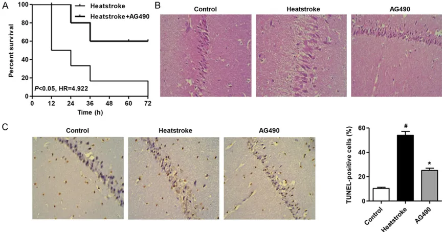
We compared the survival time in heatstroke
rats. The cumulative survival rate was signifi -cantly lower in heatstroke rats than that in heatstroke rats with AG490 treatment (Figure 1A). These results indicated that AG490 could represent a new prognostic factor in heatstroke rats. As shown in Figure 1B, after heatstroke in rats, the hippocampal neuron damage scores
were significantly increased in rats with heat -stroke compared with the control group.
Histopathological verification revealed edema
[image:3.612.90.524.72.299.2]JAK2/STAT3 and inflammation in heatstroke rats
tective effects. Figure 1C showed absence of or only a few scattered TUNEL-positive cells of hippocampal neuron in control group. In severe heatstroke, TUNEL-positive staining indicative of apoptotic cell death was extensive in hippo-campal neuron compared with the control group (Figure 1C). However, the extensive apoptotic cells of hippocampal neuron in
heat-stroke-induced rats were significantly attenu -ated by AG490 tre-ated rats.
AG490 treatment inhibits of heatstroke-in-duced up-regulation of MDA, iNOS, ROS levels and down-regulation of SOD level
The comparisons of the MDA, iNOS, ROS and SOD levels in rat with heatstroke were shown in Figure 2. The MDA, iNOS and ROS levels of hip-pocampus tissue in rats with heatstroke were
significantly higher than that in the control
group (Figure 2A-D), and the SOD level of hip-pocampus tissue in rats with heatstroke were
significantly lower than that in the control group
(Figure 2B). However, the MDA, iNOS, ROS and
SOD levels were reverse in the AG490 treat-ment group respectively.
AG490 treatment represses
heatstroke-in-duced inflammation factor secretions
Next we measured TNF-α, IL-1β, IL-6 and IL-8
secretions in response to heatstroke. After
exposure of rats to heat stress for 1 h, TNF-α, IL-1β, IL-6 and IL-8 secretions were signi-ficantly increased, respectively (Figure 3A-D). Pretreatment with AG490 for 2 h before
expo-sure to heat stress markedly inhibited TNF-α, IL-1β, IL-6 and IL-8 secretions from rats, respec -tively. These results suggest that AG490
pos-sesses an anti-inflammatory effect in heat -stroke-induced rats.
Inhibition of injury is involved in the protection
of AG490 against inflammation in
heatstroke-induced rats
[image:4.612.92.521.71.192.2]Since inhibitor AG490 was implicated in the inhibitory effect on JAK2, we further explored the role of AG490 against heatstroke-induced Figure 2. Effect of AG490 on the levels of MDA, iNOS, ROS and SOD. AG490 treatment inhibition of up-regulation of MDA (A), iNOS (C), ROS levels (D) and down-regulation of SOD level (B) induced by heatstroke. #P < 0.01 compared
[image:4.612.93.523.251.368.2]with the control group. *P < 0.01 compared with the heatstroke group.
Figure 3. AG490 inhibits heatstroke-induced TNF-α, IL-1β, IL-6 and IL-8 secretions. Rats were exposed to heatstroke for 1 h in the absence of presence of AG490. ELISA was performed to detect the levels of TNF-α, IL-1β, IL-6 and IL-8
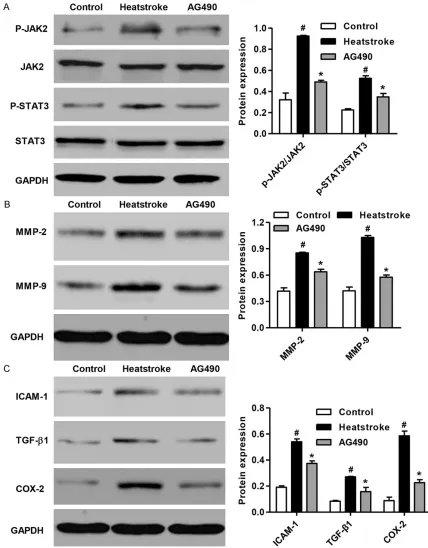
inflammatory responses. As shown in Figure 4A, pretreatment of rats with AG490 sup-pressed p-JAK2/JAK2 and p-STAT3/STAT3
lev-els. Similar to the anti-inflammatory effect of
[image:5.612.90.518.72.620.2]JAK2/STAT3, pretreatment of rats with AG490 suppressed heatstroke-induced increase of Figure 4. Effect of AG490 on JAK2/STAT3 pathway and inflammation. AG490 treatment suppressed activation of JAK2/STAT3 pathway and increase of inflammation related protein, MMP2 and MMP-9. And AG490 treatment also inhibition of inflammation factors secretions induced by heatstroke. #P < 0.01 compared with the control group. *P
JAK2/STAT3 and inflammation in heatstroke rats
MMP2 and MMP-9 protein levels (Figure 4B). In addition, compared with the control group, heatstroke rats had higher levels of ICAM-1,
TGF-β1, COX-2 after the onset of heatstroke
(Figure 4C). The increase in the protein levels of these three markers caused by heatstroke
were significantly reduced by AG490
pre-treatment. Discussion
Heatstroke is defined as a condition in which
the core temperature is elevated to a critical level that induced multi-organ damage and dys-function [17]. Evidence has accumulated to
suggest that thermoregulatory deficits may
occur during heatstroke. For example, unanes-thetized, unrestrained heatstroke mice dis-played hypothermia when exposed to room temperature [11, 18-20]. The hypothermia that occurred after onset of heatstroke may have resulted from neuronal apoptosis and systemic
imflammation in the hypothalamus [21]. In the
current study, we demonstrated the protective effects of JAK2 inhibitor AG490 in reducing heatstroke-induced hippocampal neuronal
apoptosis and inflammation responses.
In terms of heatstroke-induced nervous system injury, the administration of dexamethasone and mannitol in combination was associated nervous system damage in rats with heatstroke [22]. Consistent with the previous study, our data showed that the hippocampal neuron in rats with heatstroke resulted in appearance of edema and disappearance of the nuclear (Figure 1A). However, AG490 treatment attenu-ated the brain injury induced by heatstroke in rats. Studies in cell lines and animal models suggest that heat directly induces not only tis-sue injury but also cell death [17]. Rat models subjected to moderate whole body hyperther-mia showed that accelerated apoptosis also contributes to cell death, but whether apopto-sis is an important cause of cell death in patients with heatstroke is not known [23]. A
major finding in this study was the dominance
of apoptosis as a mechanism of cell death in heatstroke. Using TUNEL assay we observed
significant increase in cell apoptosis of hippo -campal neuron in heatstroke-induced rats (Figure 1B). However, rats with AG490 treat-ment showed inhibition of cell apoptosis of hip-pocampal neuron with heatstroke.
In the present study, the results showed that AG490 suppressed the MDA, iNOS and ROS up-regulation and SOD down-regulation in- duced by heatstroke in rats (Figure 2A-D).
These findings were similar to those in multiple
organic damages [24. 25]. Heatstroke not only directly induces cell injury of tissue, but also
causes to release large amounts of inflamma
-tory mediators to induce a systemic inflamma
-tory response. The plasma levels of inflamma
-tory cytokines such as TNF-α and IL-1β are ele -vated in persons and animals with heatstroke [26-28]. The increase in the plasma levels of
these pro-inflammatory cytokines is associated
with the severity of heatstroke. A number of studies have shown that AG490 is able to
effec-tively reduce the levels of serum pro-inflamma
-tion cytokines, such as TNF-α, IL-1, IL-6 and IL-8
[29, 30]. The results in the present study showed that AG490 suppressed the increase
of TNF-α, IL-1, IL-6 and IL-8 in levels induced by
heatstroke in rats (Figure 3A-D). To further
investigate the inflammatory responses
in-duced by heatstroke, we also detected the pro-tein expression of MMP-2, MMP-9, ICAM-1,
TGF-β1 and COX-2. MMP-2 and MMP-9 as two novel markers of inflammation were significant -ly increased (Figure 4B) and the expression of
inflammatory molecules, including ICAM-1, TGF-β1 and COX-2 was also increased in heat -stroke-induced rats (Figure 4C). However, the rats treated with AG490 inhibited the activation of JAK2/STAT3 pathway and followed by
inhib-ited the increase of inflammation-associated
proteins expression (Figure 4A-C).
In conclusion, our results demonstrate that, heatstroke-induced hippocampal neuron injury
and apoptosis, and the systemic inflammatory response can be significantly prevented by JAK2 inhibitor AG490 treatment. These find -ings indicate that JAK2/STAT3 may improve heat tolerance by reducing the occurrence of
apoptosis and the systemic inflammatory
res-ponse.
Acknowledgements
This research did not receive any specific grant
from any funding agency in the public,
commer-cial or not-for-profit sector.
Disclosure of conflict of interest
Address correspondence to: Dr. Bing-Zhen Cao, Department of Neurology, General Hospital of Jinan Military Command, 25 Shifan Road, Tianqiao District, Jinan 250031, Shandong, China. Tel: +86-531-51665480; Fax: +86-531-51665480, E-mail: caobzwz@126.com
References
[1] Bouchama A, Knochel JP. Heat stroke. N Engl J Med 2002; 346: 1978-88.
[2] Varghese G, John G, Thomas K, Abraham O, Mathai D. Predictors of multi-organ dysfunc-tion in heatstroke. Emerg Med J 2005; 22: 185-87.
[3] Takagi M, Sakata K, Someya M, Matsumoto Y, Tauchi H, Hareyama M, Fukushima M. The combination of hyperthermia or chemotherapy with gimeracil for effective radiosensitization. Strahlenther Onkol 2012; 188: 255-61. [4] Roberts GT, Ghebeh H, Chishti MA, Al-Mohanna
F, El-Sayed R, Al-Mohanna F, Bouchama A.
Microvascular injury, thrombosis, inflamma -tion, and apoptosis in the pathogenesis of heatstroke a study in baboon model. Arterioscl Throm Vas 2008; 28: 1130-36.
[5] Yu HC, Qin HY, He F, Wang L, Fu W, Liu D, Guo FC, Liang L, Dou KF, Han H. Canonical notch pathway protects hepatocytes from ischemia/ reperfusion injury in mice by repressing reac-tive oxygen species production through JAK2/ STAT3 signaling. Hepatology 2011; 54: 979-88.
[6] Yang Y, Duan W, Jin Z, Yi W, Yan J, Zhang S, Wang N, Liang Z, Li Y, Chen W. JAK2/STAT3 ac-tivation by melatonin attenuates the mitochon-drial oxidative damage induced by myocardial ischemia/reperfusion injury. J Pineal Res 2013; 55: 275-86.
[7] Luan HF, Zhao ZB, Zhao QH, Zhu P, Xiu MY, Ji Y.
Hydrogen sulfide postconditioning protects iso -lated rat hearts against ischemia and reperfu-sion injury mediated by the JAK2/STAT3 sur-vival pathway. Braz J Med Biol Res 2012; 45: 898-905.
[8] Planas A, Gorina R, Chamorro A. Signalling
pathways mediating inflammatory responses
in brain ischaemia. Biochem Soc T 2006; 34: 1267-70.
[9] Schäbitz WR, Kollmar R, Schwaninger M, Juettler E, Bardutzky J, Schölzke M, Sommer C, Schwab S. Neuroprotective Effect of Granu- locyte Colony–Stimulating Factor After Focal Cerebral Ischemia. Stroke 2003; 34: 745-51. [10] Lu KC, Wang JY, Lin SH, Chu P, Lin YF. Role of
circulating cytokines and chemokines in exer-tional heatstroke. Crit Care Med 2004; 32: 399-403.
[11] Shen KH, Lin CH, Chang HK, Chen WC, Chen SH. Premarin can act via estrogen receptors to rescue mice from heatstroke-induced lethality. Shock 2008; 30: 668-74.
[12] Kim I, Moon SO, Kim SH, Kim HJ, Koh YS, Koh GY. Vascular endothelial growth factor expres-sion of intercellular adheexpres-sion molecule 1 (ICAM-1), vascular cell adhesion molecule 1 (VCAM-1), and E-selectin through nuclear
factor-κB activation in endothelial cells. J Biol
Chem 2001; 276: 7614-20.
[13] Yang L, Froio RM, Sciuto TE, Dvorak AM, Alon R, Luscinskas FW. ICAM-1 regulates neutrophil
adhesion and transcellular migration of TNF-α-activated vascular endothelium under flow.
Blood 2005; 106: 584-92.
[14] Hagiwara S, Iwasaka H, Matsumoto S, Noguchi T, Yoshioka H. Association between heat stress protein 70 induction and decreased
pulmo-nary fibrosis in an animal model of acute lung
injury. Lung 2007; 185: 287-93.
[15] Firstenberg M, Abel E, Blais D, Andritsos M. Delayed malignant hyperthermia after routine coronary artery bypass. Ann Thorac Surg 2010; 89: 947-48.
[16] Pişkin B, Atac MS, Konca E, Yildirim M, Avsever H, Şevketbeyoğlu H. A Suspected case of ma -lignant hyperthermia after tooth extraction: case report. J Oral Maxil Surg 2011; 69: 1331-34.
[17] Bouchama A, Roberts G, Al Mohanna F, El-Sayed R, Lach B, Chollet-Martin S, Ollivier V, Al
Baradei R, Loualich A, Nakeeb S. Inflammatory,
hemostatic, and clinical changes in a baboon experimental model for heatstroke. J Appl Physiol 2005; 98: 697-705.
[18] Leon LR, Blaha MD, DuBose DA. Time course of cytokine, corticosterone, and tissue injury responses in mice during heat strain recovery. J Appl Physiol 2006; 100: 1400-1409. [19] Chatterjee S, Premachandran S, Bagewadikar
RS, Bhattacharya S, Chattopadhyay S, Poduval T. Arginine metabolic pathways determine its
therapeutic benefit in experimental heat -stroke: Role of Th 1/Th 2 cytokine balance. Nitric Oxide 2006; 15: 408-16.
[20] Chatterjee S, Premachandran S, Sharma D, Bagewadikar RS, Poduval T. Therapeutic treat-ment with L-arginine rescues mice from heat stroke-induced death: physiological and mo-lecular mechanisms. Shock 2005; 24: 341-47. [21] Liu WS, Chen CT, Foo NH, Huang HR, Wang JJ,
Chen SH, Chen TJ. Human umbilical cord blood cells protect against hypothalamic apoptosis
and systemic inflammation response during
heatstroke in rats. Pediatr Neonatol 2009; 50: 208-16.
JAK2/STAT3 and inflammation in heatstroke rats
dexamethasone and mannitol on neuronal damage and survival in experimental heat stroke. Biol Pharm Bull 2010; 33: 1522-28. [23] Sakaguchi Y, Stephens LC, Makino M, Kaneko
T, Strebel FR, Danhauser LL, Jenkins GN, Bull JM. Apoptosis in tumors and normal tissues induced by whole body hyperthermia in rats. Cancer Res 1995; 55: 5459-64.
[24] Ge G, Zhang Q, Ma J, Qiao Z, Huang J, Cheng W, Wang H. Protective effect of Salvia miltior-rhiza aqueous extract on myocardium oxida-tive injury in ischemic–reperfusion rats. Gene 2014; 546: 97-103.
[25] Yan X, Qiu W, Jia B, Zhong H, Li X, Chen Z.
Myocardial protection by interferon-γ late pre -conditioning during cardiopulmonary bypass-associated myocardial ischemia-reperfusion in pigs. Oncol Rep 2013; 30: 2145-52.
[26] Niu K, Lin K, Yang C, Lin M. Protective effects of alpha-tocopherol and mannitol in both circu-latory shock and cerebral ischaemia injury in rat heatstroke. Clin Exp Pharmacol Physiol 2003; 30: 745-51.
[27] Hsiao SH, Chang CP, Chiu TH, Lin MT. Resuscitation from experimental heatstroke by brain cooling therapy. Resuscitation 2007; 73: 437-45.
[28] Li B, Li J, Pan X, Ding G, Cao H, Jiang W, Zheng J, Zhou H. Artesunate protects sepsis model mice challenged with Staphylococcus aureus
by decreasing TNF-α release via inhibition
TLR2 and Nod2 mRNA expressions and
transcription factor NF-κB activation. Int
Immunopharmacol 2010; 10: 344-50. [29] Agrawal S, Gollapudi S, Su H, Gupta S. Leptin
activates human B cells to secrete TNF-α, IL-6,
and IL-10 via JAK2/STAT3 and p38MAPK/ ERK1/2 signaling pathway. J Clin Immunol 2011; 31: 472-78.
[30] Vuolteenaho K, Koskinen A, Kukkonen M, Nieminen R, Päivärinta U, Moilanen T, Moilanen E. Leptin Enhances Synthesis of
Proinflammatory Mediators in Human
Osteo-arthritic Cartilage—Mediator Role of NO in Leptin-Induced, IL-6, and IL-8 Production.